. 2023 Nov 30;97(11):e0147023.
doi: 10.1128/jvi.01470-23. Epub 2023 Oct 26.

PGAM5 degrades PDCoV N protein and activates type I interferon to antagonize viral replication

Affiliations

PGAM5 degrades PDCoV N protein and activates type I interferon to antagonize viral replication

Xinyu Yang et al. J Virol. .

Abstract

As a member of the δ-coronavirus family, porcine deltacoronavirus (PDCoV) is a vital reason for diarrhea in piglets, which can contribute to high morbidity and mortality rates. Initially identified in Hong Kong in 2012, the virus has rapidly spread worldwide. During PDCoV infection, the virus employs evasion mechanisms to evade host surveillance, while the host mounts corresponding responses to impede viral replication. Our research has revealed that PDCoV infection down-regulates the expression of PGAM5 to promote virus replication. In contrast, PGAM5 degrades PDCoV N through autophagy by interacting with the cargo receptor P62 and the E3 ubiquitination ligase STUB1. Additionally, PGAM5 interacts with MyD88 and TRAF3 to activate the IFN signal pathway, resulting in the inhibition of viral replication.

Keywords: N protein; PDCoV; PGAM5; autophagy; type I interferon.


Conflict of interest statement

The authors declare no conflict of interest.

Figures

Fig 1
Fig 1
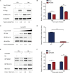
PGAM5 expression is regulated during PDCoV infection. (A) LLC-PK1 cells were subject to infection with PDCoV (MOI of 1) for 14 h and 16 h and then collected for western blot analysis. (B) The samples from the experiment described in (A) were used for qRT-PCR detection. (C AND D) LLC-PK1 cells were subject to infection with varying MOI of PDCoV for 14 h and later collected to analyze the expression of PGAM5. The data represent mean ± SD values from triplicate experiments. *, P < 0.05; **, P < 0.01; ***, P < 0.001 (two-tailed Student’s t-test).
Fig 2
Fig 2
PGAM5 inhibits PDCoV replication. (A) The PGAM5 plasmid was overexpressed in LLC-PK1 cells. When the cells reached the appropriate density, they were infected with 0.01 MOI of PDCoV. Protein samples were gathered at 12 and 14 h for western blot analysis. (B) The mRNA level of PDCoV N was determined through qRT-PCR using the samples described in (A). (C) LLC-PK1 cells were transfected with gradient concentrations of the PGAM5 plasmids and subsequently infected with 0.01 MOI of PDCoV. Protein samples were gathered for performing western blot analysis. (D) The interference efficiency of PGAM5 siRNA was identified by qRT-PCR. (E AND F) LLC-PK1 cells were transfected with PGAM5 siRNA and negative control and later infected with 0.01 MOI of PDCoV. We gathered cells and supernatants at 12 and 14 h after virus infection.
Fig 3
Fig 3
PGAM5 interacts with the N protein and degrades it via autophagy. (A) HEK 293T cells were co-transfected with PGAM5-Flag and PDCoV N-HA plasmids, and cell samples were collected for co-IP analysis. (B) Overexpression of the PDCoV N-Flag plasmid was carried out in HEK 293T cells. The interaction between endogenous PGAM5 and the N protein was verified through co-IP assay. (C) PGAM5 and PDCoV N were cloned into Pcold-TF and Pcold-GST vectors, respectively, and transformed into BL21(DE3) cells. The relationship between PGAM5 and the N protein was assessed with the use of the GST pull-down kit. (D) HeLa cells were transfected with PGAM5 and PDCoV N plasmids, followed by incubation with specific antibodies. Confocal immunofluorescence microscopy was adopted for visualizing the results. (E) HEK 293T cells were transfected with PGAM5 and PDCoV N plasmids, and protein samples were collected for western blot analysis. (F) Following co-transfection with PGAM5 and PDCoV N plasmids, the cells were exposed to the treatment with autophagy inhibitors BafA1, CQ, and 3-MA and the mitochondrial autophagy inhibitor Mdivi-1, as well as the ubiquitination inhibitor MG132.
Fig 4
Fig 4
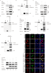
PGAM5 degrades PDCoV N by interacting with P62 and STUB1. (A) HEK 293T cells were co-transfected with PGAM5-HA and P62-Flag plasmids. Using western blot assay, we explored the interaction between PGAM5 and P62. (B) The GST pull-down assay proved that PGAM5 and P62 interact directly. (C) HEK 293T cells were subject to co-transfection with the PDCoV N plasmid and P62, and the interaction between N and P62 was investigated using western blot assay. (D) The direct interaction between PDCoV N and P62 was analyzed through the GST pull-down assay. (E AND F) The interaction between PGAM5 and STUB1 by co-IP and GST pull-down assay. (G) The direct relationship between PDCoV N and STUB1 was analyzed through the GST pull-down assay. (H) HeLa cells were transfected with PGAM5, PDCoV N, STUB1, and P62 followed by incubation with specific antibodies and staining of the nucleus with DAPI. To obtain the results, we used a confocal immunofluorescence microscope. (I) HEK 293T cells were transfected with PDCoV N, PGAM5, and P62 siRNA. Western blot analysis was adopted for assessing the abundance of the N protein.
Fig 5
Fig 5
PGAM5 up-regulates IFN via MyD88-TRAF3-IRF7. (A AND B) HEK 293T cells were transfected with IFN-β or ISRE luciferase reporter plasmids together with the elevating amounts of PGAM5-Flag plasmids, and the dual luciferase activity was measured. (C) HEK 293T cells were co-transfected with PGAM5, IFN-β luciferase reporter, and plasmids encoding various signaling molecules, including RIG-I, MDA5, MAVS, MyD88, TRAF3, TRAF6, TBK1, IKK, and IRF3, to examine the dual luciferase activity. (D) Immunofluorescence assay was made to explore the co-localization of PGAM5, MyD88, and TRAF3 through laser confocal experiments. (E AND F) The interaction between PGAM5 and MyD88 was investigated through Co-IP and GST pull-down assays. (G AND H) Co-IP and GST pull-down assays were adopted for analyzing the interaction between PGAM5 and TRAF3. (I) The mRNA levels of interferon-related molecules were identified using qRT-PCR after overexpression of PGAM5. (J) Western blot analysis was performed with the aim of determining the expression of IRF7 in HEK 293T cells overexpressing MyD88 and TRAF3. (K) Western blot analysis was conducted to assess the protein levels of IRF7 and phosphorylated IRF7 after overexpression of PGAM5. (L) HEK 293T cells were co-transfected with IFN-β luciferase reporter, PGAM5 plasmid, and P62 siRNA. The dual luciferase activity was measured.
Fig 6
Fig 6
PDCoV N degrades PGAM5 through autophagy. (A AND B) HEK 293T or LLC-PK1 cells were co-transfected with the elevating amounts of N-HA plasmid, and the expression of PGAM5 was explored with the use of western blot. (C AND D) HEK 293T cells or LLC-PK1 cells were transfected with PDCoV N plasmid and then treated with autophagy inhibitor, ubiquitination inhibitor, and mitochondrial autophagy inhibitor. The cells were subsequently explored using western blot.
Fig 7
Fig 7
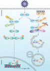
The antiviral mechanism of PGAM5 inhibits PDCoV replication. During PDCoV infection, PGAM5 interacts with the cargo receptor P62 and the E3 ubiquitinating ligase STUB1 to degrade N protein through the autophagy pathway. Meanwhile, PDCoV also utilized viral N protein to degrade host protein PGAM5 through autophagy process to facilitate virus replication. Additionally, PGAM5 activates type I IFN signaling through targeting MyD88 and TRAF3 to up-regulate IRF7 expression.

References

    1. Woo PC, Lau SK, Lam CS, Lau CC, Tsang AK, Lau JH, Bai R, Teng JL, Tsang CC, Wang M, Zheng BJ, Chan KH, Yuen KY. 2012. Discovery of seven novel mammalian and avian coronaviruses in the genus deltacoronavirus supports bat coronaviruses as the gene source of alphacoronavirus and betacoronavirus and avian coronaviruses as the gene source of gammacoronavirus and deltacoronavirus. J Virol 86:3995–4008. doi:10.1128/JVI.06540-11 - DOI - PMC - PubMed
    1. Li G, Chen Q, Harmon KM, Yoon KJ, Schwartz KJ, Hoogland MJ, Gauger PC, Main RG, Zhang J. 2014. Full-length genome sequence of porcine deltacoronavirus strain USA/IA/2014/8734. Genome Announc 2:e00278-14. doi:10.1128/genomeA.00278-14 - DOI - PMC - PubMed
    1. Lee S, Lee C. 2014. Complete genome characterization of Korean porcine deltacoronavirus strain KOR/Knu14-04/2014. Genome Announc 2. doi:10.1128/genomeA.01191-14 - DOI - PMC - PubMed
    1. Suzuki T, Shibahara T, Imai N, Yamamoto T, Ohashi S. 2018. Genetic characterization and pathogenicity of Japanese porcine deltacoronavirus. Infect Genet Evol 61:176–182. doi:10.1016/j.meegid.2018.03.030 - DOI - PMC - PubMed
    1. Madapong A, Saeng-Chuto K, Lorsirigool A, Temeeyasen G, Srijangwad A, Tripipat T, Wegner M, Nilubol D. 2016. Complete genome sequence of porcine deltacoronavirus isolated in Thailand in 2015. Genome Announc 4:e00408-16. doi:10.1128/genomeA.00408-16 - DOI - PMC - PubMed

Publication types

MeSH terms