. 2024 Apr 15;30(8):1567-1581.
doi: 10.1158/1078-0432.CCR-23-1079.

A B7-H4-Targeting Antibody-Drug Conjugate Shows Antitumor Activity in PARPi and Platinum-Resistant Cancers with B7-H4 Expression

Affiliations

A B7-H4-Targeting Antibody-Drug Conjugate Shows Antitumor Activity in PARPi and Platinum-Resistant Cancers with B7-H4 Expression

Sarah B Gitto et al. Clin Cancer Res. .

Abstract

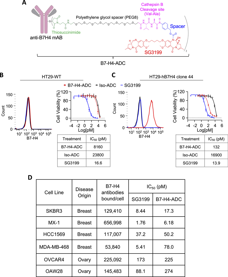
Purpose: Platinum and PARP inhibitors (PARPi) demonstrate activity in breast and ovarian cancers, but drug resistance ultimately emerges. Here, we examine B7-H4 expression in primary and recurrent high-grade serous ovarian carcinoma (HGSOC) and the activity of a B7-H4-directed antibody-drug conjugate (B7-H4-ADC), using a pyrrolobenzodiazepine-dimer payload, in PARPi- and platinum-resistant HGSOC patient-derived xenograft (PDX) models.

Experimental design: B7-H4 expression was quantified by flow cytometry and IHC. B7-H4-ADC efficacy was tested against multiple cell lines in vitro and PDX in vivo. The effect of B7-H4-ADC on cell cycle, DNA damage, and apoptosis was measured using flow cytometry.

Results: B7-H4 is overexpressed in 92% of HGSOC tumors at diagnosis (n = 12), persisted in recurrent matched samples after platinum treatment, and was expressed at similar levels across metastatic sites after acquired multi-drug resistance (n = 4). Treatment with B7-H4-ADC resulted in target-specific growth inhibition of multiple ovarian and breast cancer cell lines. In platinum- or PARPi-resistant ovarian cancer cells, B7-H4-ADC significantly decreased viability and colony formation while increasing cell-cycle arrest and DNA damage, ultimately leading to apoptosis. Single-dose B7-H4-ADC led to tumor regression in 65.5% of breast and ovarian PDX models (n = 29), with reduced activity in B7-H4 low or negative models. In PARPi and platinum-resistant HGSOC PDX models, scheduled B7-H4-ADC dosing led to sustained tumor regression and increased survival.

Conclusions: These data support B7-H4 as an attractive ADC target for treatment of drug-resistant HGSOC and provide evidence for activity of an ADC with a DNA-damaging payload in this population. See related commentary by Veneziani et al., p. 1434.


Conflict of interest statement

Competing interests: F.S. serves on a scientific advisory board and has received funding for clinical trials from AstraZeneca. E.J.B. serves on the scientific advisory board of Atrin Pharmaceuticals and has been an advisor for Sierra Oncology. D.J.P serves on the advisory board for AstraZeneca and holds patents on isolated B7-H4 specific compositions and methods of use thereof. G.D., K.K., A.L., S.M., J.A., M.A., G.R., S.C., and R.W. are employees of AstraZeneca or were employees of AstraZeneca while they were involved with this work and have stock and/or stock interests in AstraZeneca. The remaining authors declare no potential conflicts of interest.

Figures

Figure 1:
Figure 1:. B7-H4 is expressed in heavily pretreated PARPi- and platinum-resistant ovarian cancer.
A, Quantitative IHC performed on patient-matched samples collected pre- and post- chemotherapy (n = 12). Two additional samples were also collected after neoadjuvant therapy. Staining was analyzed for proportion score (0 to 5+). Representative IHC for pre- and post-treatment samples where B7-H4 expression increased (red), remained consistent (orange), or decreased (blue). B, Quantitative IHC performed on rapid autopsy samples collected from primary tumor and distant metastatic sites (n = 4 donors). Staining was analyzed for proportion score (0 to 5+) and B7-H4 intensity (0 to 3+). Cellular localization was also assessed; m = cell membrane B7-H4 expression only, c = cell cytoplasm B7-H4 expression only, m>c = membrane and cytoplasm B7-H4 expression, where membrane expression is greater.
Figure 2:
Figure 2:. A B7-H4 targeting PBD ADC that specifically binds to and kills cells expressing B7-H4.
A, Schematic of B7-H4-ADC. B and C, Flow cytometric analysis of B7-H4 expression on (B) HT29-WT or (C) HT29-hB7H4 cells stably overexpressing human B7-H4. Cells were incubated with staining buffer alone, AF647-labelled anti-B7-H4 or isotype control antibody HT29-WT or HT29-hB7H4 cells were incubated with increasing concentrations of B7-H4-ADC, an isotype control ADC (Iso-ADC) or free warhead (SG3199) for 6 days. The cell viability was measured by a CellTiter-Glo assay, and the IC50 values were calculated. Data are presented as the mean ± SEM. D, Anti-B7-H4 antibodies bound/cell (quantitative flow cytometry) and B7-H4-ADC or SG3199 cytotoxicity (CellTiter-Glo assay) was measured in in breast and ovarian cancer cell lines.
Figure 3:
Figure 3:. B7-H4-ADC has bystander killing activity.
A, Cytotoxicity induced on HT29-WT (B7-H4-), HT29-hB7H4 (B7-H4+), and co-cultures with 100 ng/mL of B7-H4-ADC or Iso-ADC. Viability was measured after six days of treatment with MTT assay. Blue lines indicate expected cell death according to the ratio of HT29-WT cells in the co-cultures. B, HT29 B7-H4+ tumor cells were treated with B7-H4-ADC or Iso-ADC for 6 days. The cell viability was measured by a CellTiter-Glo assay (left). Conditioned media was collected and transferred to either B7-H4+ or B7-H4- HT29 cells (middle and right, respectively). Cells were cultured for 6 days and viability was measured. Cells were also treated with B7-H4-ADC representing a benchmark for activity. Data are presented as the mean ± SD. C, Mice were inoculated with either B7-H4+, B7-H4- HT29, or a 1:1 ratio of both lines. Fourteen days post inoculation (dotted lines), and when tumors reached 200 mm3, mice were treated with a single intravenous dose of B7-H4-ADC (1 mg/kg), Iso-ADC (1 mg/kg), or vehicle control. Values are mean ± SEM tumor volumes for n = 6–8 animals per group. Data for groups treated with the B7-H4-ADC are plotted together (bottom right). The mixed B7-H4 +/− mice have similar tumor reduction as the B7-H4+ HT29 barring mice, with only two time points reaching significant differences (2-way ANOVA).
Figure 4:
Figure 4:. B7-H4-ADC demonstrates activity in a cell line-derived xenograft and PDX tumors representing a range of B7-H4 expression.
(A) MX-1, (B) HCC1569, MDA-MB-468, (C) MDA-MB-468, or (D) OVCAR4 xenografts were grown in SCID mice and treated with a single intravenous dose of either vehicle control, B7-H4-ADC, or Iso-ADC as indicated. Values are mean ± SEM tumor volumes for n = 5–8 animals per group. The dotted line denotes the day of dosing. E, Breast and ovarian cancer PDXs were grown from fragments in athymic nude mice. Digital image analysis of IHC staining for B7-H4 expression in PDX tumors. Values are the percentage of tumor cells with positive membrane staining of B7-H4 (IHC intensity score 0 to 3+), n = 3–6 animals per group. F, When tumor size reached 200 mm3, mice were treated with a single intravenous dose of either vehicle control or 0.3 mg/kg B7-H4-ADC. For each model, percentage tumor growth of ADC-treated mice was compared to vehicle control treated mice. Models with mutations in BRCA1 (orange) and BRCA2 (gold) are labeled (data from whole exome sequencing, provided by the vendor). G, Box and whisker plots of H-score of responders (percent tumor growth reduced) and non-responders (percent tumor growth increased), two-tailed Mann-Whiney test.
Figure 5:
Figure 5:. B7-H4-ADC results in DNA damage and cell cycle arrest in PARPi resistant and CCNE1 amplified HGSOC cells.
A, Cell surface expression of B7-H4 by flow cytometry. B, B7-H4 receptor density was calculated using quantitative flow cytometry and B7-H4-ADC (ADC) or Iso-ADC viability (CellTiter-Glo assay) was measured in indicated cell lines. Cell lines were incubated with increasing concentrations (4 μg/mL–16 pg/mL) of B7-H4-ADC or Iso-ADC for 6 days. The cell viability was expressed as a percentage of mock-treated control cells and then the IC50 values were calculated. C, Representative images of cell colonies and their quantification show the effects of B7-H4-ADC, Iso-ADC, olaparib (PARPi) and carboplatin on colony formation after 11–14 days of treatment at indicated doses. D-F, Cell lines were plated at 2 × 10⁵ cells in 6-well dishes and allowed to adhere overnight, then incubated with PARPi (1μM), Iso-ADC (10 ng/mL and 100 ng/mL), and B7-H4-ADC (10 ng/mL and 100 ng/mL) for 48 hrs (JHOS4, JHOS4-PR and OVCAR3) and 96 hrs (OVKATE). D, BrdU flow cytometry analysis shows cell cycle phase distribution. E, Non-adherent and adherent cells were collected, counted, then fixed in 2% paraformaldehyde. 3 × 10⁵ cells were stained and analyzed for PI and γH2AX staining by flow cytometry. Single cells were analyzed for their expression of γH2AX in G0/G1, S and G2/M phase. F, The apoptotic cells were analyzed by Annexin V PI staining after six days of treatment. All data is represented as mean ± SD, n = 3. One-way ANOVA with Tukey’s multiple comparisons test was used to calculate the p-values. *P<0.05, **P<0.01, ***P<0.001, ****P<0.0001, ns = not significant.
Figure 6:
Figure 6:. B7-H4-ADC reduces tumor burden in vivo in PARPi and platinum resistant HGSOC models.
A, Parent tumor compared to PDX PARPi naïve, and PARPi-R PDX models stained for B7-H4 protein by IHC. B7-H4 proportion score of matched primary tumors and PARPi-resistant PDX models. B-D, Ovarian cancer patient-derived xenograft tissue fragments were transplanted onto the ovaries of NSG mice. When tumor volume reached 50 mm3 mice were dosed with MTD Olaparib (75 mg/kg) D1–6 until tumors reached an average volume of 100 mm3. Mice were then randomized to groups and administered vehicle control, PARPi (75 mg/kg, D 1–6), Carbo (0.3 mg/kg, weekly), Iso-ADC (0.3 or 0.1 mg/kg) or B7-H4-ADC (0.3 or 0.1 mg/kg). ADCs were dosed IP every 28 days. Tumor volumes and mouse weights were measured weekly. Values are represented as mean ± SEM for n = 3–8 animals per group.

References

    1. Ozols RF, Bundy BN, Greer BE, Fowler JM, Clarke-Pearson D, Burger RA, et al. Phase III trial of carboplatin and paclitaxel compared with cisplatin and paclitaxel in patients with optimally resected stage III ovarian cancer: a Gynecologic Oncology Group study. J Clin Oncol 2003;21:3194–200 - PubMed
    1. Bookman MA, Brady MF, McGuire WP, Harper PG, Alberts DS, Friedlander M, et al. Evaluation of new platinum-based treatment regimens in advanced-stage ovarian cancer: a Phase III Trial of the Gynecologic Cancer Intergroup. J Clin Oncol 2009;27:1419–25 - PMC - PubMed
    1. Ledermann JA, Pujade-Lauraine E. Olaparib as maintenance treatment for patients with platinum-sensitive relapsed ovarian cancer. Ther Adv Med Oncol 2019;11:1758835919849753 - PMC - PubMed
    1. Rodriguez-Garcia A, Minutolo NG, Robinson JM, Powell DJ. T-cell target antigens across major gynecologic cancers. Gynecol Oncol 2017;145:426–35 - PubMed
    1. Stewart D, Cristea M. Antibody-drug conjugates for ovarian cancer: current clinical development. Curr Opin Obstet Gynecol 2019;31:18–23 - PubMed

MeSH terms

Substances