. 2023 Nov 15;150(22):dev201709.
doi: 10.1242/dev.201709. Epub 2023 Nov 23.

Cell-type-specific interacting proteins collaborate to regulate the timing of Cyclin B protein expression in male meiotic prophase

Affiliations

Cell-type-specific interacting proteins collaborate to regulate the timing of Cyclin B protein expression in male meiotic prophase

Catherine C Baker et al. Development. .

Abstract

During meiosis, germ cell and stage-specific components impose additional layers of regulation on the core cell cycle machinery to set up an extended G2 period termed meiotic prophase. In Drosophila males, meiotic prophase lasts 3.5 days, during which spermatocytes upregulate over 1800 genes and grow 25-fold. Previous work has shown that the cell cycle regulator Cyclin B (CycB) is subject to translational repression in immature spermatocytes, mediated by the RNA-binding protein Rbp4 and its partner Fest. Here, we show that the spermatocyte-specific protein Lut is required for translational repression of cycB in an 8-h window just before spermatocytes are fully mature. In males mutant for rbp4 or lut, spermatocytes enter and exit meiotic division 6-8 h earlier than in wild type. In addition, spermatocyte-specific isoforms of Syncrip (Syp) are required for expression of CycB protein in mature spermatocytes and normal entry into the meiotic divisions. Lut and Syp interact with Fest independent of RNA. Thus, a set of spermatocyte-specific regulators choreograph the timing of expression of CycB protein during male meiotic prophase.

Keywords: Drosophila; Cyclin B; Meiosis; RNA; Spermatogenesis; Translation.


Conflict of interest statement

Competing interests The authors declare no competing or financial interests.

Figures
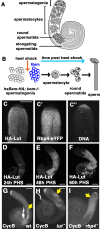
Fig. 1.
Fig. 1.
Lutin is expressed early in spermatocyte development and is required for repression of cycB translation in still-immature spermatocytes. (A) Illustration of testis with major germline cell types labeled. (B) Schematic of the hs-Bam differentiation time-course. (C-C″) Immunofluorescence image of the apical tip of a testis from a male carrying HA-Lut and Rbp4-eYFP transgenes, stained with anti-HA (C), anti-GFP (C′) and DAPI (C″). (D-F) Anti-HA staining of apical tips of testes from flies expressing HA-Lut in the hs-Bam time-course collected at 24 h post-heat-shock (PHS) (D), 48 h PHS (E) and 60 h PHS (F). (G-I) Anti-CycB staining of wild-type (wt) (G), lut (H) and rbp4 (I) mutant testes. Yellow arrows indicate onset of CycB protein expression in spermatocytes. Scale bars: 100 µm.
Fig. 2.
Fig. 2.
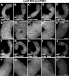
Lutin interacts with Fest and with Rbp4 dependent on Fest. (A) Western blots probed with anti-GFP or anti-HA showing proteins immunoprecipitated with anti-GFP from testis extracts from flies expressing HA-Lut and either Rbp4-eYFP (lanes 2-5) or eYFP-Fest (lanes 6-9). Negative control: HA-Lut alone (lane 1). RNAse A or RNAse inhibitor was added as indicated. Flies used in lanes 4-5 were fest mutants. Flies used in lanes 8-9 were rbp4 mutants. (B,C) Testes from the hs-Bam time-course immunostained with anti-CycB. (B) 72 h post-heat-shock (PHS). Segmented line: region of the testis containing spermatocytes. (C) 104 h PHS. Segmented line: CycB-positive spermatocytes. (D,E) smFISH with cycB probes on 72 h PHS (D) and 104 h PHS testes (E). (F) Western blots probed with anti-GFP or anti-HA of proteins immunoprecipitated with anti-GFP from testis extracts of flies expressing eYFP-Fest and Rbp4-HA. Samples were dissected at 72 h or 104 h PHS as indicated, and all samples had RNAse A added. Negative control: Rbp4-HA alone. (G) Western blots probed with anti-GFP or anti-HA of proteins immunoprecipitated with anti-GFP from testis extracts of flies expressing eYFP-Fest and HA-Lut. Samples were dissected at 72 h and 104 h PHS, and all samples had RNAse A added. Negative control: HA-Lut alone. Scale bars: 100 µm.
Fig. 3.
Fig. 3.
Ectopic expression of CycB occurs later in lut mutants than it does in rbp4 mutants. (A-L) Testes from the hs-Bam time-course stained with anti-CycB. (A-D) wild type (E-H) lut (I-L) rbp4. Segmented lines: CycB-positive spermatocytes. PHS, post-heat-shock. Scale bars: 100 µm.
Fig. 4.
Fig. 4.
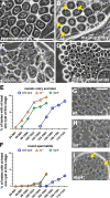
lut and rbp4 mutant spermatocytes enter and exit the meiotic divisions earlier than in wild type. (A-D) Phase-contrast imaging of unfixed wild-type (wt) testis squashes. (A) Mature spermatocytes. (B) Spermatocytes initiating meiotic entry, showing rounded nuclei (arrows) and crumbly nucleoli. (C) Cells in the first meiotic division. (D) Round spermatids, each with a small phase-light nucleus and phase-dark mitochondrial derivative. (E,F) Graphs showing percentage of testes from wt, lut mutants and rbp4 mutants with at least one cyst at meiotic entry or later (i.e. including stages shown in B-D) (E) or at least one cyst with round spermatids (F). N=100 for each timepoint/genotype combination. (G-I) Phase-contrast imaging of testis squashes with round spermatids from wild type (G), lut mutants (H) and rbp4 mutants (I). Arrows in I show larger-than-normal nebenkern, indicating at least one failed meiotic cytokinesis. Scale bars: 50 µm (A-D); 25 µm (G-I).
Fig. 5.
Fig. 5.
Loss of function of isoforms of Syp upregulated in testis leads to loss of CycB expression in spermatocytes. (A) The syp genomic locus, showing four promoters and four possible C-terminal ends, based on the FlyBase annotation. Different colors of the C-terminal exons indicate differences in protein isoforms as in Fig. S2E, due to different C-terminal splice forms and/or shifts in reading frame in shared exons. The exons common to all syp transcripts are shown above. Asterisks indicate location of the two CRISPR-induced microdeletions that together make the sypdub allele. The complete syp-RD transcript is shown in full. (B-B″) Apical tip of a testis carrying eYFP-Syp-PD and Rbp4-HA transgenes, immunostained with anti-GFP (B) and anti-HA (B′). (B″) DAPI (DNA). (C) Western blots probed with anti-Syp (top) and anti-Tubulin (bottom) of lysate from wild-type (wt) testes, syp mutant testes, wild-type heads and syp mutant heads The syp mutant here and throughout the paper is sypdub/Df. (D-F) Phase-contrast images of wild-type testes (D), syp mutant testes (E) and testes from syp mutants carrying eYFP-Syp-PD (F). Arrows indicate elongating spermatids. (G-I) Anti-CycB on wild-type testes (G), syp mutant testes (H), and testes from syp mutants carrying eYFP-Syp-PD (I). (J-L) smFISH with probes against cycB on wild-type testes (J), syp mutant testes (K) and testes from syp mutants carrying eYFP-Syp-PD (L). (M) Quantification of smFISH signal in spermatocytes from the genotypes in J-L. Values represent the mean signal measurement for three testes per genotype. Brackets denote standard deviation. Wild type versus syp, *P=0.0190; wild type versus syp;eYFP-Syp-PD, P=0.9526 (ns, not significant) (Welch's t-test). (N) Diagram of numbered biotin probes tiled across the cycB coding sequence and 3′ UTR showing probes that brought down Syp (black) and probes that did not (gray). (O) Western blots probed with anti-Syp of proteins isolated by biotin pulldown from wild-type testis extract, with probe number indicated. (P) Western blot probed with anti-GFP of biotin pulldowns with probes 8 and 9, from testis extract from eYFP-Syp-expressing flies. (Q) Integrated Genomics Viewer snapshot of the cycB locus, with annotated transcript isoform, gene transcribed from right to left. Tracks from top to bottom: CLIP signal from two replicates of wild-type (no-epitope) controls [CLIP (no Epi)]; CLIP signal from two replicates eYFP-Syp [CLIP (Syp)]; wt RNA-seq signal; CAGE signal marking the transcription start site at 72 h post-heat-shock (PHS) (Lu et al., 2020); 3′-seq signal marking where the spermatocyte transcript ends at 72 h PHS (Berry et al., 2022). Note that the 3′ UTR of the transcript isoform expressed in spermatocytes is shorter than the annotated isoform, as indicated by the 3′ seq peak. Scale bars: 100 µm (B-B″,G-L); 200 µm (D-F).
Fig. 6.
Fig. 6.
cycB RNA levels are lower but detectable through 108 h PHS in syp mutants. (A-J) Testes from time points in the hs-Bam time-course probed for cycB RNA by smFISH. (A-E) Wild-type time-course testes. (A′-E′) Higher magnification of spermatocyte regions of A-E. (F-J) syp mutant time-course testes. (F′-J′) Higher magnification of spermatocyte regions of F-J. 100 h, 104 h, 108 h, 112 h and 116 h post-heat-shock (PHS) as indicated for both genotypes. Scale bars: 100 µm (A-J); 50 µm (A′-J′).
Fig. 7.
Fig. 7.
Spermatocytes mutant for syp arrest before prometaphase. (A-D′) Phase-Hoechst images of spermatocytes in squashed preparations of testes from the hs-Bam time-course on wild type (A-B′) and syp mutants (C-D′) at 72 h or 110 h post-heat-shock (PHS) as indicated. (A,B,C,D) Phase-contrast. (A′,B′,C′,D′) Hoechst (DNA). Arrows in A′, B′, C′ and D′ point roughly to the outer edge of the nucleus of interest. Arrows in A′ and C′ indicate nuclei with chunky, crescent-shaped chromosomes; arrows in B′ indicate nuclei with condensed chromosomes; arrows in D′ indicate nuclei with partially condensed chromosomes. Arrowheads in A, C and D indicate intact nucleoli; Arrowheads in B indicate broken-down nucleoli. Scale bar: 25 µm.
Fig. 8.
Fig. 8.
Syp interacts with Fest and co-immunoprecipitates with Rbp4 and Lut in the presence of Fest. (A) Western blots probed with anti-GFP or anti-HA of anti-GFP immunoprecipitates from testes expressing eYFP-Syp and HA-Fest, with either RNAse A or RNAse inhibitor, as indicated. Negative control: HA-Fest alone. Flies were either wild type (wt; first three lanes) or mutant for rbp4 (lanes 4 and 5). (B) Western blots probed with anti-GFP or anti-HA of anti-GFP immunoprecipitates from hs-Bam time-course testes expressing eYFP-Syp and HA-Fest, collected at 72 h and 104 h post-heat-shock (PHS), as indicated. All samples included RNAse A. Negative controls: HA-Fest only. (C) Western blots probed with anti-GFP or anti-HA of anti-GFP immunoprecipitations from testes expressing eYFP-Syp and Rbp4-HA, with either RNAse A or RNAse inhibitor, as indicated. Negative control: Rbp4-HA alone. Flies were wt (first three lanes) or mutant for fest (lanes 4 and 5). (D) Western blots probed with anti-GFP or anti-HA of anti-GFP immunoprecipitates from testes also expressing eYFP-Syp and HA-Lut. All samples included RNAse A. Flies were wt or mutant for fest, as indicated. Negative control: HA-Lut alone.
Fig. 9.
Fig. 9.
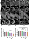
In the absence of Rbp4, Syp is not required for cycB translation in mid-stage spermatocytes. (A-F) Immunofluorescence images of whole testes stained with anti-CycB showing wild type (wt; A) and lut (B), rbp4 (C), syp (D), lut syp (E) and rbp4 syp (F) mutants. Yellow bar in F highlights a puff of CycB expression. (G-L) Whole testes probed for cycB RNA by smFISH showing wild type (wt; G) and lut (H), rbp4 (I), syp (J), lut syp (K) and rbp4 syp (L) mutants. (G′-L′) High magnification views of spermatocyte regions in (G-L). (G″-L″) The same testes probed for loopin-1 RNA by smFISH. (M,N) Quantification of cycB (M) and loopin-1 (N) smFISH signal in wt and lut, rbp4, syp, lut syp and rbp4 syp mutant spermatocyte regions. Values represent the mean signal measurement for three testes per genotype. Error bars denote standard deviation. P-values for mutant versus wild type shown where P<0.05 (Welch's t-test). Scale bars: 100 µm (A-L;G″-L″); 50 µm (G′-L′).
Fig. 10.
Fig. 10.
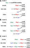
Three models for Lut, Syp and Rbp4 function in regulating cycB translation and stability. Solid lines indicate active promotion or repression; dotted lines indicate abrogated function. Not shown, but contributing to all three models: Syp and Rbp4 act in parallel to stabilize the cycB RNA. See Discussion.

Update of

References

    1. Alphey, L., Jimenez, J., White-Cooper, H., Dawson, I., Nurse, P. and Glover, D. M. (1992). twine, a cdc25 homolog that functions in the male and female germline of Drosophila. Cell 69, 977-988. 10.1016/0092-8674(92)90616-K - DOI - PubMed
    1. Arvola, R. M., Chang, C.-T., Buytendorp, J. P., Levdansky, Y., Valkov, E., Freddolino, P. L. and Goldstrohm, A. C. (2020). Unique repression domains of Pumilio utilize deadenylation and decapping factors to accelerate destruction of target mRNAs. Nucleic Acids Res. 48, 1843-1871. 10.1093/nar/gkz1187 - DOI - PMC - PubMed
    1. Baker, C. C. and Fuller, M. T. (2007). Translational control of meiotic cell cycle progression and spermatid differentiation in male germ cells by a novel eIF4G homolog. Development 134, 2863-2869. 10.1242/dev.003764 - DOI - PMC - PubMed
    1. Baker, C. C., Gim, B. S. and Fuller, M. T. (2015). Cell type-specific translational repression of Cyclin B during meiosis in males. Development 142, 3394-3402. 10.1242/dev.122341 - DOI - PMC - PubMed
    1. Bassett, A. R., Tibbit, C., Ponting, C. P. and Liu, J. L. (2013). Highly efficient targeted mutagenesis of Drosophila with the CRISPR/Cas9 system. Cell Rep. 4, 220-228. 10.1016/j.celrep.2013.06.020 - DOI - PMC - PubMed

Publication types