. 2023 Oct 26;21(10):e3002341.
doi: 10.1371/journal.pbio.3002341. eCollection 2023 Oct.

Mammalian cells internalize bacteriophages and use them as a resource to enhance cellular growth and survival

Affiliations

Mammalian cells internalize bacteriophages and use them as a resource to enhance cellular growth and survival

Marion C Bichet et al. PLoS Biol. .

Abstract

There is a growing appreciation that the direct interaction between bacteriophages and the mammalian host can facilitate diverse and unexplored symbioses. Yet the impact these bacteriophages may have on mammalian cellular and immunological processes is poorly understood. Here, we applied highly purified phage T4, free from bacterial by-products and endotoxins to mammalian cells and analyzed the cellular responses using luciferase reporter and antibody microarray assays. Phage preparations were applied in vitro to either A549 lung epithelial cells, MDCK-I kidney cells, or primary mouse bone marrow derived macrophages with the phage-free supernatant serving as a comparative control. Highly purified T4 phages were rapidly internalized by mammalian cells and accumulated within macropinosomes but did not activate the inflammatory DNA response TLR9 or cGAS-STING pathways. Following 8 hours of incubation with T4 phage, whole cell lysates were analyzed via antibody microarray that detected expression and phosphorylation levels of human signaling proteins. T4 phage application led to the activation of AKT-dependent pathways, resulting in an increase in cell metabolism, survival, and actin reorganization, the last being critical for macropinocytosis and potentially regulating a positive feedback loop to drive further phage internalization. T4 phages additionally down-regulated CDK1 and its downstream effectors, leading to an inhibition of cell cycle progression and an increase in cellular growth through a prolonged G1 phase. These interactions demonstrate that highly purified T4 phages do not activate DNA-mediated inflammatory pathways but do trigger protein phosphorylation cascades that promote cellular growth and survival. We conclude that mammalian cells are internalizing bacteriophages as a resource to promote cellular growth and metabolism.


Conflict of interest statement

JJB has a patent application related to this work (WO2018129536A1).

Figures

Fig 1
Fig 1. Uptake of T4 phage by mammalian cells does not trigger a pro-inflammatory immune response.
(A) A549 cells and (B) MDCK-I cells incubated with T4 phages for 2 hours. Images were taken with a confocal microscope; the plasma membrane is shown in magenta, T4 phage DNA in green, and the cell nucleus in blue. (C) A549 cells transfected with NF-κB-dependent luciferase reporter plasmid, or (D) IFN-β promoter-dependent luciferase reporter plasmid, followed by 48 hours incubation with 109 T4 phages/mL or a Filter control. Differentiated WT (E) or STING KO (F) BMDM cells were incubated for 18 hours 107 T4 phages/mL, Filter control, Capsid-only or transfected with phage DNA using Lipofectamine 2000. Raw data can be found in S1 Data; each set of data follows the normality law; P values between the different groups were calculated from a one-way ANOVA with multiple comparisons, shown as stars (P < 0.0001 = ****; A: F (3, 12) = 31.06; B: F (3, 12) = 2.812; C: F (4, 25) = 5.7; D: F (4, 25) = 0.8181).
Fig 2
Fig 2. Network analysis of mammalian cells treated with T4 phages.
(A) Kinexus KAM-1325 antibody microarray with MDCK-I cells after 8 hours of incubation with T4 phages. (B) Kinexus KAM-2000 antibody microarray with A549 cells after 8 hours of incubation with T4 phages. Figures report major cellular pathways of the main up- and down-regulated leads from the network analysis. Boxes highlighted in red are proteins discussed in this manuscript. The color gradient and arrow width indicate the Log2 fold change values.
Fig 3
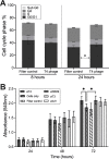
Fig 3. Phage application to in vitro mammalian cells leads to enhanced growth and proliferation.
(A) Cell cycle stage repartition within the A549 cell population after 8- or 24-hour incubation with phages or Filter control (data are mean with error bars representing 95% CI, n = 3 independent replicates with 100,000 cells analyzed). Not all the cells are included in a cell cycle phase (S4 and S5 Figs). P values of each cell cycle stage between the Filter control and T4 phage were calculated using a two-way ANOVA, shown as stars (F (3, 32) = 2.237). (B) Cell proliferation assay as measured via absorbance (540 nm) using a modified MTT colorimetric assay with A549 cells incubated with phages for 24, 48, or 72 hours (data are mean with error bars representing 95% CI, n = 3 independent replicates). Raw data can be found in S2 Data; P values were calculated using a two-way ANOVA, shown as stars (F (2, 224) = 1,015).
Fig 4
Fig 4. Overview of the effect of exogenous phages on cellular pathways.
(A) Innate immune pathways in BMDM and A549 cells. Phage DNA is protected by the phage capsid and is not detected by the TLR9 or cGAS-STING. (B) The effect of phages on MDCK-I and A549 cells after 8 hours. The AKT pathway on the left and the CDK1 pathway on the right show the major cellular changes detected in response to T4 phage.

References

    1. Barr JJ. A bacteriophages journey through the human body. Immunol Rev. 2017;279(1):106–122. doi: 10.1111/imr.12565 - DOI - PubMed
    1. Merril CR. Bacteriophage interactions with higher organisms. Trans N Y Acad Sci. 1974;36:265–272. doi: 10.1111/j.2164-0947.1974.tb01573.x - DOI - PubMed
    1. Dabrowska K, Switała-Jelen K, Opolski A, Weber-Dabrowska B, Gorski A. Bacteriophage penetration in vertebrates. J Appl Microbiol. 2005;98(1):7–13. doi: 10.1111/j.1365-2672.2004.02422.x - DOI - PubMed
    1. Shkoporov AN, Hill C. Bacteriophages of the Human Gut: The “Known Unknown” of the Microbiome. Cell Host Microbe. 2019;25:195–209. doi: 10.1016/j.chom.2019.01.017 - DOI - PubMed
    1. Clokie MR, Millard AD, Letarov A V, Heaphy S. Phages in nature. Bacteriophage. 2011. Jan;1(1):31–45. - PMC - PubMed