Review
. 2023 Nov 1;47(6):fuad060.
doi: 10.1093/femsre/fuad060.

Pseudomonas aeruginosa biofilm exopolysaccharides: assembly, function, and degradation

Affiliations
Review

Pseudomonas aeruginosa biofilm exopolysaccharides: assembly, function, and degradation

Andreea A Gheorghita et al. FEMS Microbiol Rev. .

Abstract

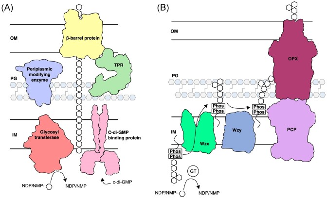
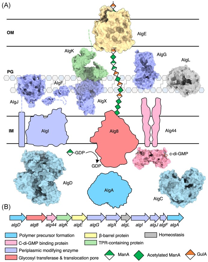
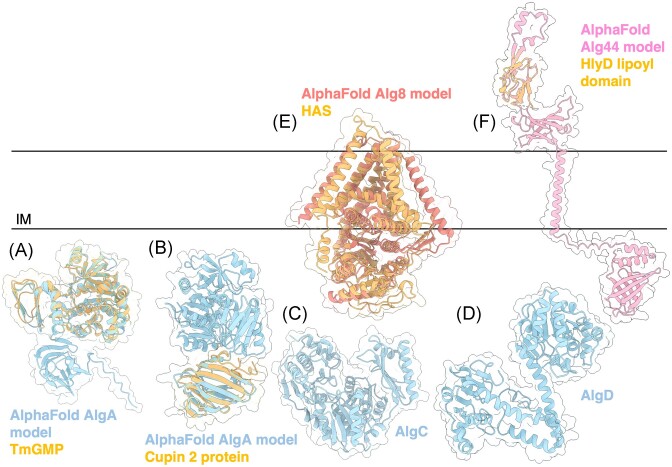
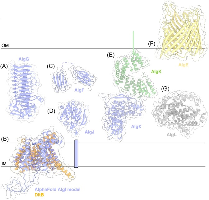
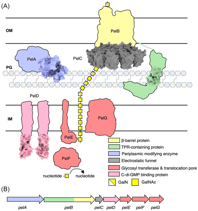
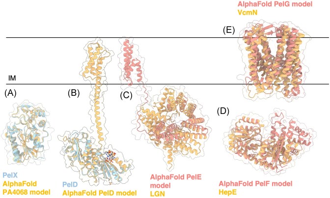
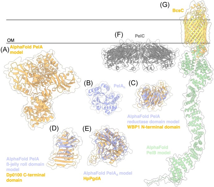
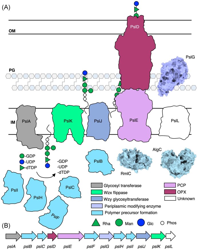
The biofilm matrix is a fortress; sheltering bacteria in a protective and nourishing barrier that allows for growth and adaptation to various surroundings. A variety of different components are found within the matrix including water, lipids, proteins, extracellular DNA, RNA, membrane vesicles, phages, and exopolysaccharides. As part of its biofilm matrix, Pseudomonas aeruginosa is genetically capable of producing three chemically distinct exopolysaccharides - alginate, Pel, and Psl - each of which has a distinct role in biofilm formation and immune evasion during infection. The polymers are produced by highly conserved mechanisms of secretion, involving many proteins that span both the inner and outer bacterial membranes. Experimentally determined structures, predictive modelling of proteins whose structures are yet to be solved, and structural homology comparisons give us insight into the molecular mechanisms of these secretion systems, from polymer synthesis to modification and export. Here, we review recent advances that enhance our understanding of P. aeruginosa multiprotein exopolysaccharide biosynthetic complexes, and how the glycoside hydrolases/lyases within these systems have been commandeered for antimicrobial applications.

Keywords: Pseudomonas aeruginosa; Pel; Psl; alginate; biofilm; biologics; exopolysaccharide; exopolysaccharide secretion system; glycoside hydrolases.


Conflict of interest statement

The authors declare no competing interests.

Figures

Figure 1.
Figure 1.
Synthase- and Wzx/Wzy-dependent secretion systems in Gram-negative bacteria. (A) In the synthase-dependent pathway, sugars are polymerized and translocated across the IM by a GT (salmon) in response to c-di-GMP binding a c-di-GMP receptor protein (pink). Once in the periplasm, the sugar polymer can be modified by periplasmic modifying enzymes (periwinkle). Transport across the OM is facilitated by a ß-barrel protein (yellow) and TPR-containing protein (green). (B) The Wzx/Wzy-dependent pathway, a sugar polymer repeat unit is assembled on a undecaprenyl-phosphate lipid carrier by GTs. A Wzx flippase (bright green) translocates the repeat unit across the IM where a Wzy glycosyltransferase (dark blue) assembles repeat units to form the polymer. The polymer is transported across the periplasm and OM by a PCP protein and an OPX protein. IM, inner membrane; PG, peptidoglycan; OM, outer membrane; NDP, nucleotide disphosphate; NMP, nucleotide monophosphate; and Phos, phosphate molecule.
Figure 2.
Figure 2.
Model of the alginate biosynthetic complex in P. aeruginosa. (A) Model of the alginate biosynthetic complex colour coded by function. Structures of AlgD (PDB: 1MFZ, 1MUU, 1MV8) (Snook et al. 2003), AlgC (PDB: 1K2Y, 1K35) (Regni et al. 2002), the cytoplasmic domain of Alg44 (PDB: 4RT0, 4RT1,) (Whitney et al. 2015), cytoplasmic domain of AlgJ (PDB: 4O8V) (Baker et al. 2014), AlgF (PDB: 6D10, 6CZT), AlgX (PDB: 4KNC) (Riley et al. 2013), AlgG (PDB: 4NK8, 4OZZ, 4OZY) (Wolfram et al. 2014), AlgL (PDB: 4OZV, 4OZW, 7SA8) (Gheorghita et al. 2022b), AlgK (PDB: 3E4B) (Keiski et al. 2010), and AlgE (PDB: 3RBH, 4AZL, 4B61, 4AFK) (Whitney et al. , Tan et al. 2014) have been experimentally determined and are drawn to scale and shown in a surface representation. (B) Alginate operon in P. aeruginosa colour coded by proposed function. IM, inner membrane; PG, peptidoglycan; and OM, outer membrane.
Figure 3.
Figure 3.
Structures and predicted AlphaFold models of P. aeruginosa alginate biosynthesis proteins. (A) Superimposition of P. aeruginosa AlgA AlphaFold model (light blue) and the crystal structure of T. maritima GMP TmGMP monomer (orange) (PDB: 2 × 65) with a Cα RMSD of 1.179 Å. (B) Superimposition of P. aeruginosa AlgA AlphaFold model (light blue) and the crystal structure of Shewanella frigidimarina Cupin 2 conserved barrel domain protein (orange) (PDB: 2PFW) with a Cα RMSD of 0.843 Å. (C) Crystal structure of P. aeruginosa AlgC (PDB: 1K2Y). (D) Crystal structure of a monomer of P. aeruginosa AlgD (PDB: 1MV8). (E) Superimposition of P. aeruginosa Alg8 AlphaFold model (salmon) with the crystal structure of the hyaluronan synthase (HAS) from Paramecium bursaria Chlorella virus CZ-2 (orange) (PDB: 7SP8) with a Cα RMSD of 2.932 Å. (F) Superimposition of a monomer of P. aeruginosa Alg44 AlphaFold model (pink) with the crystal structure of the lipoyl domain from the membrane fusion protein HlyD from E. coli (orange) (PDB: 5C22) with a Cα RMSD of 0.774 Å. IM; inner membrane.
Figure 4.
Figure 4.
Structures and predicted AlphaFold models of P. aeruginosa alginate modification and export proteins. (A) Crystal structure of P. syringae pv. Tomato AlgG (PDB: 4NK6). (B) Superimposition of P. aeruginosa AlgI AlphaFold model (periwinkle) and the crystal structure of Streptococcus thermophilus O-acyltransferase DltB (orange) (PDB: 6BUG) with a Cα RMSD of 1.788 Å. (C) CS-Rosetta determined structure of P. aeruginosa AlgF (N-terminus PDB: 6CZT; C-terminus: 6D10). (D) Crystal structure of P. aeruginosa AlgJ (PDB: 4O8V). (E) Crystal structure of P. aeruginosa AlgL (4OZV). (F) Crystal structure of the P. putida AlgKX complex (PDB: 7ULA). (G) Crystal structure of P. aeruginosa AlgE (PDB: 3RBH). (H) Crystal structure of P. aeruginosa AlgL (PDB: 4OZV). IM, inner membrane; OM, outer membrane.
Figure 5.
Figure 5.
Model of the Pel biosynthetic complex in P. aeruginosa. (A) Model of the Pel biosynthetic complex colour coded by function. Structures of the cytoplasmic domain of PelD (PDB: 4DN0) (Whitney et al. 2012), the hydrolase domain PelA (PDB: 5TCB), PelC (PDB: 5T11, 5T10, 5T0Z) (Marmont et al. 2017a), and the complete TPR motifs R9 and R10 of PelB (PDB: 5WFT) (Marmont et al. 2017b) have been experimentally determined and are drawn to scale and shown in a surface representation. (B) Pel operon in P. aeruginosa colour coded by function. IM, inner membrane; PG, peptidoglycan; and OM, outer membrane.
Figure 6.
Figure 6.
Structures and predicted AlphaFold models of P. aeruginosa Pel biosynthesis proteins. (A) Superimposition of P. aeruginosa PA4068 AlphaFold model (blue) with the crystal structure of PelX from P. protogens (orange) (PDB: 6WJB) with a Ca RMSD of 0.345 Å. (B) Crystal structure of a monomer of P. aeruginosa PelD (blue) bound to c-di-GMP (PDB: 4DN0) superimposed with the AlphaFold model of PelD (orange). (C) Superimposition of P. aeruginosa PelE AlphaFold model (salmon) with the crystal structure of LGN from Mus musculus (orange) (PDB: 4JHR) with a Ca RMSD of 4.023 Å. (D) Superimposition of P. aeruginosa PelF AlphaFold model (salmon) with the crystal structure of HepE from Anabaena sp. (orange) (PDB: 4XSO) with a Ca RMSD of 1.251 Å. (E) Superimposition of P. aeruginosa PelG AlphaFold model (salmon) with the crystal structure of VcmN from Vibrio cholerae (orange) (PDB: 6IDS) with a Ca RMSD of 5.077 Å. IM, inner membrane.
Figure 7.
Figure 7.
Structures and predicted AlphaFold models of P. aeruginosa Pel modification and export proteins. (A) AlphaFold model of P. aeruginosa PelA. (B) Crystal structure of the P. aeruginosa PelA hydrolase domain (PelAh) (PDB: 5TCB). (C) Superimposition of P. aeruginosa PelA reductase domain AlphaFold model (periwinkle) with the CryoEM structure of WBP1 from Saccharomyces cerevisiae (orange) (PDB: 6EZN) with a Cα RMSD of 4.650 Å. (D) Superimposition of P. aeruginosa PelA β-jelly roll AlphaFold model (periwinkle) with the crystal structure of the C-terminal domain of Dp0100 alginate lyase from Defluviitalea phaphyphila (orange) (PDB: 6JP4) with a Cα RMSD of 4.379 Å. (E) Superimposition of P. aeruginosa PelA deacetylase (PelAd) domain AlphaFold model (periwinkle) with the crystal structure of HpPgdA (PDB: 4LY4) peptidoglycan deacetylase from Helicobacter pylori (orange) with a Cα RMSD of 3.100 Å. F, Crystal structure of Paraburkholderia phytofirmans PelC (PDB: 5T10). G, Superimposition of P. aeruginosa PelB AlphaFold model (yellow and green) with the crystal structure of BcsC (orange) (PDB: 6TZK) from E. coli with a Cα RMSD of 3.119 Å. OM, outer membrane.
Figure 8.
Figure 8.
Model of the Psl biosynthetic complex in P. aeruginosa. (A) Model of the Psl biosynthetic complex colour coded by function. The structures of PslG (PDB: 5BXA, 5BX9) (Baker et al. 2015), AlgC (PDB: 1K2Y, 1K35) (Regni et al. 2002), and RmlC (PDB: 2IXJ, 2IXK, 2IXH, 2IXI) (Dong et al. 2007) have been experimentally determined and are drawn to scale and shown in surface representations. (B) Psl operon in P. aeruginosa colour coded by proposed function. IM, inner membrane; PG, peptidoglycan; and OM, outer membrane.
Figure 9.
Figure 9.
Structures and predicted AlphaFold models of P. aeruginosa enzymes involved in Psl precursor formation. (A) Superimposition of the P. aeruginosa PslB AlphaFold model (blue) with the crystal structure of T. maritima GMP TmGMP monomer (orange) (PDB: 2 × 65) with a Cα RMSD of 1.403 Å and the crystal structure of S. frigidimarina Cupin 2 conserved barrel domain protein (dark orange) (PDB: 2PFW) with a Cα RMSD of 0.801 Å. (B) Crystal structure of P. aeruginosa AlgC (PDB: 2FKF). (C) P. aeruginosa GalU AlphaFold model. (D) Crystal structure of a monomer of P. aeruginosa RmlC (PDB: 2IXH).
Figure 10.
Figure 10.
Structures and predicted AlphaFold models of P. aeruginosa Psl biosynthesis proteins. (A) Superimposition of the P. aeruginosa PslF AlphaFold model (blue) with crystal structure of TeSPS from T. elongatus (orange) (PDB: 6KIH) with a Cα RMSD of 2.54 Å. (B) Superimposition of the P. aeruginosa PslF (blue), PslH (periwinkle), and PslI (dark blue) AlphaFold models. Superimposition of PslH and PslI with PslF generates a Cα RMSD of 3.618 Å and 3.290 Å, respectively. (C) Superimposition of the P. aeruginosa PslC AlphaFold model (blue) with the cryoEM structure of HAS from P. bursaria Chlorella virus CZ-2 (orange) (PDB: 7SP8) with Cα RMSD of 2.836 Å. IM, inner membrane.
Figure 11.
Figure 11.
Structures and predicted AlphaFold models of P. aeruginosa Psl biosynthesis proteins, continued. (A) P. aeruginosa PslA AlphaFold model. (B) Superimposition of the P. aeruginosa PslA AlphaFold model (grey) with the crystal structure of the N-terminal domain of UDP-d-quinovosamine 4-dehydrogenase from V. fischeri (orange) (PDB: 3NKL) with a CαRMSD of 1.337 Å. (C) Superimposition of the C-terminus of P. aeruginosa PslA AlphaFold model (grey) with the crystal structure of the phosphoglycosyl transferase PglC from C. concisus (orange) (PDB: 5W7L) with a Cα RMSD of 1.603 Å. IM, inner membrane. (D) Superimposition of the P. aeruginosa PslJ AlphaFold model (navy) with the cryoEM structure of O-antigen ligase WaaL from Cupriavidus metallidurans (orange) (PDB: 7TPG) with a Cα RMSD of 4.42 Å. (D) Superimposition of the P. aeruginosa PslK AlphaFold model (green) with the crystal structure of the lipid II flippase MurJ from Thermosipho africanus (orange) (PDB: 6NC6) with a Cα RMSD of 2.04 Å. (F) AlphaFold model of P. aeruginosa PslL. (G) Crystal structure of P. aeruginosa PslG (PDB: 5BXA). IM, inner membrane.
Figure 12.
Figure 12.
Structures and predicted AlphaFold models of P. aeruginosa Psl export proteins. (A) Superimposition of P. aeruginosa PslE AlphaFold model (purple) with the tyrosine-protein kinase Wzc from E. coli (orange) (PDB: 7NII) with a Cα RMSD of 5.40 Å. (B) Superimposition of P. aeruginosa PslD AlphaFold model (burgundy) with the crystal structure of Wza from E. coli (orange) (PDB: 2J58) with a Cα RMSD of 2.81 Å. IM, inner membrane; OM, outer membrane.
None
None
None

References

    1. Acheson JF, Derewenda ZS, Zimmer J. Architecture of the cellulose synthase outer membrane channel and its association with the periplasmic TPR domain. Structure. 2019;27:1855–61.e3. - PMC - PubMed
    1. Adams JL, McLean RJ. Impact of rpoS deletion on Escherichia coli biofilms. Appl Environ Microbiol. 1999;65:4285–87. - PMC - PubMed
    1. Alipour M, Suntres ZE, Omri A. Importance of DNase and alginate lyase for enhancing free and liposome encapsulated aminoglycoside activity against Pseudomonas aeruginosa. J Antimicrob Chemother. 2009;64:317–25. - PubMed
    1. Alkawash MA, Soothill JS, Schiller NL. Alginate lyase enhances antibiotic killing of mucoid Pseudomonas aeruginosa in biofilms. Apmis. 2006;114:131–8. - PubMed
    1. Armbruster CR, Wolter DJ, Mishra Met al. . Staphylococcus aureus protein A mediates interspecies interactions at the cell surface of Pseudomonas aeruginosa. mBio. 2016;7:1–9. - PMC - PubMed

Publication types

MeSH terms

Substances