. 2023 Oct 26;11(1):57.
doi: 10.1038/s41413-023-00295-4.

Genetic interactions between polycystin-1 and Wwtr1 in osteoblasts define a novel mechanosensing mechanism regulating bone formation in mice

Affiliations

Genetic interactions between polycystin-1 and Wwtr1 in osteoblasts define a novel mechanosensing mechanism regulating bone formation in mice

Zhousheng Xiao et al. Bone Res. .

Abstract

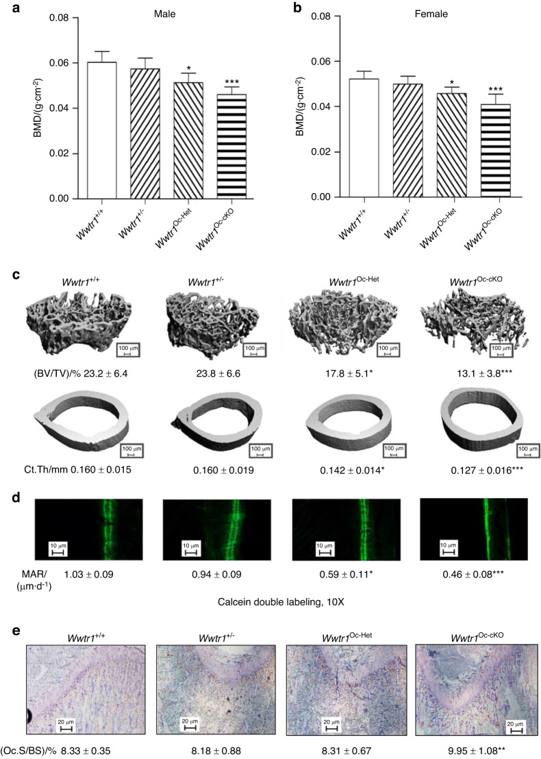
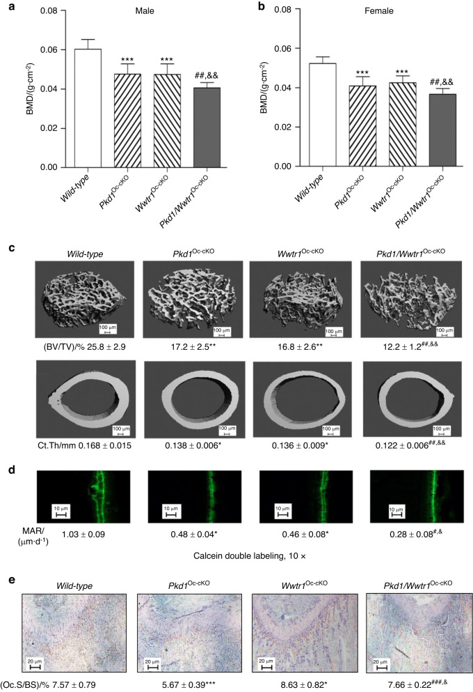
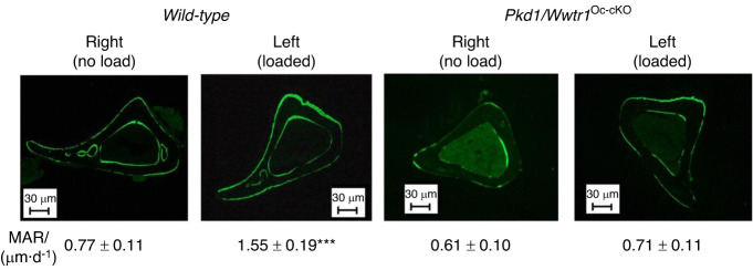
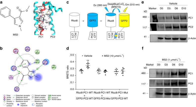
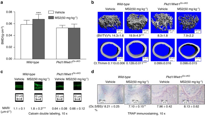
Molecular mechanisms transducing physical forces in the bone microenvironment to regulate bone mass are poorly understood. Here, we used mouse genetics, mechanical loading, and pharmacological approaches to test the possibility that polycystin-1 and Wwtr1 have interdependent mechanosensing functions in osteoblasts. We created and compared the skeletal phenotypes of control Pkd1flox/+;Wwtr1flox/+, Pkd1Oc-cKO, Wwtr1Oc-cKO, and Pkd1/Wwtr1Oc-cKO mice to investigate genetic interactions. Consistent with an interaction between polycystins and Wwtr1 in bone in vivo, Pkd1/Wwtr1Oc-cKO mice exhibited greater reductions of BMD and periosteal MAR than either Wwtr1Oc-cKO or Pkd1Oc-cKO mice. Micro-CT 3D image analysis indicated that the reduction in bone mass was due to greater loss in both trabecular bone volume and cortical bone thickness in Pkd1/Wwtr1Oc-cKO mice compared to either Pkd1Oc-cKO or Wwtr1Oc-cKO mice. Pkd1/Wwtr1Oc-cKO mice also displayed additive reductions in mechanosensing and osteogenic gene expression profiles in bone compared to Pkd1Oc-cKO or Wwtr1Oc-cKO mice. Moreover, we found that Pkd1/Wwtr1Oc-cKO mice exhibited impaired responses to tibia mechanical loading in vivo and attenuation of load-induced mechanosensing gene expression compared to control mice. Finally, control mice treated with a small molecule mechanomimetic, MS2 that activates the polycystin complex resulted in marked increases in femoral BMD and periosteal MAR compared to vehicle control. In contrast, Pkd1/Wwtr1Oc-cKO mice were resistant to the anabolic effects of MS2. These findings suggest that PC1 and Wwtr1 form an anabolic mechanotransduction signaling complex that mediates mechanical loading responses and serves as a potential novel therapeutic target for treating osteoporosis.


Conflict of interest statement

The authors declare no competing interests.

Figures

Fig. 1
Fig. 1
Conditional deletion of Wwtr1 in mature osteoblasts on postnatal bone homeostasis. a, b Bone mineral density by DEXA scan in male and female mice. c Bone structure by micro-CT 3D images analysis from both male and female mice. d Periosteal mineral apposition rate (MAR) by calcein double labeling. There was a significant reduction in periosteal MAR in single Wwtr1Oc-Het heterozygous mice compared with age-matched control mice and an even greater decrement in Wwtr1Oc-cKO null mice, indicating a gene-dosage effect of Wwtr1 on osteoblast-mediated bone formation. e TRAP staining (red color) for osteoclast activity. Data are expressed as the mean ± S.D. from serum samples of individual mice (n = 6). *P < 0.05, **P < 0.01, ***P < 0.001 compared with wild-type control mice. P values were determined by 1-way ANOVA with Tukey’s multiple-comparisons test
Fig. 2
Fig. 2
Conditional deletion of Pkd1 and Wwtr1 in mature osteoblasts on postnatal bone homeostasis. a, b Bone mineral density by DEXA scan in male and female mice. c Bone structure by micro-CT 3D images analysis from male mice. d Periosteal mineral apposition rate (MAR) by Calcein double labeling. There was a significant reduction in periosteal MAR in single Pkd1Oc-cKO or TAZOc-cKO mice compared with age-matched control mice and an even greater decrement in double Pkd1/TAZOc-cKO null mice, indicating an additive effect of PC1 and TAZ on osteoblast-mediated bone formation. e TRAP staining (red color) for osteoclast activity. Data are expressed as the mean ± S.D. from serum samples of individual mice (n = 6). *P < 0.05, **P < 0.01, ***P < 0.001 compared with wild-type mice, #P < 0.05, ##P < 0.01, ###P < 0.001 compared with Wwtr1Oc-cKO mice, and &P < 0.05, &&P < 0.01, &&&P < 0.001 compared with Pkd1Oc-cKO mice, respectively. P values were determined by 1-way ANOVA with Tukey’s multiple-comparisons test
Fig. 3
Fig. 3
An impairment of anabolic response to mechanical loading in conditional Pkd1 and Wwtr1 deletion in bone. Representative images of midshaft tibia cross sections from no-load and loaded ulnae of wild-type control and compound Pkd1/Wwtr1Oc-cKO null mice after loading. Data are mean ± S.D. from 6 tibias of wild-type control and Pkd1/Wwtr1Oc-cKO mice. ***P < 0.001 compared with wild-type mice. P values were determined by 1-way ANOVA with Tukey’s multiple-comparisons test
Fig. 4
Fig. 4
Cell-based BRET2 assays for MS2-target engagement assays. a Chemical structure of MS2 and an example of predicted 3D binding mode for MS2 (ball-and-sticks rendering in CPK colors) in PC2 (light blue). PC1 (red) as bound to PC2 in the homology structure is superimposed. b An example of calculated 2D binding mode and residues for MS2 in PC1/PC2 C-tails. c A diagram of BRET2 constructs and reactions in the presence of DeepBlue C with or without MS2 stimulation. d BRET2 signal changes from wild-type and mutant constructs with or without MS2 incubation. e, f Time-dependent changes of PC1 and PC2 proteins with or without MS2 incubation during osteogenic differentiation cultures in MC3T3-E1 cell line. Incubation Data are presented as the mean ± SD from 3 independent experiments (n = 3). **P < 0.01 compared with vehicle control
Fig. 5
Fig. 5
Effects of MS2 on bone formation in wild-type and compound Pkd1/Wwtr1Oc-cKO null mice. a Bone mineral density by DEXA scan. b Bone structure by micro-CT 3D images analysis. c Periosteal mineral apposition rate (MAR) by Calcein double labeling. d Osteoclast activities by TRAP immunostaining after MS2 (50 mg·kg−1) treatment for 4 weeks compared to vehicle control. Data are mean ± S.D. from 6 tibias of wild-type control and compound Pkd1/Wwtr1Oc-cKO null mice. *P < 0.05, **P < 0.01, ***P < 0.001 compared with wild-type control mice. P values were determined by 1-way ANOVA with Tukey’s multiple-comparisons test

Update of

References

    1. Qin L, Liu W, Cao H, Xiao G. Molecular mechanosensors in osteocytes. Bone Res. 2020;8:23. doi: 10.1038/s41413-020-0099-y. - DOI - PMC - PubMed
    1. Xiao Z, et al. Polycystin-1 interacts with TAZ to stimulate osteoblastogenesis and inhibit adipogenesis. J. Clin. Investig. 2018;128:157–174. doi: 10.1172/JCI93725. - DOI - PMC - PubMed
    1. Xiao Z, et al. Conditional deletion of Pkd1 in osteocytes disrupts skeletal mechanosensing in mice. FASEB J. 2011;25:2418–2432. doi: 10.1096/fj.10-180299. - DOI - PMC - PubMed
    1. Xiao Z, et al. Osteoblast-specific deletion of Pkd2 leads to low-turnover osteopenia and reduced bone marrow adiposity. PLoS One. 2014;9:e114198. doi: 10.1371/journal.pone.0114198. - DOI - PMC - PubMed
    1. Qiu N, Xiao Z, Cao L, David V, Quarles LD. Conditional mesenchymal disruption of pkd1 results in osteopenia and polycystic kidney disease. PLoS One. 2012;7:e46038. doi: 10.1371/journal.pone.0046038. - DOI - PMC - PubMed

Publication types

MeSH terms

Substances