. 2023 Oct 26;11(1):56.
doi: 10.1038/s41413-023-00296-3.

Trim21 depletion alleviates bone loss in osteoporosis via activation of YAP1/β-catenin signaling

Affiliations

Trim21 depletion alleviates bone loss in osteoporosis via activation of YAP1/β-catenin signaling

Ri-Xu Liu et al. Bone Res. .

Abstract

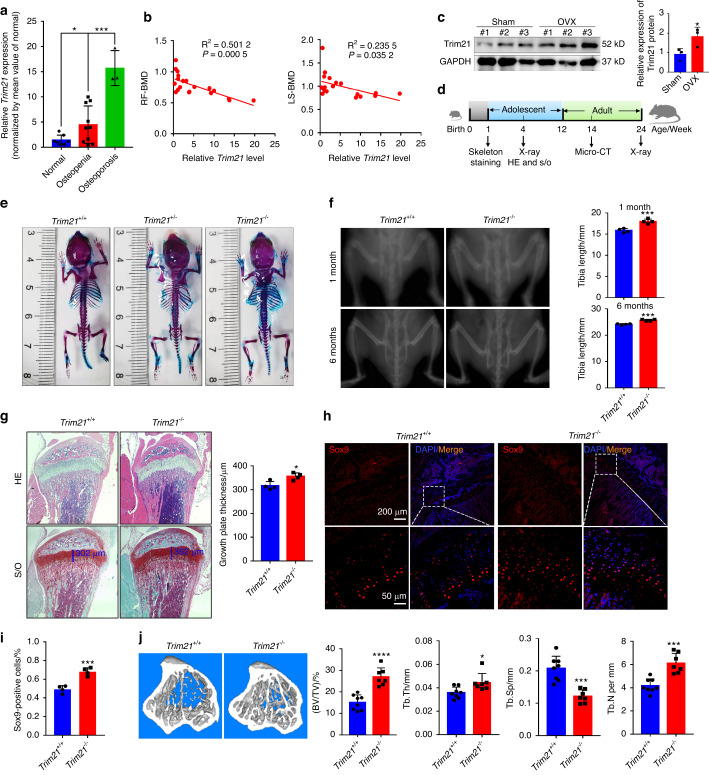
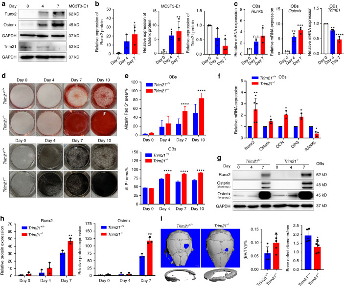
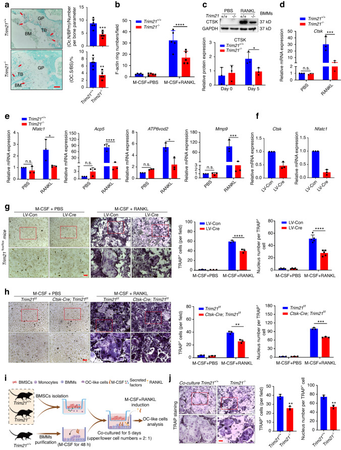
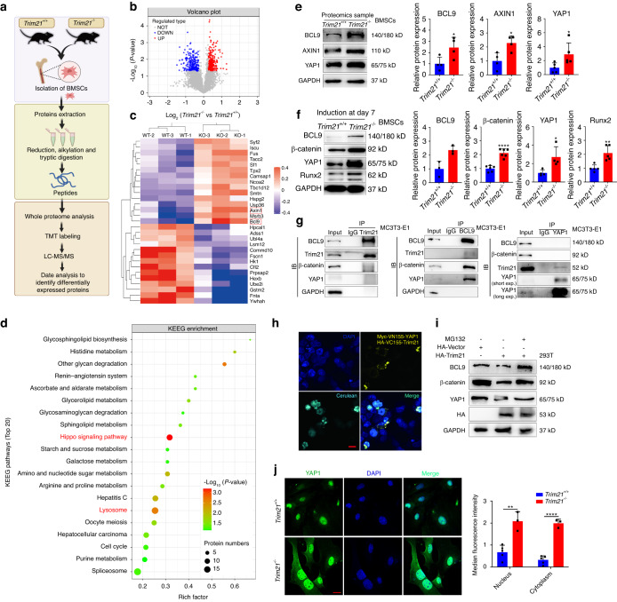
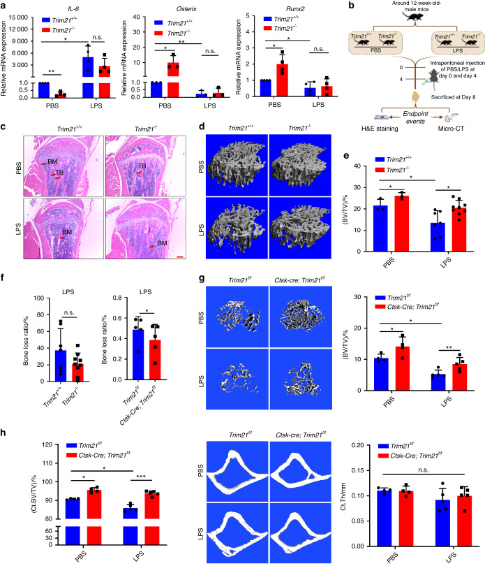
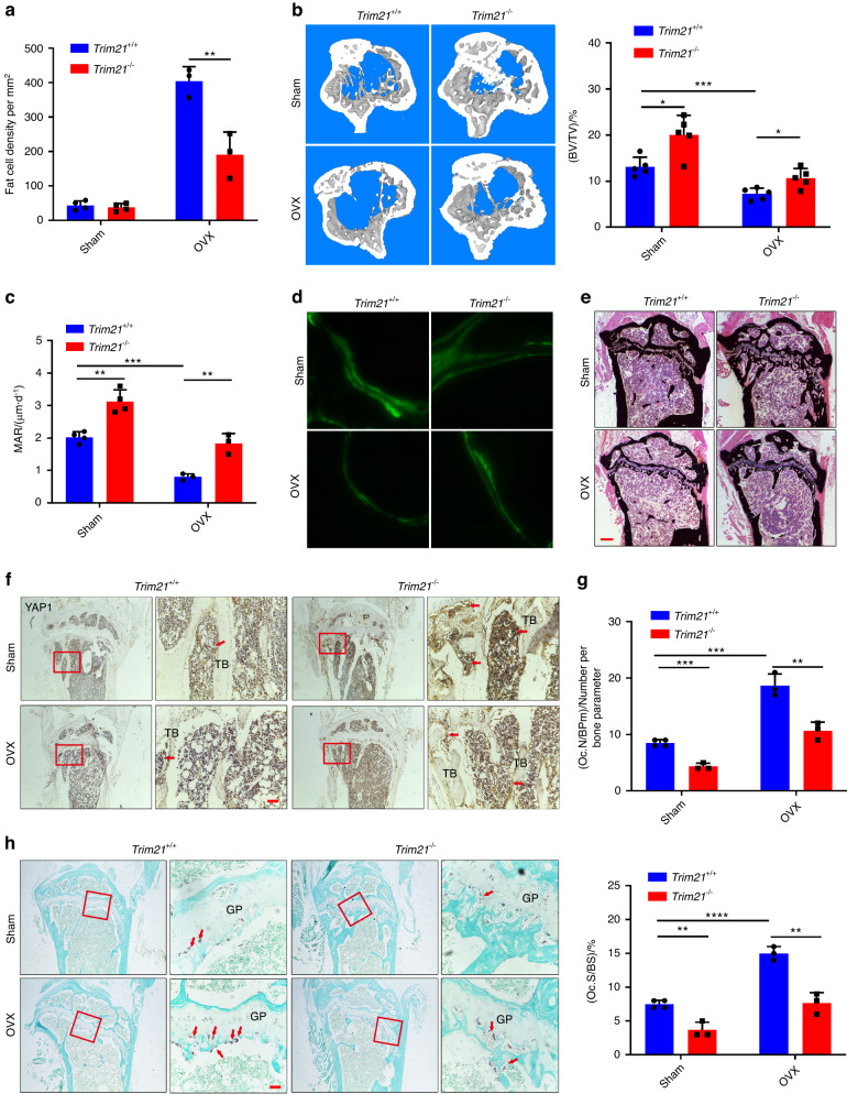
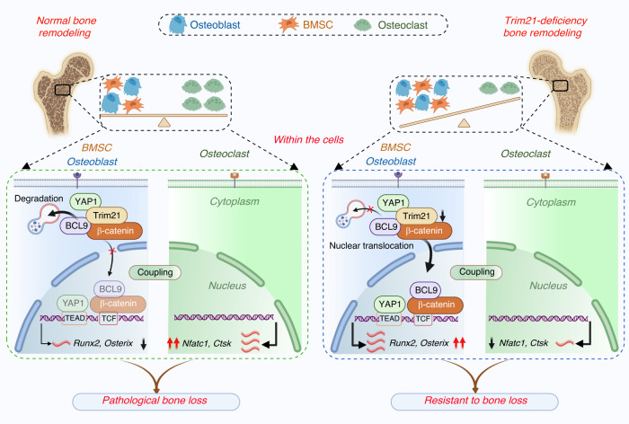
Despite the diverse roles of tripartite motif (Trim)-containing proteins in the regulation of autophagy, the innate immune response, and cell differentiation, their roles in skeletal diseases are largely unknown. We recently demonstrated that Trim21 plays a crucial role in regulating osteoblast (OB) differentiation in osteosarcoma. However, how Trim21 contributes to skeletal degenerative disorders, including osteoporosis, remains unknown. First, human and mouse bone specimens were evaluated, and the results showed that Trim21 expression was significantly elevated in bone tissues obtained from osteoporosis patients. Next, we found that global knockout of the Trim21 gene (KO, Trim21-/-) resulted in higher bone mass compared to that of the control littermates. We further demonstrated that loss of Trim21 promoted bone formation by enhancing the osteogenic differentiation of bone marrow mesenchymal stem cells (BMSCs) and elevating the activity of OBs; moreover, Trim21 depletion suppressed osteoclast (OC) formation of RAW264.7 cells. In addition, the differentiation of OCs from bone marrow-derived macrophages (BMMs) isolated from Trim21-/- and Ctsk-cre; Trim21f/f mice was largely compromised compared to that of the littermate control mice. Mechanistically, YAP1/β-catenin signaling was identified and demonstrated to be required for the Trim21-mediated osteogenic differentiation of BMSCs. More importantly, the loss of Trim21 prevented ovariectomy (OVX)- and lipopolysaccharide (LPS)-induced bone loss in vivo by orchestrating the coupling of OBs and OCs through YAP1 signaling. Our current study demonstrated that Trim21 is crucial for regulating OB-mediated bone formation and OC-mediated bone resorption, thereby providing a basis for exploring Trim21 as a novel dual-targeting approach for treating osteoporosis and pathological bone loss.


Conflict of interest statement

The authors declare no competing interests.

Figures

Fig. 1
Fig. 1
Trim21 is elevated in osteoporotic patients, and its deficiency leads to high bone mass. a Quantitative RT‒PCR analysis of Trim21 mRNA expression in bone specimens from patients with different bone mineral densities (BMDs), which were defined as normal, osteopenia, and osteoporosis. b Correlation analysis between Trim21 mRNA expression and RF-BMD and LS-BMD. RF, right femur; LS, lumbar spine. c Immunoblotting analysis of Trim21 protein expression in the lumbar vertebra of 5-month-old sham-operated or ovariectomized mice. d Schematic diagram showing the analysis of skeletal parameters of mice at different ages. e Alcian blue/Alizarin Red staining of the whole skeleton of 1-week-old Trim21+/+, Trim21+/−, and Trim21−/− littermates. f X-ray images of Trim21+/+ and Trim21−/− mice at 1 month and 6 months (left panel). Quantitative analysis of the tibia length of mice at different ages (right panel). g Representative H&E and S/O staining images of tibial sections from 1-month-old Trim21+/+ and Trim21−/− mice (left panel). Quantitative analysis of the growth plate thickness of the indicated mice (right panel). h, i Representative immunofluorescence images (h) showing the expression of Sox9+ cells (i) in growth plates of tibial sections in 1-month-old Trim21+/+ and Trim21−/− mice. j Representative micro-CT images of the proximal tibia bone of 14-week-old mice. Quantitative measurements of bone volume per tissue volume (BV/TV), trabecular thickness (Tb. Th), trabecular number (Tb. N), and trabecular separation (Tb.Sp). All bar graphs are presented as the mean ± SD. *P < 0.05; ***P < 0.001; ****P < 0.000 1; n.s. not significant by Student’s t test
Fig. 2
Fig. 2
Loss of Trim21 enhances osteoblast activity and favors bone formation. a, b Representative immunoblotting analysis (a) and quantification of Runx2, Osterix, and Trim21 in MC3T3-E1 cells (b) treated with osteogenic medium for 0, 4, and 7 days. c Quantitative RT‒PCR analysis of osteogenic biomarker genes (Osterix, Runx2, and Trim21) in OBs with osteogenic induction. d, e Alizarin Red S (upper panel) and ALP (lower panel) staining of primary osteoblasts (OBs) after induction with osteogenic medium for different times (d). The percentage of Alizarin Red S- (n ≥ 3) and ALP- (n ≥ 3) stained area (e). f Quantitative RT‒PCR detection of osteogenic biomarker genes (Runx2, Osterix, OCN, OPG, and RANKL) in OBs derived from Trim21−/ and Trim21+/+ mice upon osteogenic induction for 7 days. g, h Representative immunoblotting analysis (g) and quantification of Runx2 and Osterix in OBs (h) after osteogenic induction for 0, 4, and 7 days. i Representative micro-CT images of calvarial bone defects in 2-month-old Trim21+/+ and Trim21−/ mice after surgical induction for 1 month (left panel). Quantitative measurements of bone volume per tissue volume (BV/TV) and bone defect diameter (right panel). All bar graphs are presented as the mean ± SD. *P < 0.05; **P < 0.01; ***P < 0.001; ****P < 0.000 1; n.s. not significant by Student’s t test
Fig. 3
Fig. 3
Loss of Trim21 inhibits osteoclast formation and differentiation. a Representative image of histological sections of the tibia that were stained with TRAP (left panel). Bone marrow (BM) and trabecular bone (TB) are indicated in black. TRAP-stained osteoclasts (OCs) are denoted by the red arrow. OC. N/BPm (OC number per bone parameter) and OC. S/BS (OC surface per bone surface) were determined (right panel). Scale bar: 100 μm. b Quantification of F-actin ring number in BMM-derived OCs from immunofluorescence staining of Fig. S6e. c Representative immunoblot analysis and quantification of Ctsk expression in BMM-derived OCs from Trim21+/+ and Trim21−/− mice. d, e Quantitative RT‒PCR detection of OC differentiation genes (Ctsk, Nfatc1, Acp5, ATP6vod2, and Mmp9) in BMM-derived OCs from Trim21+/+ and Trim21/ mice. PBS indicates PBS containing 30 ng·mL−1 M-CSF, while RANKL indicates induction with 30 ng·mL−1 M-CSF and 100 ng·mL−1 RANKL f, g Quantitative RT‒PCR detection (f) of OC differentiation genes (Ctsk and Nfatc1) and TRAP staining (g) in BMM-derived OCs from Trim21f/f mice treated with 30 ng·mL−1 M-CSF plus 100 ng·mL−1 RANKL or PBS containing 30 ng·mL−1 M-CSF for 5 days and infected with lentivirus expressing EGFP or Cre recombinase (defined as LV-Con or LV-Cre, respectively). Quantification of TRAP-positive OCs and the number of nuclei per TRAP+ cell (right panel) (g). Scale bar: 50 μm. h BMMs derived from 4-week-old Trim21f/f and Ctsk-cre; Trim21f/f mice were induced for OC differentiation with either 30 ng·mL−1 M-CSF plus 100 ng·mL−1 RANKL or PBS containing 30 ng·mL−1 M-CSF for 5 days (left panel). Quantification of TRAP-positive OCs and the number of nuclei per TRAP+ cell (right panel). Scale bar: 50 μm. i, j Schematic diagram illustrating the coculture model of OCs with BMSCs from Trim21+/+ and Trim21/ mice (i). Representative images (left panel) and quantification data of TRAP-positive OCs and nucleus number per TRAP+ cell (right panel) (j). Scale bar: 50 μm. All bar graphs are presented as the mean ± SD. *P < 0.05; **P < 0.01; ***P < 0.001; ****P < 0.000 1; n.s. not significant by Student’s t test
Fig. 4
Fig. 4
YAP1/β-catenin signaling is essential for Trim21-mediated osteogenic differentiation. a Schematic diagram showing TMT-based quantitative proteomics for the identification of differentially expressed proteins (DEPs) in bone marrow mesenchymal stem cells (BMSCs) derived from Trim21+/+ and Trim21−/− mice. b Volcano plots of DEPs in BMSCs from Trim21+/+ and Trim21−/− mice. c Heatmap analysis of DEPs in BMSCs. Three replicates of each group were included, and the top 29 DEPs are shown. d KEGG enrichment analysis of the DEPs in the BMSCs. e Representative immunoblotting analysis and quantification of BCL9, AXIN1, and YAP1 in BMSCs; proteomics sample: part of the samples subjected to proteomics analysis. f Representative immunoblotting analysis and quantification of BCL9, β-catenin, YAP1, and Runx2 protein expression in BMSCs after osteogenic induction for 7 days. g The endogenous interaction between Trim21, BCL9, β-catenin, and YAP1 was evaluated using a co-IP assay. h Protein‒protein interaction of YAP1 and Trim21 in living cells. The two BiFC plasmids encoding Myc-VN155-YAP1 and HA-VC155-Trim21 along with HA-cerulean were cotransfected into HEK293T cells for 24 h. Representative images showing transfected cells (cerulean) and the interaction between YAP1 and Trim21 (Venus). Nuclei were stained with DAPI. Scale bar: 20 μm. i Immunoblotting analysis of BCL9, β-catenin, YAP1, and HA-Trim21 protein expression in HEK293T cells treated with or without MG132. j Representative images showing the expression of YAP1+ OBs derived from Trim21+/+ and Trim21−/ mice. Scale bar: 20 μm. All bar graphs are presented as the mean ± SD. *P < 0.05; **P < 0.01; ***P < 0.001; ****P < 0.000 1; n.s. not significant by Student’s t test
Fig. 5
Fig. 5
Loss of Trim21 protects mice from lipopolysaccharide (LPS)-induced bone loss. a Quantitative RT‒PCR determination of IL-6, Osterix, and Runx2 mRNA expression in OBs with or without lipopolysaccharide (LPS) treatment during osteogenic induction. b Schematic diagram showing the H&E staining and micro-CT analysis of Trim21+/+ and Trim21/− mice induced by PBS or LPS. c Representative images of H&E staining of tibia sections of 13-week-old Trim21+/+ and Trim21−/− mice induced by PBS or LPS. Bone marrow (BM) and trabecular bone (TB) are labeled with red arrows. d, e Representative micro-CT images (d) and BV/TV (e) of proximal tibia trabecular bone of 13-week-old Trim21+/+ and Trim21−/− mice induced by PBS or LPS. f The bone loss ratio after LPS treatment in global knockout mice (left panel) and conditional knockout mice (right panel). g Representative micro-CT images and BV/TV of proximal tibia trabecular bone of 13-week-old Trim21f/f and Ctsk-cre; Trim21f/f mice induced by PBS or LPS. h Representative micro-CT images of cortical bones and quantification of BV/TV (left panel) and thickness (right panel) of Trim21f/f and Ctsk-cre; Trim21f/f mice induced by either PBS or LPS. All bar graphs are presented as the mean ± SD. *P < 0.05; **P < 0.01; ***P < 0.001; n.s. not significant by Student’s t test
Fig. 6
Fig. 6
Trim21 orchestrates ovariectomy (OVX)-induced bone metabolism by targeting YAP1 signaling. a Determination of fat cell density in the proximal tibia of 20-week-old Trim21+/+ and Trim21−/− mice induced by sham operation or OVX. b Representative micro-CT images and quantitative data (BV/TV) of proximal tibial bone of 20-week-old Trim21+/+ and Trim21−/− mice induced by sham operation or OVX. c, d Calcein double labeling of mineral layers of tibial trabecular bone of 5-month-old mice. e Representative images of von Kossa staining of the undecalcified proximal tibia of 5-month-old mice. Scale bar: 50 μm. f IHC staining images of the proximal tibia of 5-month-old mice using an antibody against YAP1. The YAP1-stained positive cells are denoted by the red arrow. Scale bar: 50 μm. g, h Representative images of histological sections of the tibia that were stained with TRAP in Trim21+/+ and Trim21−/− mice induced by sham operation or OVX. TRAP-stained osteoclasts (OCs) are denoted by the red arrow. OC. N/BPm (OC number per bone parameter) and OC. S/BS (OC surface per bone surface) was determined. Scale bar: 100 μm. All bar graphs are presented as the mean ± SD. *P < 0.05; **P < 0.01; ***P < 0.001; ****P < 0.000 1; n.s. not significant by Student’s t test
Fig. 7
Fig. 7
A schematic of Trim21 in the regulation of bone remodeling via YAP1/β-catenin signaling. Normal bone remodeling is maintained by the balance of MSC/osteoblast-mediated bone formation and osteoclast-mediated bone resorption. Trim21, by interacting with the protein complex formed by YAP1/β-catenin/BCL9, dictates the degradation of this protein complex, which in turn inactivates YAP1 and β-catenin signaling, which is essential for osteoblast differentiation. However, Trim21 is critical for maintaining the basic expression of osteoclast biomarkers, including Nfatc1 and Ctsk. Therefore, the coupling of osteoblasts with osteoclasts is attributed to dynamic changes in bone metabolism (left panel). In contrast, the loss of Trim21 causes disassociation with the YAP1/β-catenin/BCL9 complex, which then enters the nucleus for subsequent activation of osteogenic genes, including Runx2 and Osterix. In addition, the loss of Trim21 suppresses the maturation of osteoclasts. Together, these results indicate that Trim21 deficiency alleviates pathological bone loss by activating YAP1/β-catenin signaling

References

    1. Weivoda MM, et al. Identification of osteoclast-osteoblast coupling factors in humans reveals links between bone and energy metabolism. Nat. Commun. 2020;11:87. doi: 10.1038/s41467-019-14003-6. - DOI - PMC - PubMed
    1. Rachner TD, Khosla S, Hofbauer LC. Osteoporosis: now and the future. Lancet. 2011;377:1276–1287. doi: 10.1016/S0140-6736(10)62349-5. - DOI - PMC - PubMed
    1. Gregson CL, et al. UK clinical guideline for the prevention and treatment of osteoporosis. Arch. Osteoporos. 2022;17:58. doi: 10.1007/s11657-022-01061-5. - DOI - PMC - PubMed
    1. Minisola S, et al. Update on the safety and efficacy of teriparatide in the treatment of osteoporosis. Ther. Adv. Musculoskelet. Dis. 2019;11:1759720X19877994. doi: 10.1177/1759720X19877994. - DOI - PMC - PubMed
    1. Langdahl BL. Overview of treatment approaches to osteoporosis. Br. J. Pharmacol. 2021;178:1891–1906. doi: 10.1111/bph.15024. - DOI - PubMed

Publication types