

Prevention of Pernicious Vascular Event: Acetabular Component Screw Impinging on External Iliac Vessels - A Unique Case Report
- PMID: 37885624
- PMCID: PMC10599367
- DOI: 10.13107/jocr.2023.v13.i10.3960
Prevention of Pernicious Vascular Event: Acetabular Component Screw Impinging on External Iliac Vessels - A Unique Case Report
Abstract
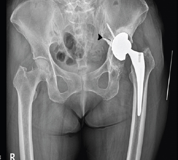
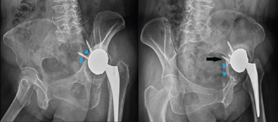
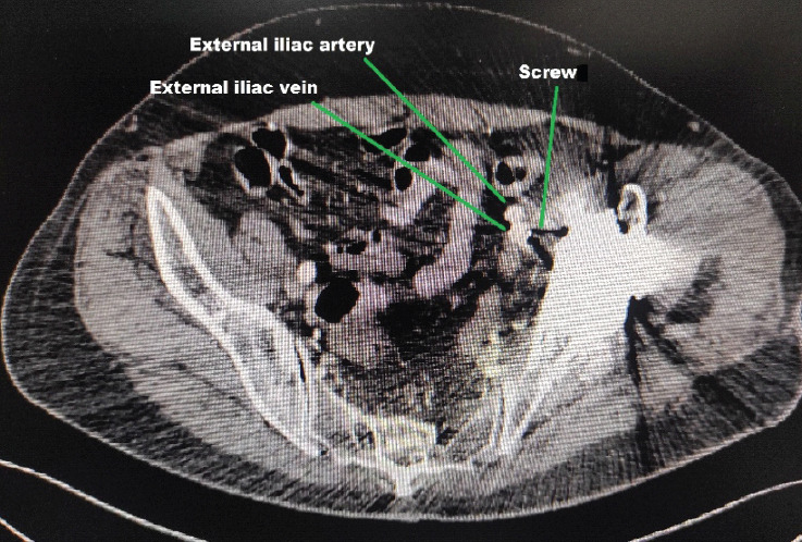
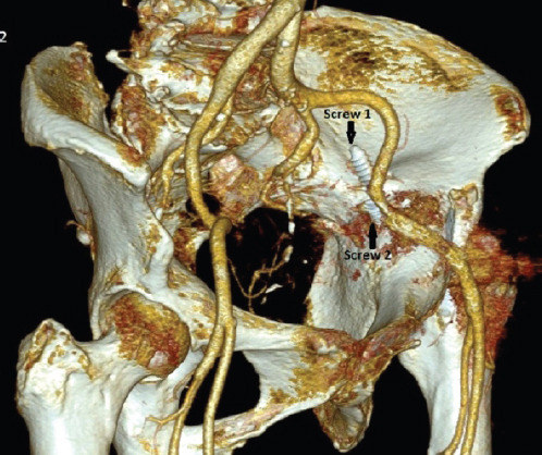
Introduction: Revision total hip arthroplasty requires meticulous planning and execution to achieve the desired outcome. Pelvic vessel injury following total hip arthroplasty is rare, but a well-known and serious complication, having a very high morbidity (15%) and mortality (7%). This case demonstrates the rare occurrence of acetabular screw abutment to the external iliac vessels, which if removed during revision hip surgery without releasing the adhesions around it, will lead to avulsion injury of the vessels and a catastrophic event.
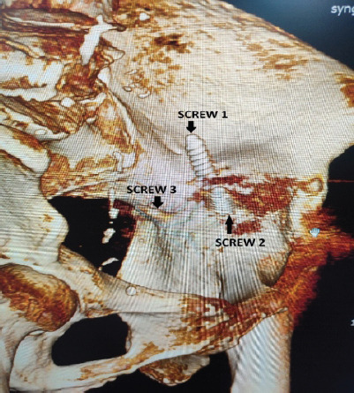
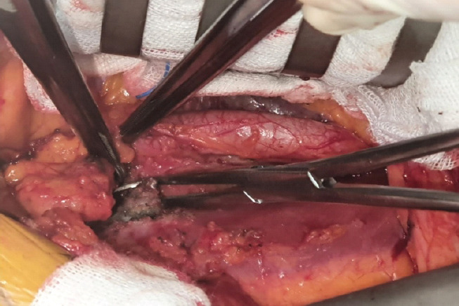
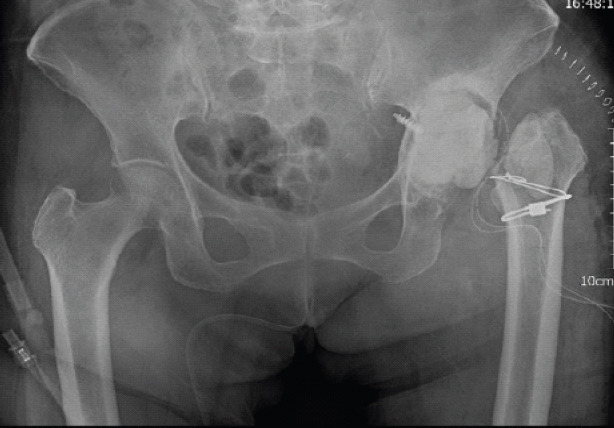
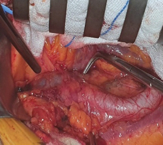
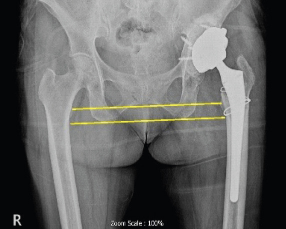
Case report: We present this challenging and unique case of a 64-year-old female patient where the acetabular component screw was found to be adherent to the external iliac vessels, with vascular injury imminent. During revision surgery, the iliac vessels were first released free of all adhesions with the intrapelvis screw using an ilioinguinal incision and retroperitoneal approach. The prosthesis was removed using a posterior approach to the hip joint. Definitive surgery was performed after 2 weeks.
Conclusion: Surgeons should be cognizant of the possibility of an avulsion vascular injury in revision cases having intrapelvic screws or implants. The proximity of such an implant with the intrapelvic vasculature must be confirmed preoperatively. Management should be individualized. Dual approach and staged procedure help in a favorable outcome. Vascular injury, revision total hip arthroplasty, screw abutting iliac vessel, external iliac vessel, computed tomographic angiography.
Keywords: Vascular injury; computed tomographic angiography; external iliac vessel; revision total hip arthroplasty; screw abutting iliac vessel.
Copyright: © Indian Orthopaedic Research Group.
Conflict of interest statement
Conflict of Interest: Nil
Figures
References
-
- Street MW, Howard LC, Neufeld ME, Masri BA. Vascular injuries during hip and knee replacement. Orthop Clin North Am. 2022;53:1–12. - PubMed
-
- Wasielewski RC, Cooperstein LA, Kruger MP, Rubash HE. Acetabular anatomy and the transacetabular fixation of screws in total hip arthroplasty. J Bone Joint Surg Am. 1990;72:501–8. - PubMed
-
- Jenkins DR, Odland AN, Sierra RJ, Hanssen AD, Lewallen DG. Minimum five-year outcomes with porous tantalum acetabular cup and augment construct in complex revision total hip arthroplasty. J Bone Joint Surg Am. 2017;99:e49. - PubMed
Publication types
LinkOut - more resources
Full Text Sources