. 2023 Oct 11:14:1257046.
doi: 10.3389/fimmu.2023.1257046. eCollection 2023.

Leishmania major- derived lipophosphoglycan influences the host's early immune response by inducing platelet activation and DKK1 production via TLR1/2

Affiliations

Leishmania major- derived lipophosphoglycan influences the host's early immune response by inducing platelet activation and DKK1 production via TLR1/2

Olivia C Ihedioha et al. Front Immunol. .

Abstract

Background: Platelets are rapidly deployed to infection sites and respond to pathogenic molecules via pattern recognition receptors (TLR, NLRP). Dickkopf1 (DKK1) is a quintessential Wnt antagonist produced by a variety of cell types including platelets, endothelial cells, and is known to modulate pro-inflammatory responses in infectious diseases and cancer. Moreover, DKK1 is critical for forming leukocyte-platelet aggregates and induction of type 2 cell-mediated immune responses. Our previous publication showed activated platelets release DKK1 following Leishmania major recognition.

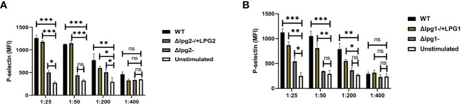
Results: Here we probed the role of the key surface virulence glycoconjugate lipophosphoglycan (LPG), on DKK1 production using null mutants deficient in LPG synthesis (Δlpg1- and Δlpg2-). Leishmania-induced DKK1 production was reduced to control levels in the absence of LPG in both mutants and was restored upon re-expression of the cognate LPG1 or LPG2 genes. Furthermore, the formation of leukocyte-platelet aggregates was dependent on LPG. LPG mediated platelet activation and DKK1 production occurs through TLR1/2.

Conclusion: Thus, LPG is a key virulence factor that induces DKK1 production from activated platelets, and the circulating DKK1 promotes Th2 cell polarization. This suggests that LPG-activated platelets can drive innate and adaptive immune responses to Leishmania infection.

Keywords: Leishmaniasis; P-selectin; innate response; leukocyte-platelet aggregates; platelet.


Conflict of interest statement

The authors declare that the research was conducted in the absence of any commercial or financial relationships that could be construed as a potential conflict of interest.

Figures

Figure 1
Figure 1
Δlpg1- and Δlpg2- induce minimal P-selectin expression in activated platelets. BALB/c mice were challenged with infective metacyclic promastigote (2 x 10⁶ parasites, n = 5) of WT, Δlpg1-, Δlpg2-, Δlpg1-/+LPG1 and Δlpg2-/+LPG2 strains via the footpad. Control mice (n = 5) were given 0.9% NaCl saline. Blood was collected via retro-orbital sinus at days 3, 14 and 42 PI. Isolated platelet samples were analyzed by flow cytometry for P-selectin expression. In all the experiments, WT-infected and non-infected mice served as positive and negative controls, respectively. Each dot indicates the expression of P-selectin by CD41+ cells (A–F). Results are presented as mean (± SEM) and are representative of 3 independent experiments. One-way ANOVA with Bonferroni’s post hoc test was performed to analyze the data *p < 0.05, **p < 0.01, ***p < 0.001, ‘ns’ indicates not significant (p > 0.05).
Figure 2
Figure 2
Less efficient expression of P-selectin from Δlpg1- and Δlpg2- SLAG-activated platelets. Platelets isolated from naïve mice were incubated with various concentrations of SLAG (derived from WT, Δlpg1-, Δlpg2-, Δlpg1-/+LPG1 and Δlpg2-/+LPG2 strains) for 1hr. Platelet samples were analyzed by Flow cytometry for P-selectin expression. In all the experiments, WT-SLAG activated and non-activated platelets served as a positive and negative control, respectively. Column graphs (A, B) indicate the expression of P-selectin by CD41+ cells. Results are presented as mean +/- SEM of replicate wells and represent 3 independent experiments. Data are presented by comparing DKK1 production from the non-SLAG and SLAG (derived from WT, Δlpg1-, Δlpg2-, Δlpg1-/+LPG1 and Δlpg2-/+LPG2 strains) activated platelets. One-way ANOVA with Bonferroni’s post hoc test was performed to analyze the data *p < 0.05, **p < 0.01, ***p < 0.001, ‘ns’ indicates not significant (p > 0.05).
Figure 3
Figure 3
Δlpg1- and Δlpg2- parasites induce less plasma DKK1 production from the platelets. Six-week-old female BALB/c mice were challenged with infective metacyclic promastigote (2 x 10⁶ parasites, n = 5) of WT, Δlpg1-, Δlpg2-, Δlpg1-/+LPG1 and Δlpg2-/+LPG2 strains via the footpad. Control mice (n = 5) were given 0.9% NaCl Saline. Blood was collected via retro-orbital sinus at days 3, 14 and 42 PI. Plasma samples were analyzed by ELISA For DKK1 production (A–F). In all experiments, WT-infected and non-infected mice served as positive and negative controls, respectively. Results are presented as mean +/- SEM of replicate wells and are representative of 3 independent experiments. One-way ANOVA with Bonferroni’s post hoc test was performed to analyze the data *p < 0.05, **p < 0.01, ***p < 0.001, ‘ns’ indicates not significant (p > 0.05).
Figure 4
Figure 4
Poor induction of DKK1 from Δlpg1- and Δlpg2- SLAG-activated platelets. Platelets from naïve mice were incubated with various concentrations of SLAG (derived from WT, Δlpg1-, Δlpg2-, Δlpg1-/+LPG1 and Δlpg2-/+LPG2 strains) for 1hr. Cell culture supernatant samples were analyzed by ELISA for DKK1 production as shown in the column graphs (A, B). In all experiments, WT-SLAG activated, and non-activated platelets served as positive and negative controls, respectively. Results are presented as mean +/- SEM of replicate wells and represent 3 independent experiments. Data are presented by comparing the DKK1 production from non-SLAG and SLAG (derived from WT, Δlpg1-, Δlpg2-, Δlpg1-/+LPG1 and Δlpg2-/+LPG2 strains) activated platelets. One-way ANOVA with Bonferroni’s post hoc test was performed to analyze the data *p < 0.05, **p < 0.01, ***p < 0.001, ‘ns’ indicates not significant (p > 0.05).
Figure 5
Figure 5
Δlpg1- and Δlpg2- parasites differ from specific add-backs and WT parasites in their capacity to induce lesions and increase the parasite burdens. The infected foot from each mouse in WT, Δlpg1-, Δlpg2-, Δlpg1-/+LPG1 and Δlpg2-/+LPG2 parasite-infected groups were measured for lesion size weekly using a vernier caliper (A, B), and parasite burden (at day 42 PI) was determined by limiting dilution assay (C, D). Results are presented as mean +/- SEM. For Figures (A, B), mice in all the infected groups were compared with the non-infected group and data analysis was done using one-way ANOVA with Bonferroni’s post hoc test *p < 0.05, **p < 0.01, ***p < 0.001, ‘ns’ indicates not significant (p > 0.05).
Figure 6
Figure 6
Anti-TLR1/2 antibody and PKC-alpha inhibitor significantly decreased DKK1 production in SLAG-activated platelets. Inhibition of DKK1 by neutralizing antibodies (10 μg/ml) following 1hr incubation with SLAG (derived from WT, Δlpg1-, Δlpg2-, Δlpg1-/+LPG1 and Δlpg2-/+LPG2 strains) activated platelets. SLAG concentration is 1:50. In all experiments, Pam2CSK4 (10 μg/ml), Pam3CSK4 (10 μg/ml) and unstimulated samples served as a positive and negative control, respectively (A, B). Inhibition of DKK1 by neutralizing antibodies (10 μg/ml) following 1hr incubation with WT SLAG (1:50) activated platelets (C). Inhibition of DKK1 by PKC-alpha inhibitor (100nM, 500nM and 1000nM) following 1hr incubation with SLAG (derived from WT, Δlpg1-, Δlpg2-, Δlpg1-/+LPG1 and Δlpg2-/+LPG2 strains) activated platelets (D, E). Results are presented as mean +/- SEM of replicate wells and represent 3 independent experiments. For Figures (A, B), Student’s t-test was performed, while one-way ANOVA with Bonferroni’s post hoc test was performed to analyze the data in Figures (C–E). *p < 0.05, **p < 0.01, ***p < 0.001, ‘ns’ indicates not significant (p > 0.05). Note that the DKK1 production from Δlpg1- and Δlpg2- SLAG-stimulated platelets was found to be background platelet DKK1, as non-stimulated platelets showed comparable levels of DKK1 release/PKC-alpha inhibition ( Figure S2 ).
Figure 7
Figure 7
Δlpg1- and Δlpg2- parasites are ineffectual in inducing LPA formation. BALB/c mice were challenged with infective metacyclic promastigote (2 x 10⁶ parasites, n = 5) of WT, Δlpg1-, Δlpg2-, Δlpg1-/+LPG1 and Δlpg2-/+LPG2 strains via the footpad. Control mice (n = 5) were given 0.9% NaCl saline. Blood was collected via retro-orbital sinus at day 3 PI. Blood samples were analyzed by flow cytometry for LPA. Representative flow cytometry dot plots showing the analyses of LPA performed on day 3 PI (A). Representative dot plots shown in (B, C) are from concatenated samples of each experimental group, and the corresponding graphs (D, E) indicate the percentage of LPA molecules by CD45+ cells. A dot plot of each sample in all the experimental groups is presented in Figure S4 (A, B). In all the experiments, WT-infected and non-infected mice served as a positive and negative control, respectively. Results are presented as mean (± SEM). One-way ANOVA with Bonferroni’s post hoc test was performed to analyze the data *p < 0.05, **p < 0.01, ‘ns’ indicates not significant (p > 0.05).

Similar articles

Cited by

References

    1. Castro M, Rode J, MaChado PR, Llanos-Cuentas A, Hueb M, Cota G, et al. . Cutaneous leishmaniasis treatment and therapeutic outcomes in special populations: A collaborative retrospective study. PloS Negl Trop Dis (2023) 17(1):e0011029. doi: 10.1371/journal.pntd.0011029 - DOI - PMC - PubMed
    1. Sheet WF. WHO Fact Sheet on Leishmaniasis . Geneva, Switzerland: World Health Organization; (Accessed July. 2021;10:2021).
    1. Cowan R, Varadarajan S, Wei A, Salim T, DallaPiazza M. Microbial perils of the tropics: A case of cutaneous leishmaniasis in an immigrant from South America. IDCases (2023) 31:e01669. doi: 10.1016/j.idcr.2022.e01669 - DOI - PMC - PubMed
    1. Barry MA, Koshelev MV, Sun GS, Grekin SJ, Stager CE, Diwan AH, et al. . Case report: cutaneous leishmaniasis in Cuban immigrants to Texas who traveled through the Darien Jungle, Panama. Am J Trop Med Hyg (2014) 91(2):345. doi: 10.4269/ajtmh.14-0124 - DOI - PMC - PubMed
    1. Horev A, Sagi O, Zur E, Ben-Shimol S. Topical liposomal amphotericin B gel treatment for cutaneous leishmaniasis caused by Leishmania major: a double-blind, randomized, placebo-controlled, pilot study. Wiley Online Library; (2023) 62:40–7. doi: 10.1111/ijd.16407 - DOI - PubMed

Publication types

LinkOut - more resources