. 2023 Oct 11:14:1250154.
doi: 10.3389/fphar.2023.1250154. eCollection 2023.

Hypoxia releases S-nitrosocysteine from carotid body glomus cells-relevance to expression of the hypoxic ventilatory response

Affiliations

Hypoxia releases S-nitrosocysteine from carotid body glomus cells-relevance to expression of the hypoxic ventilatory response

James M Seckler et al. Front Pharmacol. .

Abstract

We have provided indirect pharmacological evidence that hypoxia may trigger release of the S-nitrosothiol, S-nitroso-L-cysteine (L-CSNO), from primary carotid body glomus cells (PGCs) of rats that then activates chemosensory afferents of the carotid sinus nerve to elicit the hypoxic ventilatory response (HVR). The objective of this study was to provide direct evidence, using our capacitive S-nitrosothiol sensor, that L-CSNO is stored and released from PGCs extracted from male Sprague Dawley rat carotid bodies, and thus further pharmacological evidence for the role of S-nitrosothiols in mediating the HVR. Key findings of this study were that 1) lysates of PGCs contained an S-nitrosothiol with physico-chemical properties similar to L-CSNO rather than S-nitroso-L-glutathione (L-GSNO), 2) exposure of PGCs to a hypoxic challenge caused a significant increase in S-nitrosothiol concentrations in the perfusate to levels approaching 100 fM via mechanisms that required extracellular Ca2+, 3) the dose-dependent increases in minute ventilation elicited by arterial injections of L-CSNO and L-GSNO were likely due to activation of small diameter unmyelinated C-fiber carotid body chemoafferents, 4) L-CSNO, but not L-GSNO, responses were markedly reduced in rats receiving continuous infusion (10 μmol/kg/min, IV) of both S-methyl-L-cysteine (L-SMC) and S-ethyl-L-cysteine (L-SEC), 5) ventilatory responses to hypoxic gas challenge (10% O2, 90% N2) were also due to the activation of small diameter unmyelinated C-fiber carotid body chemoafferents, and 6) the HVR was markedly diminished in rats receiving L-SMC plus L-SEC. This data provides evidence that rat PGCs synthesize an S-nitrosothiol with similar properties to L-CSNO that is released in an extracellular Ca2+-dependent manner by hypoxia.

Keywords: L-S-nitrosoglutathione; S-ethyl-L-cysteine; S-nitroso-L-cysteine; Smethyl-L-cysteine; minute ventilation; primary glomus cells.


Conflict of interest statement

SB was employed by Galleon Pharmaceuticals, Inc. The remaining authors declare that the research was conducted in the absence of any commercial or financial relationships that could be construed as a potential conflict of interest.

Figures

FIGURE 1
FIGURE 1
The response coefficients of our S-nitrosothiol biosensor to a 1,000x dilution of primary glomus cell lysates (analyte), 100 fM S-nitroso-L-cysteine (L-CSNO) or 100 fM S-nitrosoglutathione (L-GSNO). Definitions: Columns designated as “Blank” refer to the electrode reaction to Dulbecco’s phosphate buffered saline (DPBS) alone for each study. Columns designated as “Analyte” refer to the electrode reaction to primary glomus cell lysates (Glomus cells), L-CSNO (100 fM) or L-GSNO (100 fM). The columns designated as “Analyte + HgCl2” refer to the electrode reactions to primary glomus cell lysates (Glomus cells), L-CSNO (100 fM) or L-GSNO (100 fM) that were exposed to HgCl2 (100 μM). The columns designated as “Analyte + HgCl2 + UVL” refer to the electrode reactions to primary glomus cell lysates (Glomus cells), L-CSNO (100 fM) or L-GSNO (100 fM) that were exposed to HgCl2 (100 μM) and ultraviolet light (UVL). The data are shown as mean ± SEM from 6-7 samples. *p < 0.05, significant change from control conditions (Analyte columns).
FIGURE 2
FIGURE 2
The response coefficient of the S-nitrosothiol biosensors to perfusion media collected from the chamber of plated PGCs exposed to normoxia, hypoxia, or return to normoxia after hypoxic challenge. All of the electrodes were exposed to DPBS before the experiment (blank) to serve as a negative control. After each experiment, electrodes were exposed to a solution of 100 fM S-nitroso-L-cysteine (L-CSNO) to ensure that the electrodes were properly detecting S-nitrosothiols and specifically, L-CSNO. The data are presented as mean ± SEM. There were 6 samples in each of the Ca2+-present studies and 5 samples in each of the Ca2+-absent studies. *p < 0.05, significant difference from blank and normoxia values. p < 0.05, Ca2+-free versus Ca2+-values resulting from hypoxic challenge. ‡p < 0.05, Ca2+-free versus Ca2+-values upon return to room-air.
FIGURE 3
FIGURE 3
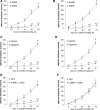
Arithmetic changes in minute ventilation during a hypoxic gas challenge (10% O2, 90% N2) for 10 min. (A) Adult sham-operated (SHAM) rats (n = 6) and those with bilateral carotid sinus nerve transection (CSNX) (n = 6). (B) Adult rats treated as neonates with vehicle (VEH; n = 9) or capsaicin (CAP; 50 mg/kg, SC; n = 9). (C) Adult rats receiving a continuous intravenous infusion of vehicle (VEH; 20 μL/min, IV; n = 9) or S-methyl-L-cysteine (L-SMC; 10 μmol/min, IV; n = 9) plus S-ethyl-L-cysteine (L-SEC; 10 μmol/min, IV; n = 9). The data are shown as mean ± SEM. *p < 0.05, significant response. †p < 0.05, CSNX versus SHAM, CAP versus VEH, L-SMC + L-SEC versus VEH.
FIGURE 4
FIGURE 4
Arithmetic changes in minute ventilation elicited by injections of L-CSNO (left-hand panels) and L-GSNO (right-hand panels). (A) and (B) Adult sham-operated (SHAM) rats (n = 6) and those with bilateral carotid sinus nerve transection (CSNX) (n = 6). (C) and (D) Adult rats treated as neonates with vehicle (VEH; n = 9) or capsaicin (CAP; 50 mg/kg, SC; n = 9). (E) and (F) Adult rats receiving a continuous intravenous infusion of vehicle (VEH; 20 μL/min, IV; n = 9) or S-methyl-L-cysteine (L-SMC; 10 μmol/min, IV; n = 9) plus S-ethyl-L-cysteine (L-SEC; 10 μmol/min, IV; n = 9). The data are shown as mean ± SEM. *p < 0.05, significant response. †p < 0.05, CSNX versus SHAM, CAP versus VEH, L-SMC + L-SEC versus VEH.
FIGURE 5
FIGURE 5
Proposed mechanism by which S-nitroso-L-cysteine (L-CSNO) is stored in primary glomus cells (PGCs) and released in response to hypoxia to act as a neurotransmitter mediating the hypoxic ventilatory response. Vesicular stores of L-CSNO within PGCs are generated by L-cysteine uptake into vesicles via membrane-bound L-amino acid transporter (L-AT) and then S-nitrosylation of L-cysteine to L-CSNO by membrane-bound nitric oxide synthase (NOS). Hypoxia causes PGCs to depolarize thereby activating voltage-gated Ca2+-channels (VGCa-channels) leading to extracellular Ca2+-dependent release of the vesicular stores of L-CSNO. The released L-CSNO then binds stereoselectively to extracellular domains of voltage-gated K+-channels (Kv-channels), such as Kvβ2 subunits, on carotid body chemoafferent nerve terminals to actively close the Kv-channels thereby preventing K+ release from the terminals. Diminished K+ release depolarizes the terminals thereby generating action potentials that activate neurons in the commissural nucleus tractus solitarius (cNTS) leading to neurotransmitter release that elicits the hypoxic ventilatory response (HVR). In addition, L-CSNO released by the PGCs is actively transported into chemoafferent terminals in the carotid body via plasma membrane-bound L-AT.

References

    1. Abrahám A. (1970). Electron microscopic studies on human carotid bodies. Z. Mikrosk. Anat. Forsch. 81, 413–453. - PubMed
    1. Abrahám A. (1981). Light- and electron-microscopic investigations on human carotid bodies. Z. Mikrosk. Anat. Forsch. 95, 33–71. - PubMed
    1. Aldossary H. S., Alzahrani A. A., Nathanael D., Alhuthail E. A., Ray C. J., Batis N., et al. (2020). G-Protein-Coupled receptor (GPCR) signaling in the carotid body: Roles in hypoxia and cardiovascular and respiratory disease. Int. J. Mol. Sci. 21, 6012. 10.3390/ijms21176012 - DOI - PMC - PubMed
    1. Anand P., Hausladen A., Wang Y. J., Zhang G. F., Stomberski C., Brunengraber H., et al. (2014). Identification of S-nitroso-CoA reductases that regulate protein S-nitrosylation. Proc. Natl. Acad. Sci. U. S. A. 111, 18572–18577. 10.1073/pnas.1417816112 - DOI - PMC - PubMed
    1. Atanasova D. Y., Dandov A. D., Dimitrov N. D., Lazarov N. E. (2020). Histochemical and immunohistochemical localization of nitrergic structures in the carotid body of spontaneously hypertensive rats. Acta histochem. 122, 151500. 10.1016/j.acthis.2019.151500 - DOI - PubMed