. 2023 Oct 18:2023:9969079.
doi: 10.1155/2023/9969079. eCollection 2023.

Irradiation Attenuates Systemic Lupus Erythematosus-Like Morbidity in NZBWF1 Mice: Focusing on CD180-Negative Cells

Affiliations

Irradiation Attenuates Systemic Lupus Erythematosus-Like Morbidity in NZBWF1 Mice: Focusing on CD180-Negative Cells

Kazuko Fujita et al. J Immunol Res. .

Abstract

Systemic lupus erythematosus (SLE) is a chronic autoimmune disease characterized by the production of autoantibodies that can induce systemic inflammation. Ultraviolet-A and X-ray irradiation have been reported to have therapeutic effects in patients with SLE. We previously demonstrated that CD180-negative cells, these are radiosensitive, contribute to the development of SLE-like morbidity in NZBWF1 mice. In this study, the effects of irradiation on SLE-like morbidity manifestations in NZBWF1 mice and on CD180-negative cells were investigated. Whole-body irradiation, excluding the head, attenuated SLE-like morbidity in vivo, as indicated by the prevention of the renal lesion development, inhibition of anti-dsDNA antibody production, reduction of urinary protein levels, and prolongation of the lifespan. Irradiation also reduced the proportion of CD180-negative cells in the spleen. Although other immune cells or molecules may be triggered because of the whole-body irradiation treatment, previous research, and the current results suggest a strong relationship between the radiation-induced decrease in CD180-negative cells and the amelioration of SLE-like morbidities. Clinical trials assessing CD180-negative cells as a therapeutic target for SLE have been hampered by the lack of validated cell markers; nonetheless, the present findings suggest that radiotherapy may be a new therapeutic strategy for managing SLE symptoms.


Conflict of interest statement

The authors declare that there is no conflict of interest regarding the publication of this paper.

Figures
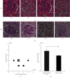
Figure 1
Figure 1
Irradiation prevents the histopathological development of renal lesions in NZBWF1 mice. (a, b) Histopathological assessment of kidney tissue samples from paired mice 15 weeks after treatment based on hematoxylin and eosin (HE) staining (a) and periodic acid-Schiff (PAS) staining (b) (Group A). Images of i and iii, and ii and iv were from sham-irradiated and 4 Gy-irradiated mice, respectively. Images of i and ii, and iii and iv were from paired mice, respectively. Scale bars indicate 50 μm. (c) Comparison of the renal lesion grades between paired sham-irradiated and irradiated mice 15 weeks after treatment (n = 13 paired mice). (d) Renal lesion grades in sham-irradiated and irradiated mice 15 weeks after treatment (n = 13 in each group). Open and solid bars indicate sham-irradiated and four irradiated mice, respectively. Data are presented as mean ± standard error. The   presents significant differences at 0.01 < p < 0.05.
Figure 2
Figure 2
Irradiation prevents proteinuria levels in NZBWF1 mice. (a) Comparison of the urinary protein levels between paired sham-irradiated and 4 Gy-irradiated mice 15 weeks after treatment (n = 13 paired mice, Group A). (b) Urinary protein levels in sham-irradiated and irradiated mice. Open and solid bars indicate sham-irradiated and 4 Gy-irradiated mice, respectively (n = 13 in each group, Group A). (c) Urinary protein levels at 6 (n = 50 in each group), 12 (n = 49 vs. 50), 24 (n = 35 vs. 49), and 36 (n = 9 vs. 24) weeks after treatment (Group B) are presented as the mean value ± standard error. (d) Duration from sham-irradiation or irradiation to the development of level 3+ urinary protein (n = 50 in each group, Group B). Open and solid bars indicate sham-irradiated and irradiated mice, respectively. Each group consisted of 50 mice (Group B). Data are presented as mean± standard error. The   presents significant difference at 0.01 < p < 0.05 and  ∗∗0.001 < p < 0.01.
Figure 3
Figure 3
Irradiation suppresses the production of anti-dsDNA autoantibody in NZBWF1 mice. (a) Comparison of anti-dsDNA antibody titers in peripheral blood between paired sham-irradiated and 4 Gy-irradiated mice 15 weeks after treatment (n = 13 pairs, Group A). (b) Anti-dsDNA antibody titers in the peripheral blood between paired sham-irradiated and 4 Gy-irradiated mice 15 weeks after treatment (n = 13 pairs, Group A). Open and solid bars indicate sham-irradiated and irradiated mice, respectively. Data are presented as the mean ± standard error. (c) Anti-dsDNA antibody mean titers in the peripheral blood from sham-irradiated and four irradiated mice at 6 (n = 8 in each group), 12 (n = 6 in each group), and 36 (n = 3 vs. 4, respectively) weeks after treatment. Open and solid bars indicate, respectively (Group B). Data are presented as the mean ± standard error.  0.01 < p < 0.05 and  ∗∗∗P < 0.001.
Figure 4
Figure 4
Irradiation prolongs the lifespan of NZBWF1 mice. (a) Comparison of the survival times in paired mice. The survival time indicates the duration from sham irradiation or irradiation to death (n = 50 paired mice, Group B). (b) Survival time of paired sham-irradiated and irradiated mice. Open and solid bars indicate sham-irradiated and 4 Gy-irradiated mice, respectively (n = 50 paired mice, Group B). (c) Survival ratios of sham-irradiated and irradiated mice (n = 50 paired mice, Group B). Blue and red lines indicate sham-irradiated and irradiated mice, respectively.  0.01 < p < 0.05 and  ∗∗∗p < 0.001.
Figure 5
Figure 5
Irradiation reduces CD180-negative cell populations in NZBWF1 mice. (a) Flow cytometric analysis of CD180 expression in splenocytes from paired NZBWF1-irradiated (right figure) and sham-irradiated (left figure) mice 15 weeks after treatments (Group A). CD180-negative B cells are shown inside the red square. (b) Frequency of CD180-negative cells population within splenic lymphocytes (up) and splenic B cells (down; n = 13 paired mice, Group A). (c) CD180-negative cell population in the spleen. Open and solid bars indicate sham-irradiated mice and 4 Gy-irradiated mice, respectively (Group A). Each group consists of 13 paired mice. Data are presented as the mean ± standard error.  ∗∗0.01 < p < 0.05.

Similar articles

References

    1. Polderman M. C. A., Huizinga T. W. J., Le Cessie S., Pavel S. UVA-1 cold light treatment of SLE: a double blind, placebo controlled crossover trial. Annals of the Rheumatic Disease . 2001;60(2):112–115. doi: 10.1136/ard.60.2.112. - DOI - PMC - PubMed
    1. McGrath H., Jr., Martínez-Osuna P., Lee F. A. Review: ultraviolet-A1 (340–400 nm) irradiation therapy in systemic lupus erythematosus. Lupus . 1996;5(4):269–274. doi: 10.1177/096120339600500405. - DOI - PubMed
    1. McGrath H., Jr Ultraviolet-A1 irradiation decreases clinical disease activity and autoantibodies in patients with systemic lupus erythematosus. Clinical and Experimental Rheumatology . 1994;12(2):129–135. - PubMed
    1. McGrath H., Jr Elimination of anticardiolipin antibodies and cessation of cognitive decline in a UV-A1-irradiated systemic lupus erythematosus patient. Lupus . 2005;14(10):859–861. doi: 10.1191/0961203305lu2164cr. - DOI - PubMed
    1. Menon Y., McCarthy K., McGrath H., Jr Reversal of brain dysfunction with UV-A1 irradiation in a patient with systemic lupus. Lupus . 2003;12(6):479–482. doi: 10.1191/0961203303lu374oa. - DOI - PubMed