[Preprint]. 2023 Oct 9:rs.3.rs-3405677.
doi: 10.21203/rs.3.rs-3405677/v1.

Activation of Cytotoxic Lymphocytes Through CD6 Enhances Killing of Cancer Cells

Affiliations

Activation of Cytotoxic Lymphocytes Through CD6 Enhances Killing of Cancer Cells

Mikel Gurrea-Rubio et al. Res Sq. .

Update in

  • Activation of cytotoxic lymphocytes through CD6 enhances killing of cancer cells.
    Gurrea-Rubio M, Wu Q, Amin MA, Tsou PS, Campbell PL, Amarista CI, Ikari Y, Brodie WD, Mattichak MN, Muraoka S, Randon PM, Lind ME, Ruth JH, Mao-Draayer Y, Ding S, Shen X, Cooney LA, Lin F, Fox DA. Gurrea-Rubio M, et al. Cancer Immunol Immunother. 2024 Jan 27;73(2):34. doi: 10.1007/s00262-023-03578-1. Cancer Immunol Immunother. 2024. PMID: 38280067 Free PMC article.

Abstract

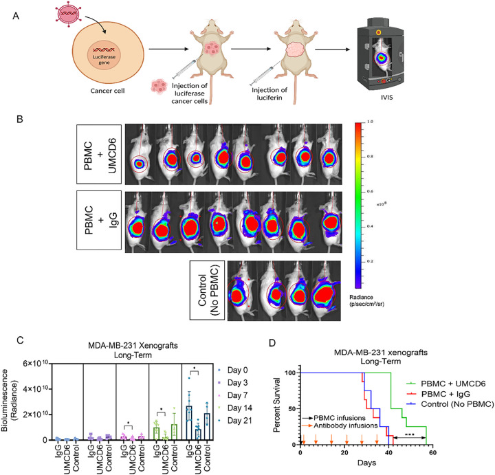
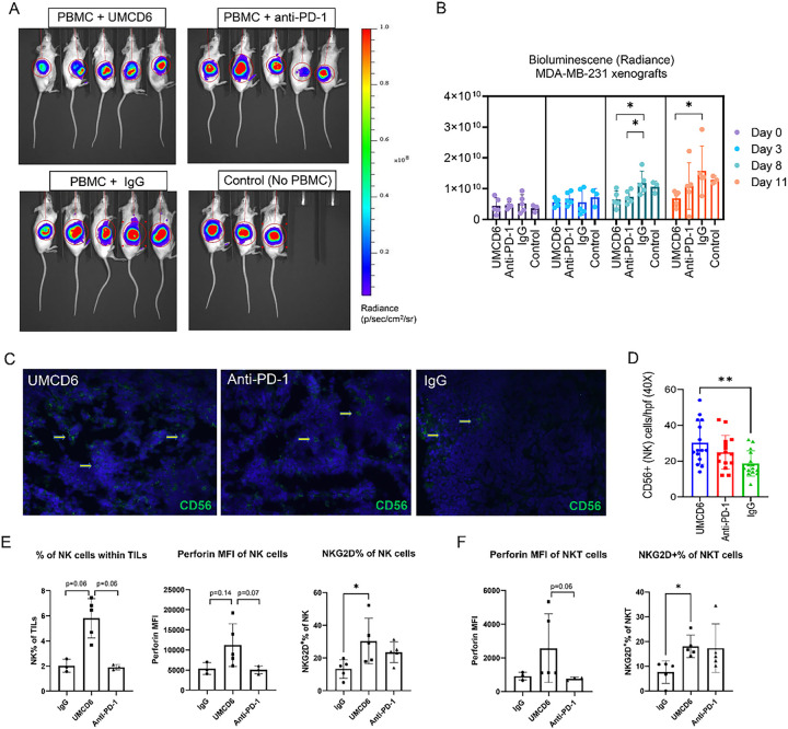
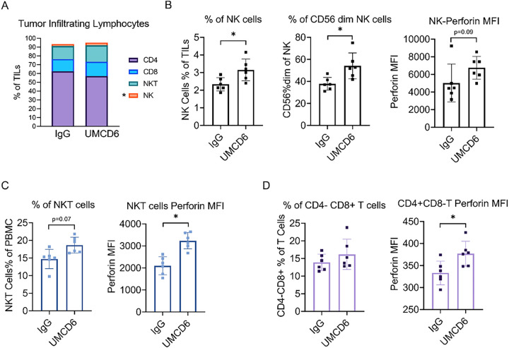
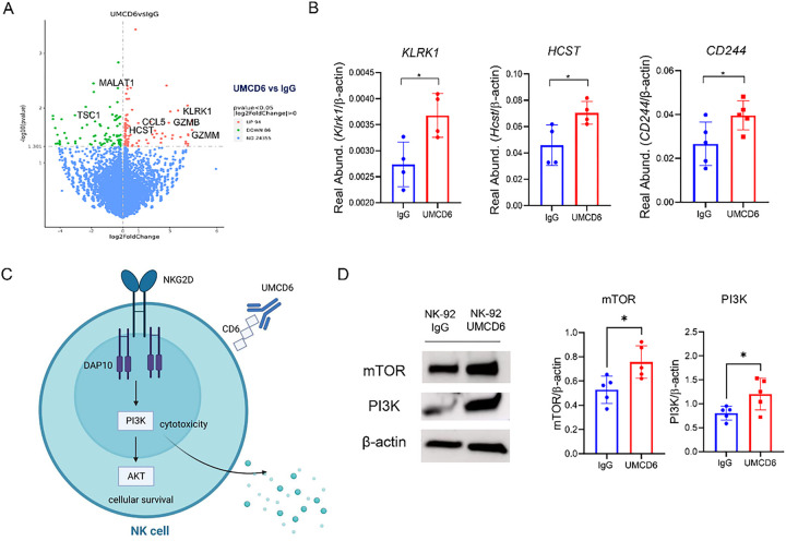
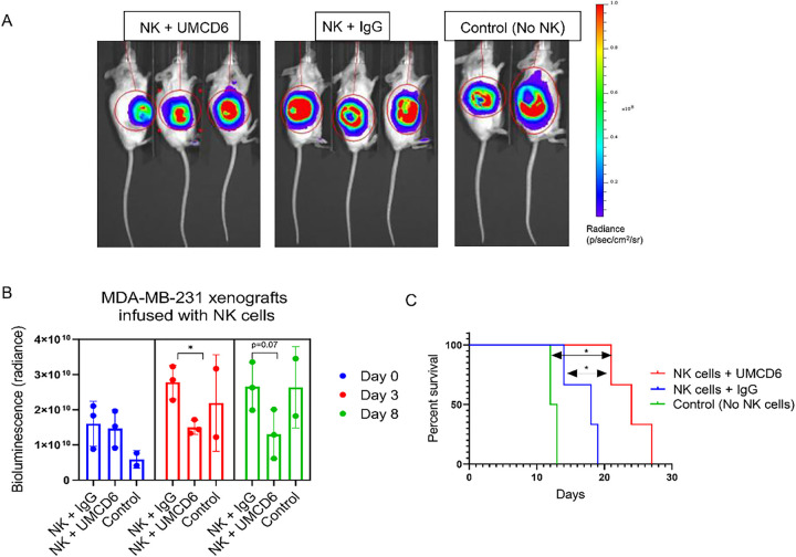
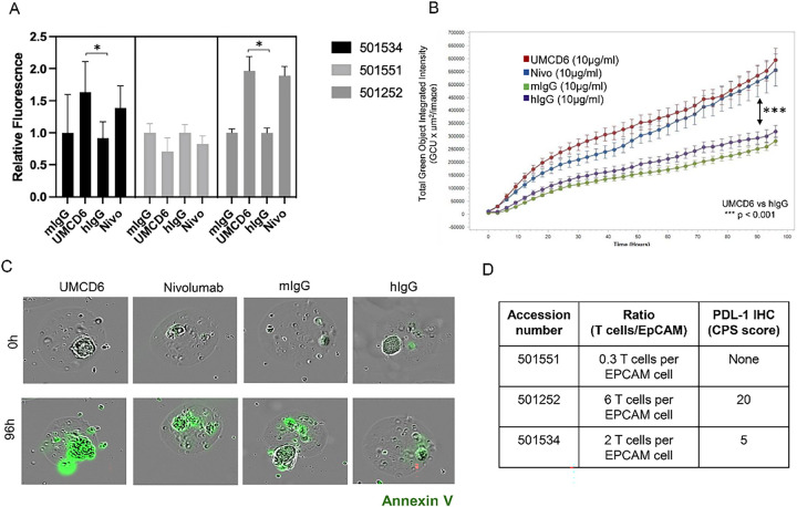
Immune checkpoint inhibitors (ICIs) have demonstrated efficacy and improved survival in a growing number of cancers. Despite their success, ICIs are associated with immune-related adverse events that can interfere with their use. Therefore, safer approaches are needed. CD6, expressed by T-lymphocytes and human NK cells, engages in cell-cell interactions by binding to its ligands CD166 (ALCAM) and CD318 (CDCP1). CD6 is a target protein for regulating immune responses and is required for the development of several mouse models of autoimmunity. Interestingly, CD6 is exclusively expressed on immune cells while CD318 is strongly expressed on most cancers. Here we demonstrate that disrupting the CD6-CD318 axis with UMCD6, an anti-CD6 monoclonal antibody, prolongs survival of mice in xenograft models of human breast and prostate cancer, treated with infusions of human lymphocytes. Analysis of tumor-infiltrating immune cells showed that augmentation of lymphocyte cytotoxicity by UMCD6 is due to effects of this antibody on NK, NKT and CD8+ T cells. Tumor-infiltrating cytotoxic lymphocytes were found in higher proportions and were activated in UMCD6-treated mice compared to controls. Similar changes in gene expression were observed by RNA-seq analysis of NK cells treated with UMCD6. Particularly, UMCD6 up-regulated the NKG2D-DAP10 complex and activated PI3K. Thus, the CD6-CD318 axis can regulate the activation state of cytotoxic lymphocytes and their positioning within the tumor microenvironment.

Keywords: CD6; NK; cytotoxic lymphocyte; immunotherapy.


Conflict of interest statement

Competing Interests: Drs. Fox and Lin hold equity in Abcon, a company created to test the use of therapeutic agents that interrupt the interactions between CD6 and CD6 ligands.

Figures

Figure 1
Figure 1. UMCD6 increases survival and augments killing by human PBMC of a breast cancer line xenotransplanted into immunodeficient mice.
A: Schematic representation of in vivo visualization of tumor growth by the IVIS imaging system. B: 2×10⁶ MDA-MB-231 cells were inoculated s.c. in the abdomen of female SCID/beige mice. Once tumors reached 1 cm, mice were administered 10×106 human PBMCs by tail vein (day 0). The next day, mice were injected with 0.1 mg control IgG or UMCD6. C: Tumor growth, measured by IVIS, showed a robust decrease in bioluminescence signal in mice treated with UMCD6 compared to IgG and control (not administered PBMCs nor antibodies). The effect of UMCD6 on tumor volume can be seen from day 7 after UMCD6 administration (**p < 0.01) and was maintained until mice were euthanized (**p < 0.01). Data represents mean of 4–8 animals ± SD. D: Survival was significantly prolonged in the UMCD6 group compared to the IgG and control groups (***p < 0.001).
Figure 2
Figure 2. The effect of UMCD6 is more sustained than pembrolizumab in the killing of MDA-MB-231-derived xenograft tumors by PBMC.
A and B: We conducted a short-term in vivo experiment in which SCID/mice were first inoculated with MDA-MB-231 breast cancer cells, then administered 10×106 human PBMCs and antibodies (UMCD6, pembrolizumab or IgG; 100 μg). Both UMCD6 and pembrolizumab enhanced tumor killing by PBMC at day 8 compared to IgG control (*p < 0.05), but only UMCD6 showed statistical significance at day 11 (*p < 0.05). Data represents mean of 4–5 animals ± SD. C: Representative pictures of tumor tissues immunostained for CD56 (human NK cell marker) at day 12 after treatment with UMCD6. D: Immunofluorescence of tumor sections stained for the presence of NK cells showed that mice administered UMCD6 had an increased number of tumor-infiltrating NK cells compared to IgG control group (40X). E: Characterization of tumor-infiltrating lymphocytes (TILs) by flow cytometry shows that tumor-infiltrating NK cells are found in higher proportions in mice treated with UMCD6. NK cells are more abundant in UMCD6-treated mice (average of 5.8% among TILs) compared with IgG (2.01%) and anti-PD-1 (1.92%). Moreover, tumor-infiltrating NK cells from UMCD6-treated xenografts express higher levels of the activating receptor NKG2D (*p < 0.05 vs IgG). F: Similarly, NKG2D expression in tumor-infiltrating NKT cells is significantly elevated upon treatment with UMCD6 (*p < 0.05 vs IgG), accompanied with an increase in perforin expression (ns).
Figure 3
Figure 3. UMCD6 increases cytotoxicity of tumor-infiltrating lymphocytes (TILs).
A: TILs from breast cancer MDA-MB-231 xenograft tumors were analyzed by flow cytometry 3 days after treatment with UMCD6 or IgG antibodies. TILs from UMCD6-treated mice had higher levels of NK cells (3.14% ± 0.6), NKT cells (18.65% ± 2.25) and CD8+ T cells (16.18% ± 4.28) compared to TILs from IgG-treated mice: NK cells (2.33% ± 0.37), NKT cells (14.72% ± 2.74) and CD8+ T cells (13.90% ± 2.33). On the contrary, CD4+ T cells from UMCD6-treated mice were found in lower proportions (57.16% ± 5.79) compared to IgG-treated mice (62.15% ± 6.28). B: We found a significant increased frequency of NK cells (*p < 0.05), specifically CD56 dim cells (*p < 0.05), in UMCD6-treated mice compared to IgG-treated mice. C: CD3+CD56+ NKT cells from UMCD6-treated mice were found to have enhanced perforin expression compared to control-treated mice (*p < 0.05). D: Perforin production was significantly up-regulated in a small portion of CD4 cells (*p< 0.05) in UMCD6-treated mice.
Figure 4
Figure 4. UMCD6 alters gene expression in human NK cells to enhance cytotoxic function.
A: RNA-seq data obtained using NK-92 cells stimulated with UMCD6 in a 6-hour culture shows widespread changes in gene expression of 180 genes. B: Up-regulation of the activating NK receptors NKG2D (Klrk1), DAP10 (Hcst) and 2B4 (CD244), shown to be involved in the activation of NK cells, was confirmed by RT-PCR. C: Schematic representation of the role of UMCD6 in the activation of NK cells. Internalization of CD6 by UMCD6 up-regulates the expression of the activating receptor NKG2D-DAP10 and PI3K pathway. D: PI3K and mTOR expression, down-stream pathways of NKG2D-DAP10 signaling complex, were found up-regulated at protein level in a 72-hour co-culture of NK-92 cells with UMCD6. Data expressed as mean +/− SD and *p<0.05.
Figure 5
Figure 5. UMCD6 enhances NK killing of human breast cancer cells in vivo.
A: Bioluminescence images of breast cancer MDA-MB-231 SCID/beige xenografts infused with 1×10⁶ human NK cells and injected with 100 μg of antibodies (UMCD6 or IgG) at day 7. Control mice were not administered NK cells or antibodies. B: Bioluminescence imaging of MDA-MB-231 mice revealed a decrease in tumor growth in mice receiving NK cells and UMCD6 at day 3 and 8 compared to mice receiving NK cells and IgG control antibody. C: Survival rate was significantly increased in the UMCD6 group compared to both the IgG and control groups. UMCD6 vs. IgG, *p = 0.0246; UMCD6 vs. untreated, *p = 0.0389.
Figure 6
Figure 6. UMCD6 enhances killing of patient-derived lung cancer micro-organospheres (MOS).
A: Lung cancer MOS generated from cancer patient tissues were used in co-cultures with PBMC pre-incubated with UMCD6, nivolumab (anti-PD-1), mouse IgG or human IgG antibodies at 10 μg/ml. UMCD6 induces apoptosis of lung tumor cells in MOS, at least as efficiently as nivolumab, a PD-1 inhibitor in 2 out of 3 samples (501534 and 501252). Tumor cell killing was measured as the number and relative fluorescence of cancer cells in each well expressing Annexin-V (green fluorescence). B: UMCD6 showed superiority to mouse and human IgG (after 12 hours; (***p < 0.001)) and anti-PD-1 (at 42 hours; (*p < 0.05); (data expressed as mean ± SD; green fluorescence, Annexin-V sensitive with y-axis linear). C: Incucyte® images from lung cancer organoids (501252) at day 4 in the presence of antibodies. D: T-cell/epithelial cell ratio (T cells/EpCAM ratio) and presence of PDL-1 were measured in each tumor MOS samples.

Similar articles

  • Activation of cytotoxic lymphocytes through CD6 enhances killing of cancer cells.
    Gurrea-Rubio M, Wu Q, Amin MA, Tsou PS, Campbell PL, Amarista CI, Ikari Y, Brodie WD, Mattichak MN, Muraoka S, Randon PM, Lind ME, Ruth JH, Mao-Draayer Y, Ding S, Shen X, Cooney LA, Lin F, Fox DA. Gurrea-Rubio M, et al. Cancer Immunol Immunother. 2024 Jan 27;73(2):34. doi: 10.1007/s00262-023-03578-1. Cancer Immunol Immunother. 2024. PMID: 38280067 Free PMC article.
  • Itolizumab regulates activating and inhibitory signals on effector cells, improving their cytotoxicity against CD318+ tumor cell lines.
    González Muñoz C, Álvarez Arzola R, Calvo Pérez A, Frometa Campanón MC, Hernández Casaña P, Fernández-Calienes Valdés A, Lorenzo-Luaces P, Mazorra Herrera Z, Crombet Ramos T, Labrada Mon M. González Muñoz C, et al. Front Immunol. 2025 May 5;16:1585597. doi: 10.3389/fimmu.2025.1585597. eCollection 2025. Front Immunol. 2025. PMID: 40391211 Free PMC article.
  • CD6 is a target for cancer immunotherapy.
    Ruth JH, Gurrea-Rubio M, Athukorala KS, Rasmussen SM, Weber DP, Randon PM, Gedert RJ, Lind ME, Amin MA, Campbell PL, Tsou PS, Mao-Draayer Y, Wu Q, Lanigan TM, Keshamouni VG, Singer NG, Lin F, Fox DA. Ruth JH, et al. JCI Insight. 2021 Mar 8;6(5):e145662. doi: 10.1172/jci.insight.145662. JCI Insight. 2021. PMID: 33497367 Free PMC article.
  • CD6 in Human Disease.
    Gurrea-Rubio M, Fox DA, Castresana JS. Gurrea-Rubio M, et al. Cells. 2025 Feb 13;14(4):272. doi: 10.3390/cells14040272. Cells. 2025. PMID: 39996744 Free PMC article. Review.
  • CD6 and Its Interacting Partners: Newcomers to the Block of Cancer Immunotherapies.
    Aragón-Serrano L, Carrillo-Serradell L, Planells-Romeo V, Isamat M, Velasco-de Andrés M, Lozano F. Aragón-Serrano L, et al. Int J Mol Sci. 2023 Dec 15;24(24):17510. doi: 10.3390/ijms242417510. Int J Mol Sci. 2023. PMID: 38139340 Free PMC article. Review.

References

    1. Martinez VG, Moestrup SK, Holmskov U, Mollenhauer J, Lozano F. The conserved scavenger receptor cysteine-rich superfamily in therapy and diagnosis. Pharmacol Rev. 2011; 63(4):967–1000. - PubMed
    1. Braun M, Muller B, ter Meer D, Raffegerst S, Simm B, Wilde S, Spranger S, Ellwart J, Mosetter B, Umansky L, Lerchl T, Schendel DJ, Falk CS. The CD6 scavenger receptor is differentially expressed on a CD56 natural killer cell subpopulation and contributes to natural killer-derived cytokine and chemokine secretion. J Innate Immun. 2011; 3(4):420–434. - PubMed
    1. Li Y, Singer NG, Whitbred J, Bowen MA, Fox DA, Lin F. CD6 as a potential target for treating multiple sclerosis. Proc Natl Acad Sci U S A. 2017; 114(10):2687–2692. - PMC - PubMed
    1. Li Y, Ruth JH, Rasmussen SM, Athukorala KS, Weber DP, Amin MA, Campbell PL, Singer NG, Fox DA, Lin F. Attenuation of Murine Collagen-Induced Arthritis by Targeting CD6. Arthritis Rheumatol. 2020; 72(9):1505–1513. - PMC - PubMed
    1. Zhang L, Li Y, Qiu W, Bell BA, Dvorina N, Baldwin WM 3rd, Singer N, Kern T, Caspi RR, Fox DA, Lin F. Targeting CD6 for the treatment of experimental autoimmune uveitis. J Autoimmun. 2018; 90:84–93. - PMC - PubMed

Publication types