[Preprint]. 2023 Oct 9:rs.3.rs-3346012.
doi: 10.21203/rs.3.rs-3346012/v1.

Neutrophil swarms containing myeloid-derived suppressor cells are crucial for limiting oral mucosal infection by C. albicans

Affiliations

Neutrophil swarms containing myeloid-derived suppressor cells are crucial for limiting oral mucosal infection by C. albicans

Mira Edgerton et al. Res Sq. .

Update in

Abstract

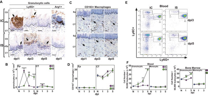
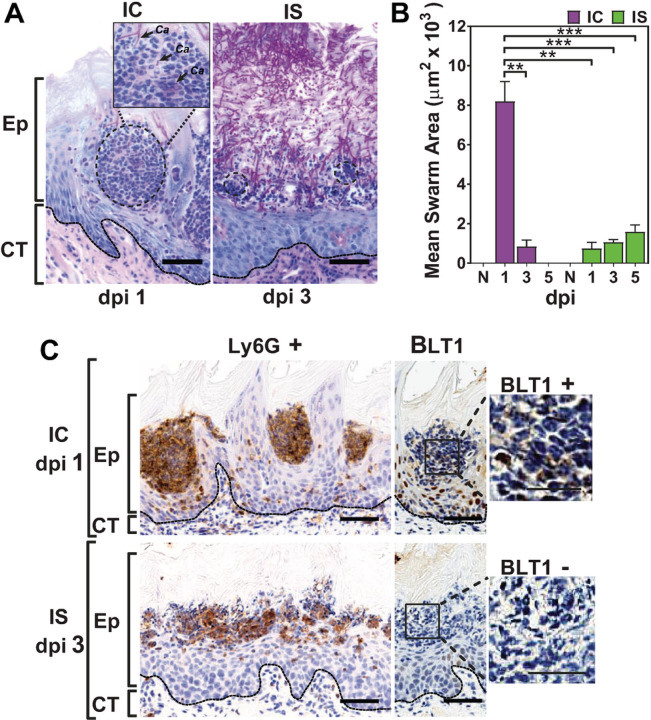
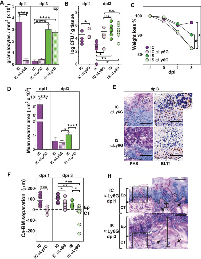
Oral mucosal colonization by C. albicans (Ca) is benign in healthy people but progresses to deeper infection known as oropharyngeal candidiasis (OPC) that may become disseminated when combined with immunosuppression. Cortisone-induced immunosuppression is a well-known risk factor for OPC, however the mechanism by which it permits infection is poorly understood. Neutrophils are the primary early sentinels preventing invasive fungal growth, and here we identify that in vivo neutrophil functional complexes known as swarms are crucial for preventing Ca invasion which are disrupted by cortisone. Neutrophil swarm function required leukotriene B4 receptor 1 (BLT1) expression, and swarms were further characterized by peripheral association of polymorphonuclear myeloid-derived suppressor cells (PMN-MDSCs) showing that OPC recruits PMN-MDSCs to this site of infection. Furthermore, PMN-MDSCs associated with Ca hyphae had no direct antifungal effect but showed prolonged survival times and increased autophagy. Thus in vivo neutrophil swarms are complex structures with spatially associated PMN-MDSCs that likely contribute immunoregulatory functions to resolve OPC. These swarm structures have an important function in preventing deep invasion by Ca within the oral mucosa and represent a mechanism for increased disease severity under immune deficient clinical settings.


Conflict of interest statement

Competing Interests. None

Figures

Figure 1
Figure 1. Immunosuppression (IS) impairs inflammatory cell recruitment and alters epithelial morphology.
Cortisone-treated mice (IS) and immunocompetent (IC) mice were infected sublingually with 107 CFU of C. albicans (Ca) and tongues were collected from 1 to 5 days post-infection (dpi) (5–7 mice/ group). A) Percentage of weight loss after Ca infection as compared to pre-infection weight. B) Number of CFU/ g tongue tissue in IC and IS mice shows rapid clearance of Ca in IC mice and a persistence of infection in IS mice. C-E) Representative co-localized tongue sections from IC and IS mice after Ca infection in IC and IS mice stained for PAS and H&E (n=3 mice/group). C) IC mice show early and robust intraepithelial recruitment of inflammatory cells to the areas of candida invasion as compared to IS mice (square) (dpi1). D) Vertical bars show differences in cellular epithelial thickness (cEp) between IC and IS mice at dpi3. E) Arrow indicates detachment of corneum stratum from underlying cEp in IS mice at dpi5. F) Changes in cEp thickness (mm) and (G) and corneum stratum thickness as compared to naive (N) mice from dpi1-3. ** p< 0.01; *** p< 0.001; **** p<0.0001 (Mann-Whitney or 2-tailed t-test). Scale bar=100 mm
Figure 2
Figure 2. Oral fungal infection leads to increased recruitment of neutrophils but not macrophages.
Representative slides showing recruitment of Ly6G+ granulocytes and a subset of Ly6G+ Arginase (Arg) 1+ granulocytes into the tongue epithelium (Ep) of IC and IS mice. B) Changes in recruitment of Ly6G+MPO+ granulocytes / mm2 (x103) from IC and IS mice as compared to naive (N) at dpi1-5 (n=3 / group). C) Representative slides showing recruitment of CD163+ macrophages into the connective tissue (CT) and not the Ep of both IC and IS mice. D) Changes in recruitment of CD163+ macrophages / mm2 in IC and IS mice as compared to N at dpi1-5. E) Representative FACS plots of CD45+CD11b+Ly6Ghigh granulocytic cells and CD45+CD11b+Ly6Glo monocytic cells in blood collected from IC and IS mice at dpi3-5 showing a significant expansion of granulocytic cells and not monocytic cells. F) Changes in monocytic and granulocytic cells in blood (dpi1-5) and bone marrow (BM) (dpi1-3) collected from IC and IS mice as compared to N (n= 3–7 mice/group, except for dpi 5 where n=2 mice/group). * p<0.05; ** p< 0.01; *** p< 0.001; **** p<0.0001 (Mann-Whitney or 2-tailed t-test). Scale bar=50 mm
Figure 3
Figure 3. Ly6G+ Arg1+ cells show immunosuppressive activity on CD4+ T cells confirming the expansion of PMN-MDSCs in response to oral candidiasis.
A) Representative slides showing epithelial infiltration of Ly6G+ Arg+ cells in IC and IS mice on dpi 1–5. B) Ly6G+ Arg+ granulocytes/ mm2 show peak recruitment on dpi1 for IC mice and dpi3-5 for IS mice (3–4 mice / group). C) Percentages of Ly6G+Arg+ granulocytes to total Ly6G+ granulocytes were higher on dpi 1 for IC and dpi 5 for IS mice. D) CD11b+ Ly6G+ enriched single cells suspensions from BM and spleen of IC and IS mice showed inhibition of spleen-derived CD4+ T cell proliferation on dpi3-5 (graphs are representative of two independent experiments). * p< 0.05; ** p< 0.01; *** p< 0.001; **** p<0.0001 (t-test). Scale bar=50 mm
Figure 4
Figure 4. Neutrophil swarming and BLT1 expression are reduced by immunosuppression.
A) PAS staining of tongue sections showing the association between neutrophil swarms (circles) and Ca. B) Measurements of mean swarm area (mm2 × 103) showed significant swarm area reduction in IS mice on dpi1-5 as compared to the swarm area of IC mice on dpi1. B) Representative tissue sections showing formation of multiple intraepithelial Ly6G+ BLT1+ neutrophil swarms in IC mice (upper panel), whereas IS mice show minimal swarm formation and absence of BLT1 expression. ** p< 0.01; *** p< 0.001; **** p<0.0001 (t-test). Scale bar=50 mm (25 mm for enlarged BLT-1 insert) (n=3 mice / group per time point).
Figure 5
Figure 5. Cortisone-induced immunosuppression delays expression of proinflammatory cytokines and chemokines in response to fungal infection.
Freshly extracted tongue tissues were dissected for separate protein isolation in epithelium (Ep) and connective tissue (CT) (n=5 mice/group per time point). A) Kinetics of MPO (ELISA) used as a surrogate marker of neutrophil and PMN-MDSC recruitment in IC and IS mice as compared to naive (N) mice. B-D) Kinetics of IL-1b (B), G-CSF (C), MIP-2 (D), and KC (E) (measured by Bio-Plex assay) in IC and IS mice as compared to N mice followed a similar pattern as MPO in dpi1-5 at Ep and CT. F) IL-17 was increased only in CT of IC mice, and not in IS mice, as compared to naïve (N) mice (dpi1-3). * p< 0.05; ** p< 0.01; *** p< 0.001; **** p<0.0001 as compared to naïve (N) mice (ANOVA and Tukey’s multiple comparisons)
Figure 6
Figure 6. Treatment with anti-Ly6G caused disruption of neutrophil swarming and increased Ca invasion into the connective tissue.
Neutrophils were depleted by i.p. injection of anti-Ly6G antibody on dpi-1 and anti-GCF antibody on dpi-1 through 2 (n=3–6 mice/ group per time point). Depletion was confirmed by flow cytometry. A) Ly6G+ cell depletion resulted in a significant reduction in total neutrophils in IC mice on dpi1-3, whereas IS mice showed increased neutrophil recruitment on dpi3 regardless of depletion. B) Number of CFU / g tongue tissue in anti-Ly6G+ treated IC and IS mice showing delayed clearance of Ca and a persistence of infection on dpi 3 comparable to isotype-treated IS mice. C) Anti-Ly6G+ treatment resulted in significant weight loss in IC and IS mice as compared to isotype-treated IC and IS mice by dpi 3. D) Ly6G+ cell-depletion in IC mice resulted in significant reduction in swarm formation on dpi1, with small swarm formation on dpi3, whereas depletion caused complete disruption of swarm formation in IS mice on dpi3. E) Representative co-localized sections for PAS and BLT1 staining, showing small BLT-1+ swarm formation (circle) in IC anti-Ly6G+ cell-depleted mice on dpi 3, however, IS anti-Ly6G+ mice showed absence of swarm formation and BLT1 expression. F) The distance (mm) was measured between Ca and the basement membrane (BM) in PAS-stained tissue sections from IC and IS mice showing that anti-Ly6G+ treatment resulted in Ca invasion of connective tissue on dpi1-3. H) Representative PAS-stained slides showing CT invasion of Ca hyphae (distance indicated by vertical bar) in anti-Ly6G+ treated IC and IS mice. * p< 0.05; ** p< 0.01; *** p< 0.001; **** p<0.0001 (t-test, ANOVA and Tukey’s multiple comparisons). Scale bar=50 mm (25 mm for enlarged images).
Figure 7
Figure 7. Anti-Ly6G+ treatment did not change recruitment of PMN-MDSCs in IC and IS mice, and human PMN-MDSCs showed absence of Ca killing and phagocytic uptake of Ca.
A) Number of PMN-MDSCs / mm2 was increased by anti-Ly6G+ treatment in IC and IS mice on dpi3 (n=3–5 mice / group per time point). B) Co-localized PAS, Arg 1+ and COX-2 stained tongue tissue sections show co-localization with Ca of Arg+ granulocytes by image deconvolution, along with expression of COX-2 in IC and IS anti-Ly6G+ treated mice. C) Human PMN-MDSCs showed significantly lower (10-fold) phagocytic index of Ca cells, compared with human neutrophils. D) Fungicidal activity was not detected in human PMN-MDSCs cells. E) In vitro Ca yeast cells showed very little contact with PMN-MDSCs, while Ca hyphae were closely associated with the surfaces of PMN-MDSCs. F) Autophagy levels of human PMN-MDSCs (MFI) incubated with Ca hyphae cells reached 70% that of rapamycin, and was significantly higher than that incubated with Ca yeast cells. G) PMN-MDSCs survival was increased by 50% after 60 min exposure to Ca hyphae. * p< 0.05; ** p< 0.01; *** p< 0.001; **** p<0.0001 (t-test, ANOVA and Tukey’s multiple comparisons). Scale bar=25 mm

References

    1. Chaffin W.L.: Candida albicans cell wall proteins. Microbiol. Mol. Biol. Rev. 72, 495–544 (2008) - PMC - PubMed
    1. Kumar R., Saraswat D., Tati S., Edgerton M.: Novel aggregation properties of Candida albicans secreted aspartyl proteinase Sap6 mediate virulence in oral candidiasis. Infect. Immun. 83, 2614–2626 (2015) - PMC - PubMed
    1. Mosci P., et al.: Involvement of IL-17A in preventing the development of deep-seated candidiasis from oropharyngeal infection. Microbes Infect. 16, 678–689 (2014) - PubMed
    1. Davidson L., Netea M.G., Kullberg B.J.: Patient susceptibility to Candidiasis-a potential for adjunctive immunotherapy. J. Fungi (Basel) 4 (2018) - PMC - PubMed
    1. Jabra-Rizk M.A., et al.: Candida albicans pathogenesis: fitting within the host-microbe damage response framework. Infect. Immun. 84, 2724–2739 (2016) - PMC - PubMed

Publication types

LinkOut - more resources