[Preprint]. 2023 Sep 25:2023.09.22.23295974.
doi: 10.1101/2023.09.22.23295974.

Cortical lesions uniquely predict motor disability accrual and form rarely in the absence of new white matter lesions in multiple sclerosis

Affiliations

Cortical lesions uniquely predict motor disability accrual and form rarely in the absence of new white matter lesions in multiple sclerosis

Erin S Beck et al. medRxiv. .

Abstract

Background and objectives: Cortical lesions (CL) are common in multiple sclerosis (MS) and associate with disability and progressive disease. We asked whether CL continue to form in people with stable white matter lesions (WML) and whether the association of CL with worsening disability relates to pre-existing or new CL.

Methods: A cohort of adults with MS were evaluated annually with 7 tesla (T) brain magnetic resonance imaging (MRI) and 3T brain and spine MRI for 2 years, and clinical assessments for 3 years. CL were identified on 7T images at each timepoint. WML and brain tissue segmentation were performed using 3T images at baseline and year 2.

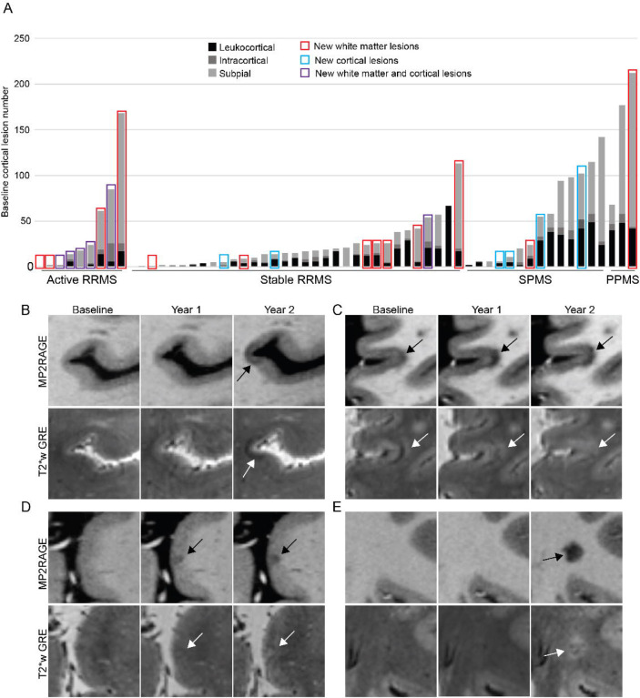
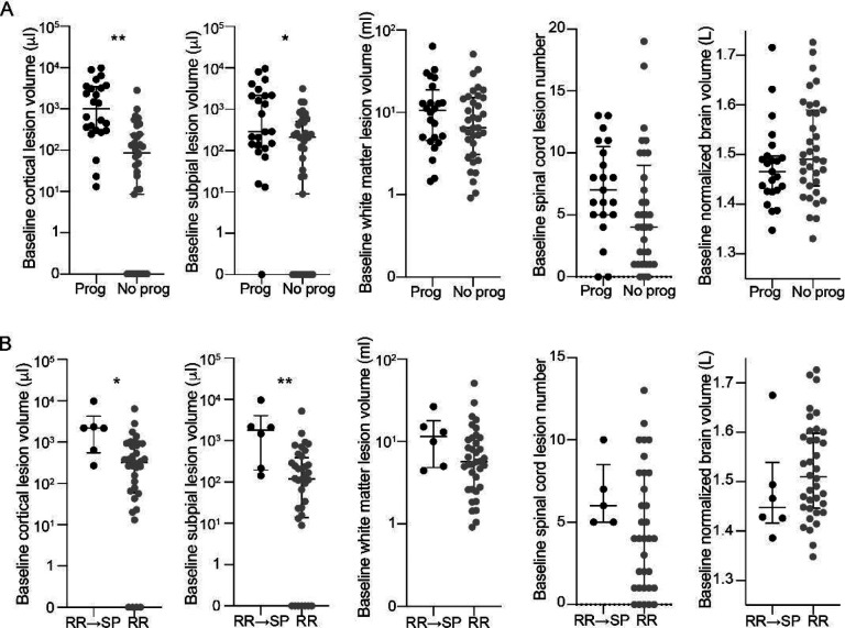
Results: 59 adults with MS had ≥1 7T follow-up visit (mean follow-up time 2±0.5 years). 9 had "active" relapsing-remitting MS (RRMS), defined as new WML in the year prior to enrollment. Of the remaining 50, 33 had "stable" RRMS, 14 secondary progressive MS (SPMS), and 3 primary progressive MS. 16 total new CL formed in the active RRMS group (median 1, range 0-10), 7 in the stable RRMS group (median 0, range 0-5), and 4 in the progressive MS group (median 0, range 0-1) (p=0.006, stable RR vs PMS p=0.88). New CL were not associated with greater change in any individual disability measure or in a composite measure of disability worsening (worsening Expanded Disability Status Scale or 9-hole peg test or 25-foot timed walk). Baseline CL volume was higher in people with worsening disability (median 1010μl, range 13-9888 vs median 267μl, range 0-3539, p=0.001, adjusted for age and sex) and in individuals with RRMS who subsequently transitioned to SPMS (median 2183μl, range 270-9888 vs median 321μl, range 0-6392 in those who remained RRMS, p=0.01, adjusted for age and sex). Baseline WML volume was not associated with worsening disability or transition from RRMS to SPMS.

Discussion: CL formation is rare in people with stable WML, even in those with worsening disability. CL but not WML burden predicts future worsening of disability, suggesting that the relationship between CL and disability progression is related to long-term effects of lesions that form in the earlier stages of disease, rather than to ongoing lesion formation.


Figures

Figure 1.
Figure 1.
New cortical lesion formation is rare in people with stable white matter lesions. (A) New cortical and white matter lesion formation was frequent in people with active RRMS (defined as having at least one new or enhancing white matter lesion in the year prior to enrollment) but was infrequent in people with radiographically stable RRMS or progressive MS. New cortical lesion formation was not related to baseline cortical lesion number (gray bars, each bar represents a single participant). (B-D) Appearance of new cortical lesions can differ from the appearance of chronic cortical lesions (cortical lesions are denoted with arrows). (B) A subpial lesion that was new on the year-2 MRI appears hypointense on T2*w GRE. (C) A leukocortical lesion that was present at baseline is initially hyperintense on T2*w GRE with a band of hypointensity at the cortex-white matter junction. This lesion expands over time and becomes more hypointense on T1w MP2RAGE, while at the same time the hypointense band on T2*w GRE disappears. (D) A subpial lesion that was new on the year-1 MRI initially appears isointense on T2*w GRE and then becomes more hyperintense at year-2. (E) A white matter lesion in the same individual as (C) was new on the year-2 MRI and is hyperintense on T2*w GRE. RRMS: relapsing remitting multiple sclerosis. SPMS: secondary progressive multiple sclerosis. PPMS: primary progressive multiple sclerosis. T2*w GRE: T2* weighted gradient recalled echo. MP2RAGE: magnetization prepared 2 rapid acquisition gradient echo.
Figure 2.
Figure 2.
People with worsening disability and transition from relapsing to progressive MS have higher cortical lesion burden at baseline. (A) Individuals with progression of disability during the study (“Prog,” defined as an increase in EDSS of ≥1 for baseline EDSS <6 or ≥0.5 for baseline EDSS ≥6 or 20% increase in 25TW or 20% increase in 9HPT) had higher baseline cortical lesion volume and subpial lesion volume, but no difference in baseline white matter lesion volume, spinal cord lesion number, or normalized brain volume, compared to people without progression of disability (“No prog”). Similar results were observed when comparing individuals who transitioned from relapsing remitting to secondary progressive MS with those who remained relapsing remitting (B). Comparisons were performed using a multivariable generalized linear model adjusted for age and sex. RR: relapsing remitting. SP: secondary progressive. * p<0.05, ** p<0.01

References

    1. Kuhlmann T, Moccia M, Coetzee T, et al. Multiple sclerosis progression: time for a new mechanism-driven framework. Lancet Neurol. Jan 2023;22(1):78–88. doi:10.1016/S1474-4422(22)00289-7 - DOI - PMC - PubMed
    1. Reich DS, Lucchinetti CF, Calabresi PA. Multiple Sclerosis. N Engl J Med. Jan 11 2018;378(2):169–180. doi:10.1056/NEJMra1401483 - DOI - PMC - PubMed
    1. Calabrese M, Agosta F, Rinaldi F, et al. Cortical lesions and atrophy associated with cognitive impairment in relapsing-remitting multiple sclerosis. Arch Neurol. Sep 2009;66(9):1144–50. doi:10.1001/archneurol.2009.174 - DOI - PubMed
    1. Roosendaal SD, Moraal B, Pouwels PJ, et al. Accumulation of cortical lesions in MS: relation with cognitive impairment. Mult Scler. Jun 2009;15(6):708–14. doi:10.1177/1352458509102907 - DOI - PubMed
    1. Nielsen AS, Kinkel RP, Madigan N, Tinelli E, Benner T, Mainero C. Contribution of cortical lesion subtypes at 7T MRI to physical and cognitive performance in MS. Neurology. Aug 2013;81(7):641–9. doi:10.1212/WNL.0b013e3182a08ce8 - DOI - PMC - PubMed

Publication types