. 2023 Sep 28;45(10):7944-7955.
doi: 10.3390/cimb45100502.

Identification of T-Cell Epitopes Using a Combined In-Silico and Experimental Approach in a Mouse Model for SARS-CoV-2

Affiliations

Identification of T-Cell Epitopes Using a Combined In-Silico and Experimental Approach in a Mouse Model for SARS-CoV-2

Noam Erez et al. Curr Issues Mol Biol. .

Abstract

Following viral infection, T-cells are crucial for an effective immune response to intracellular pathogens, including respiratory viruses. During the COVID-19 pandemic, diverse assays were required in pre-clinical trials to evaluate the immune response following vaccination against SARS-CoV-2 and assess the response following exposure to the virus. To assess the nature and potency of the cellular response to infection or vaccination, a reliable and specific activity assay was needed. A cellular activity assay based on the presentation of short peptides (epitopes) allows the identification of T cell epitopes displayed on different alleles of the MHC, shedding light on the strength of the immune response towards antigens and aiding in antigen design for vaccination. In this report, we describe two approaches for scanning T cell epitopes on the surface glycoprotein of the SARS-CoV-2 (spike), which is utilized for attachment and entry and serves as an antigen in many vaccine candidates. We demonstrate that epitope scanning is feasible using peptide libraries or computational scanning combined with a cellular activity assay. Our scans identified four CD8 T cell epitopes, including one novel undescribed epitope. These epitopes enabled us to establish a reliable T-cell response assay, which was examined and used in various experimental mouse models for SARS-CoV-2 infection and vaccination. These approaches could potentially aid in future antigen design for vaccination and establish cellular activity assays against uncharacterized antigens of emerging pathogens.

Keywords: SARS-CoV-2; T-cell; T-cell epitope; immune response; mouse.


Conflict of interest statement

The authors declare no conflict of interest.

Figures

Figure 1
Figure 1
Testing immunodominance of epitopes from in-silico screen following VSV-ΔG-spike vaccination. C57BL/6 mice were vaccinated i.m. with either 106 pfu (n = 3) or 107 pfu (n = 3) VSV-ΔG-spike and sacrificed after seven days. The pool of splenocytes from each vaccinated group was used to evaluate IFNγ secreting T-cells by ELISpot assay in response to each peptide at a concentration of 2 μg/mL. Numbers at the upper left of each well represent the number of counted spots. In the positive control column (PMA + Ionomycin (PMA+I)), asterisk near the numbers represents wells in which some part was saturated; TNTC—too numerous to count.
Figure 2
Figure 2
Quantification of T-cell response to different spike epitopes. C57BL/6 mice were vaccinated i.m. with either 106 pfu (A) or 107 pfu (B) VSV-ΔG-spike and sacrificed after seven days. T-cell response to the different epitopes was evaluated by IFNγ ELISpot assay. For each vaccination dose, groups of three animals were used. Bars indicate means ± SEM. Values are normalized to spots per 106 seeded cells.
Figure 3
Figure 3
Evaluation of T-cell response to different T-cell epitopes by ICS Following VSV-ΔG-spike vaccination. C57BL/6 mice were vaccinated i.m. with 107 pfu VSV-ΔG-spike and sacrificed after seven days. Splenocytes were used to evaluate IFNγ secreting T-cells by ICS in response to each peptide at a concentration of 2 μg/mL.
Figure 4
Figure 4
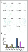
Evaluation of Spike epitopes from in-silico screen following SARS-CoV-2 infection. C57BL/6 mice were infected i.n. with 104 pfu MA10 SARS-CoV-2 and sacrificed after 21 days. T-cell response to different Spike epitopes was evaluated on a pool of splenocytes from infected animals (n = 5) by ELISpot assay (A) and quantified (B). Bars indicate means ± SEM of 3 replicates. Values were normalized to spots per 106 seeded cells.
Figure 5
Figure 5
T-cell activation following SARS-CoV-2 infection. K18-hACE2 mice were exposed to SARS-CoV-2 by i.n instillation (2 pfu) or i.m. immunization (106 pfu). T-cell activation was evaluated at day seven by ELISpot assay using peptide S539 (A) or S915 (B) at a concentration of 2 μg/mL. Bars indicate means ± SEM from 3 animals per group.

References

    1. Christensen J.P., Doherty P.C., Branum K.C., Riberdy J.M. Profound protection against respiratory challenge with a lethal H7N7 influenza A virus by increasing the magnitude of CD8+ T-cell memory. J. Virol. 2000;74:11690–11696. doi: 10.1128/JVI.74.24.11690-11696.2000. - DOI - PMC - PubMed
    1. Zhao J., Zhao J., Perlman S. T cell responses are required for protection from clinical disease and for virus clearance in severe acute respiratory syndrome coronavirus-infected mice. J. Virol. 2010;84:9318–9325. doi: 10.1128/JVI.01049-10. - DOI - PMC - PubMed
    1. Zhong W., Roberts A.D., Woodland D.L. Antibody-independent antiviral function of memory CD4+ T cells in vivo requires regulatory signals from CD8+ effector T cells. J. Immunol. 2001;167:1379–1386. doi: 10.4049/jimmunol.167.3.1379. - DOI - PubMed
    1. Tarke A., Sidney J., Methot N., Yu E.D., Zhang Y., Dan J.M., Goodwin B., Rubiro P., Sutherland A., Wang E., et al. Impact of SARS-CoV-2 variants on the total CD4+ and CD8+ T cell reactivity in infected or vaccinated individuals. Cell Rep. Med. 2021;2:100355. doi: 10.1016/j.xcrm.2021.100355. - DOI - PMC - PubMed
    1. Farber D.L., Yudanin N.A., Restifo N.P. Human memory T cells: Generation, compartmentalization and homeostasis. Nat. Rev. Immunol. 2014;14:24–35. doi: 10.1038/nri3567. - DOI - PMC - PubMed