. 2023 Oct 19;12(20):2488.
doi: 10.3390/cells12202488.

Inducing Angiogenesis in the Nucleus Pulposus

Affiliations

Inducing Angiogenesis in the Nucleus Pulposus

Sheela R Damle et al. Cells. .

Abstract

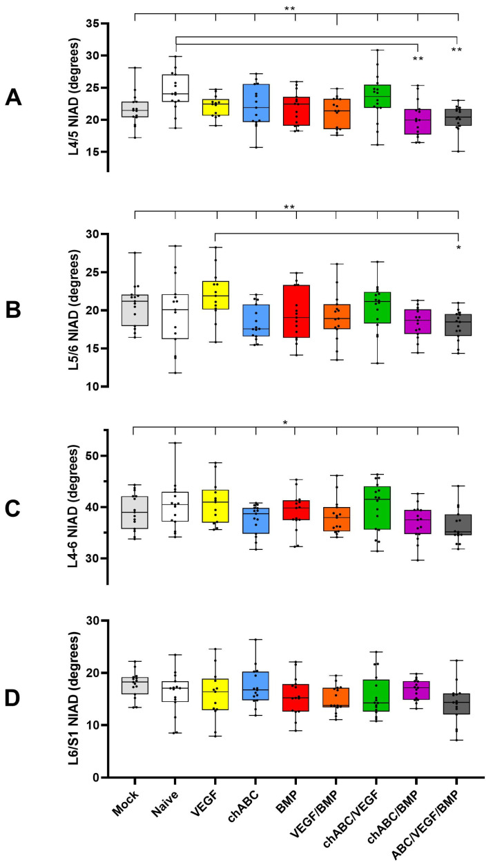
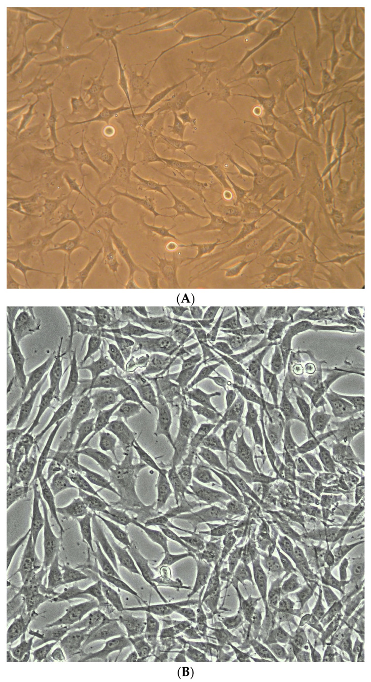
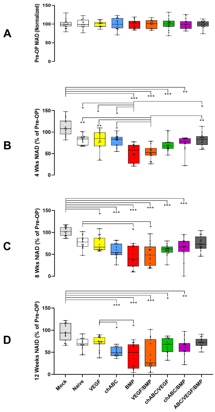
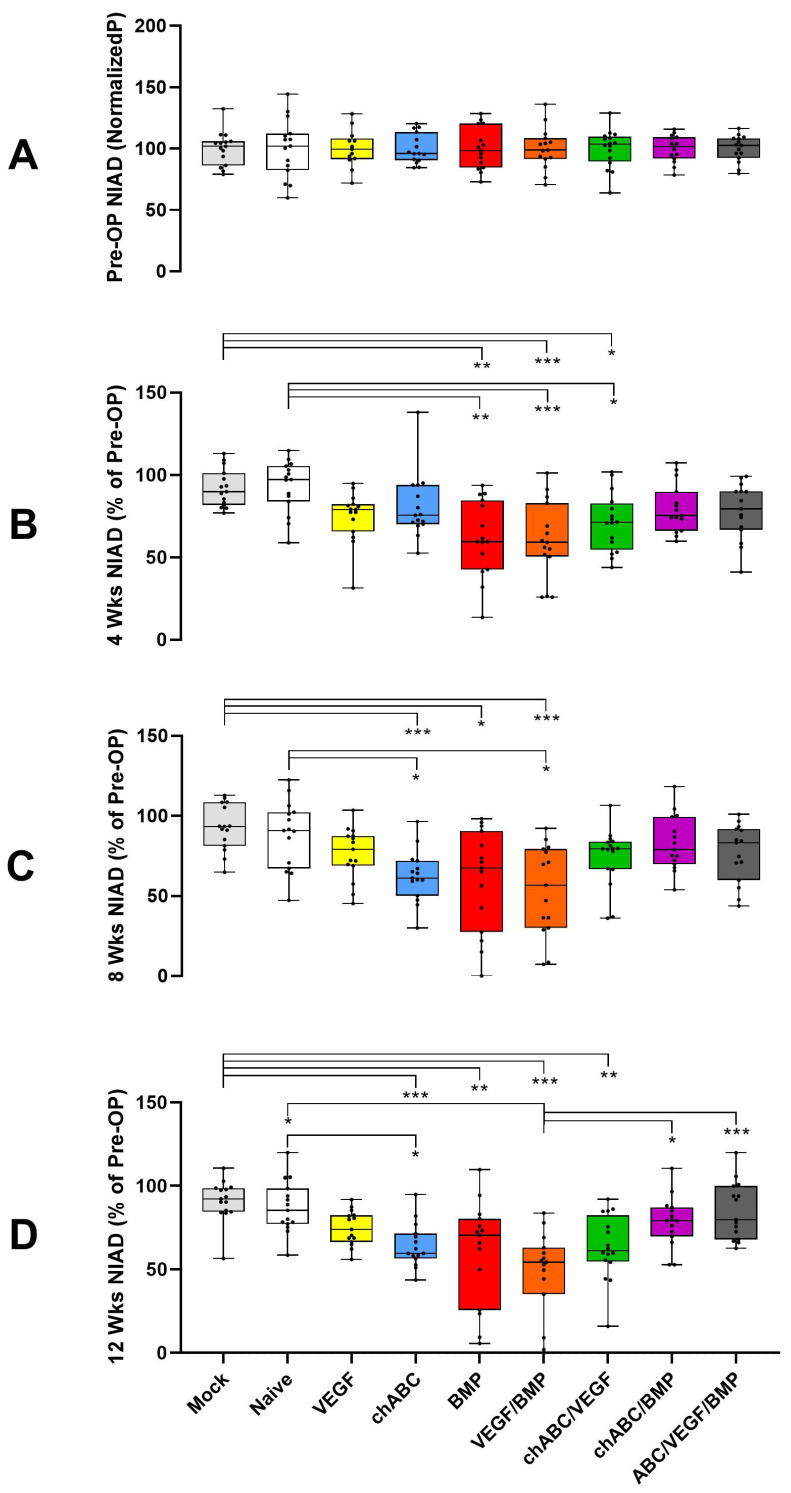
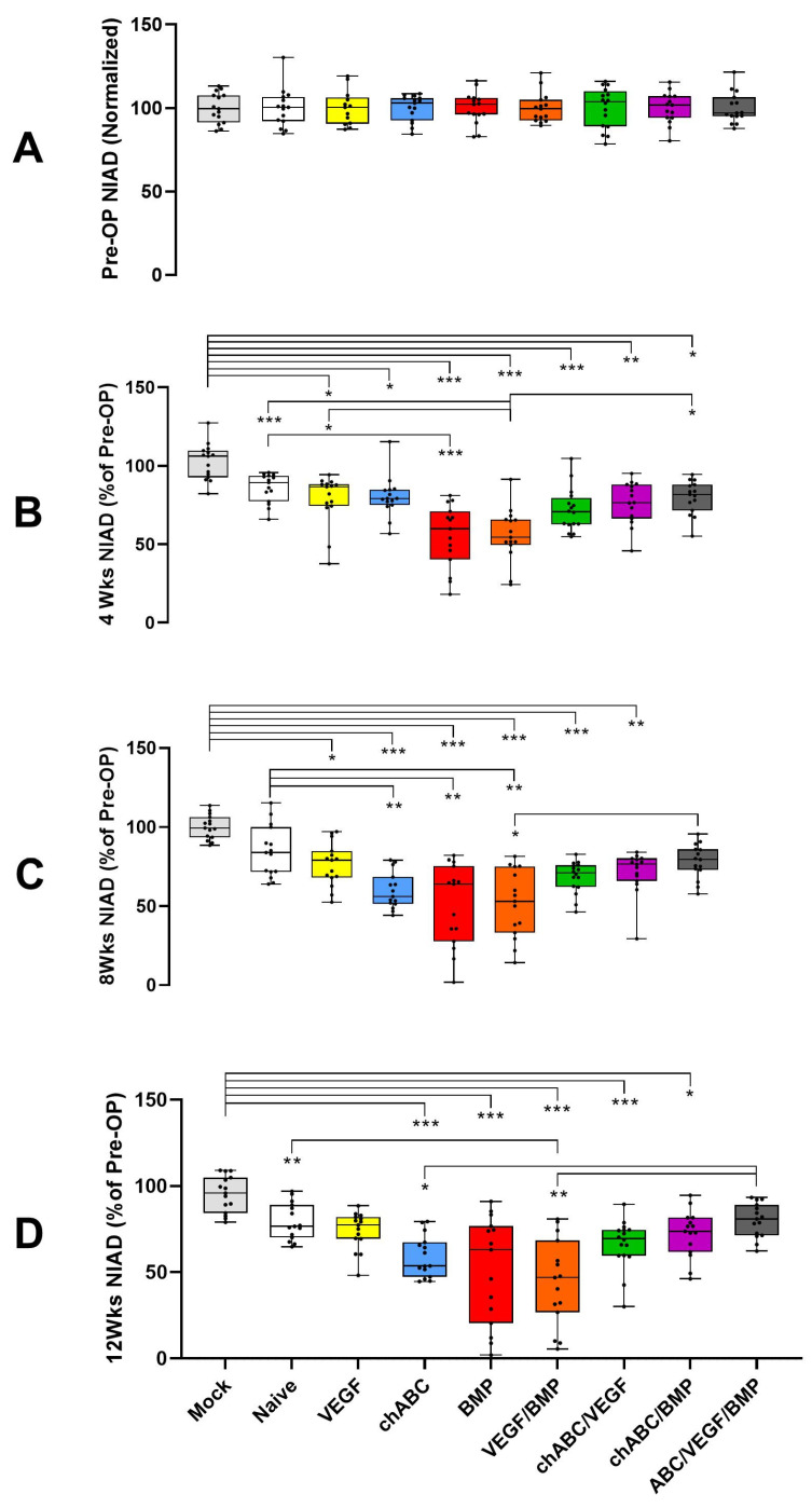
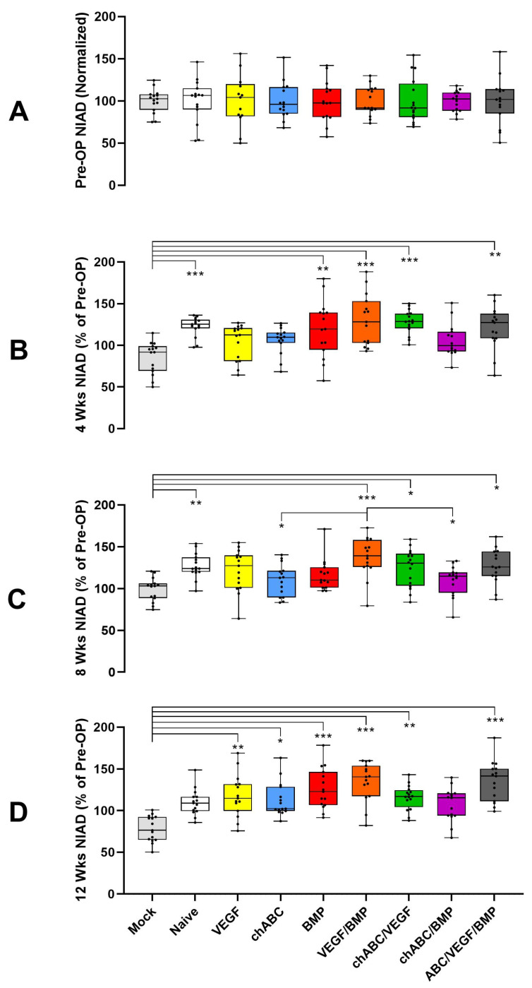
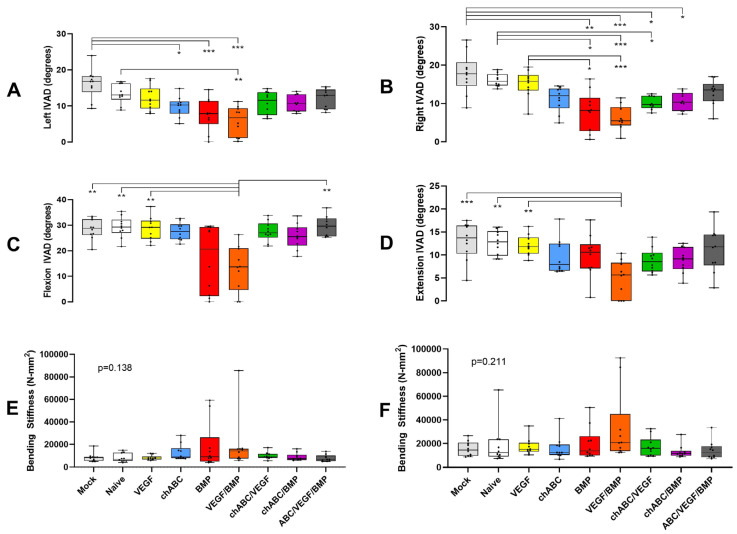
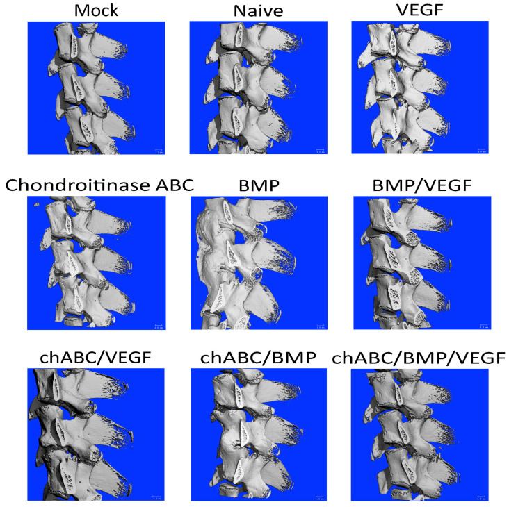
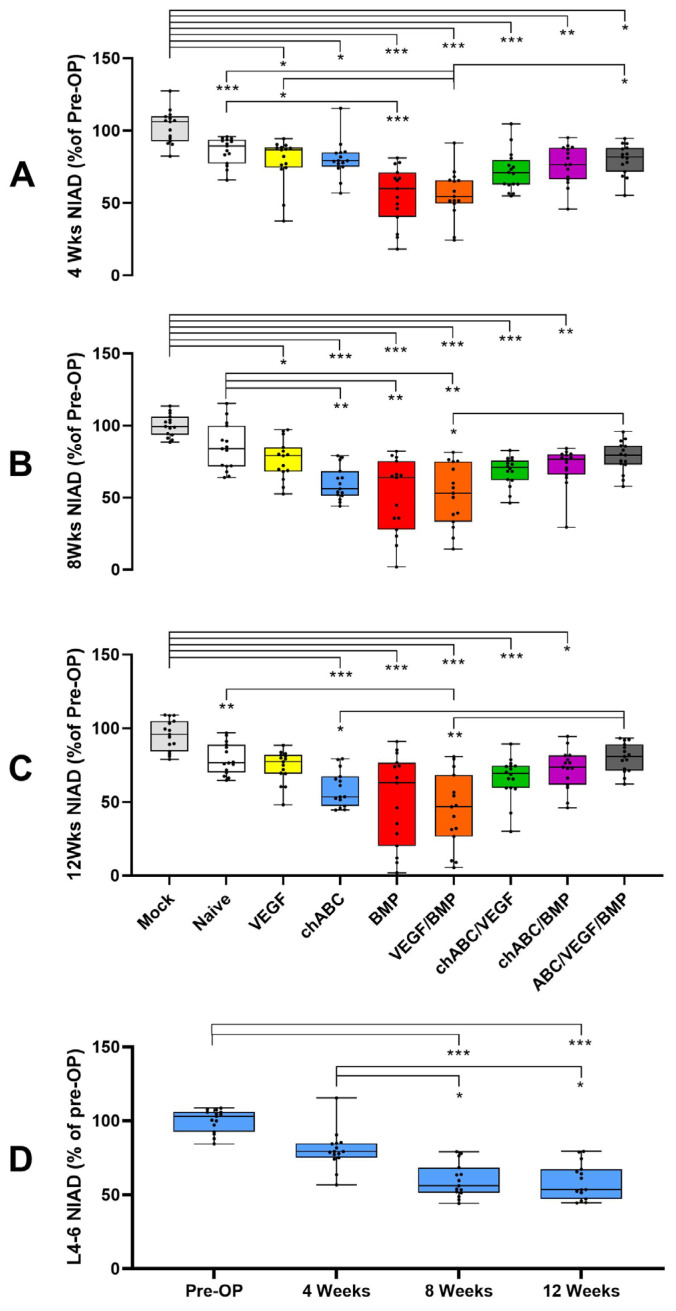
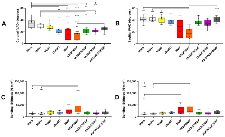
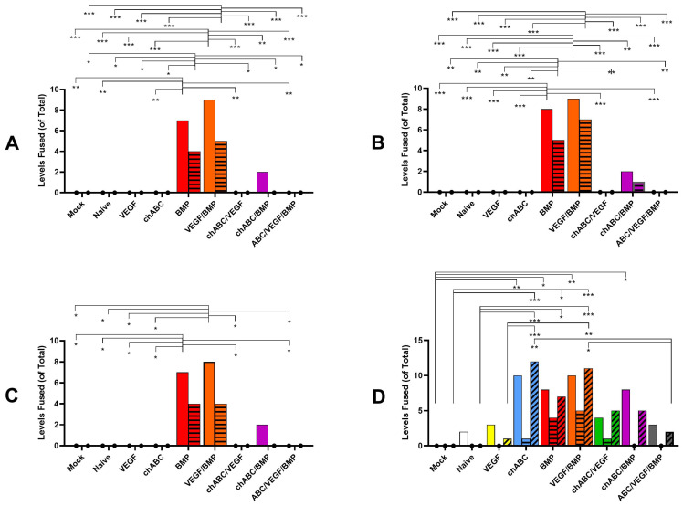
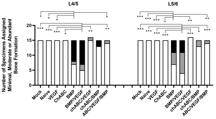
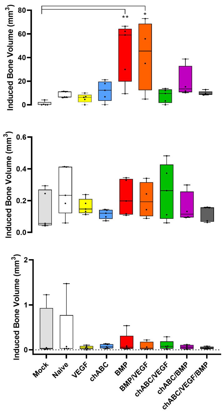
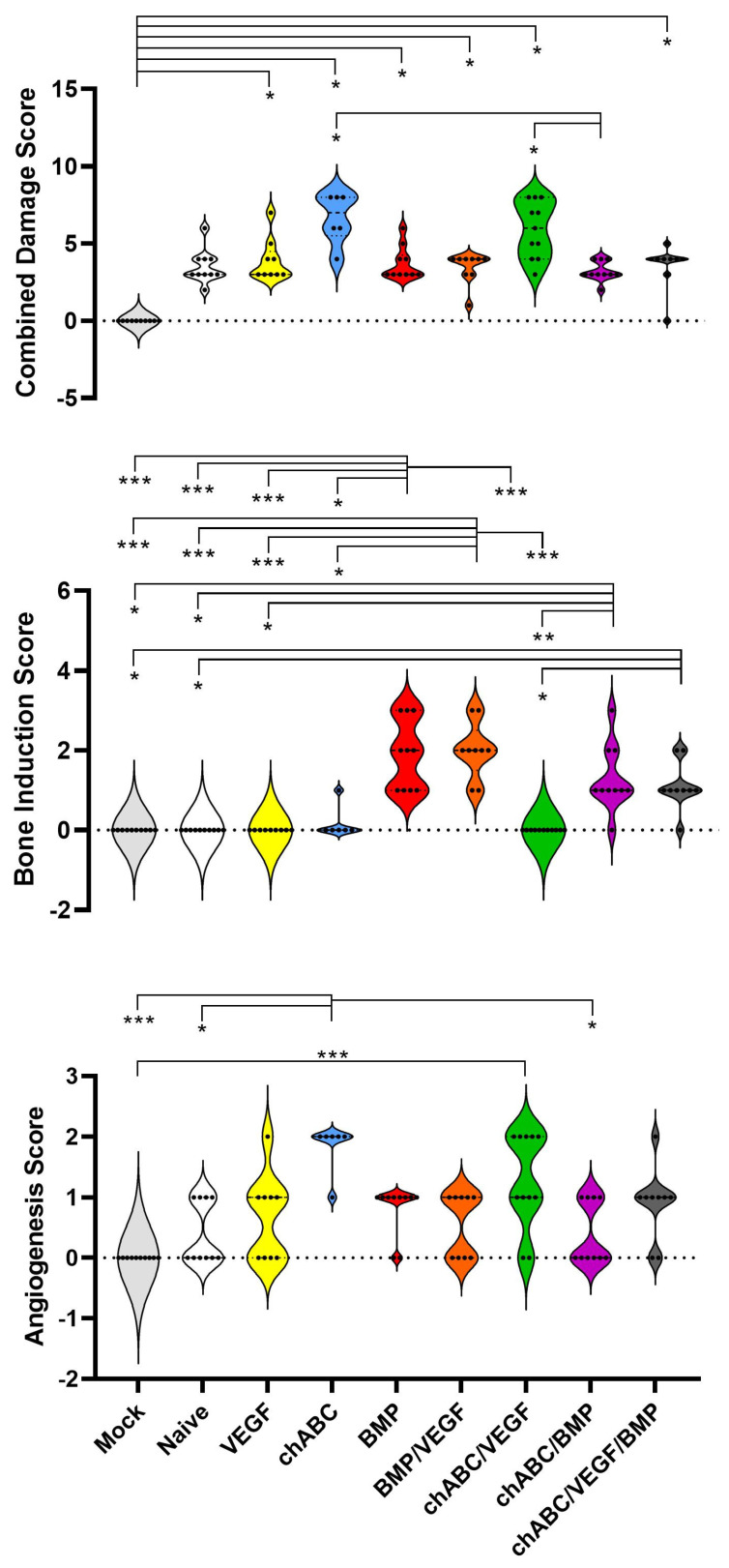
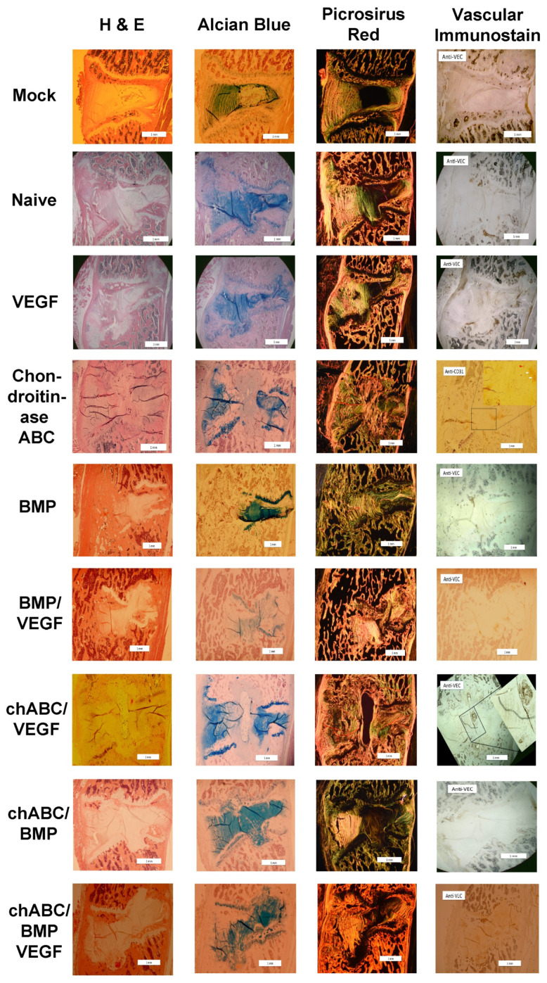
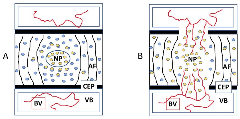
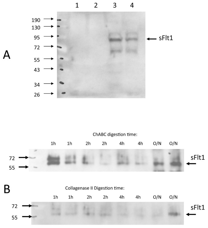
Bone morphogenetic protein (BMP) gene delivery to Lewis rat lumbar intervertebral discs (IVDs) drives bone formation anterior and external to the IVD, suggesting the IVD is inhospitable to osteogenesis. This study was designed to determine if IVD destruction with a proteoglycanase, and/or generating an IVD blood supply by gene delivery of an angiogenic growth factor, could render the IVD permissive to intra-discal BMP-driven osteogenesis and fusion. Surgical intra-discal delivery of naïve or gene-programmed cells (BMP2/BMP7 co-expressing or VEGF165 expressing) +/- purified chondroitinase-ABC (chABC) in all permutations was performed between lumbar 4/5 and L5/6 vertebrae, and radiographic, histology, and biomechanics endpoints were collected. Follow-up anti-sFlt Western blotting was performed. BMP and VEGF/BMP treatments had the highest stiffness, bone production and fusion. Bone was induced anterior to the IVD, and was not intra-discal from any treatment. chABC impaired BMP-driven osteogenesis, decreased histological staining for IVD proteoglycans, and made the IVD permissive to angiogenesis. A soluble fragment of VEGF Receptor-1 (sFlt) was liberated from the IVD matrix by incubation with chABC, suggesting dysregulation of the sFlt matrix attachment is a possible mechanism for the chABC-mediated IVD angiogenesis we observed. Based on these results, the IVD can be manipulated to foster vascular invasion, and by extension, possibly osteogenesis.

Keywords: angiogenesis; fusion; gene delivery; intervertebral disc; osteogenesis; proteoglycanase nucleus pulposus.


Conflict of interest statement

The authors declare no conflict of interest influencing the representation or interpretation of the reported research results.

Figures

Figure A1
Figure A1
Coronal noninvasive angular displacement (NIAD) assessment of rat lumbar motion segments preformed pre-operatively. Figure panels represent different lumbar levels: (A) L4/5, (B) L5/6, (C) combined L4–6, and (D) L6/S1. Small but statistically significant differences between groups were noted preoperatively, which was addressed by normalizing each specimen to its pre-operative NIAD assessment at the 4, 8 and 12 week NIAD measurements and reporting as ‘% of Pre-Operative NIAD’, as described in Material and Methods. Chondroitinase ABC is abbreviated as chABC. Indicated significance: p < 0.01 (**), and p ≤ 0.05 (*).
Figure A2
Figure A2
Bovine nucleus pulposus cultures remain stable after passaging. Primary cultures of bovine nucleus pulposus cells are shown, after harvest from caudal discs as described in Materials and Methods. High-power inverted microscope images are shown, panel (A) are cells at 60–70% confluence being grown up immediately after harvest (passage 0), and panel (B) are passage 10 cells at 80–90% confluence. Both panels show cells being cultured in normoxic conditions.
Figure A3
Figure A3
Coronal noninvasive angular displacement (NIAD) assessment of the L4/5 level. Time-course assessments were made: pre-operatively (A), 4 weeks post-OP (B), 8 weeks post-OP (C), and 12 weeks post-OP (D). It was expected that the BMP-containing samples would drive bone formation and cause stiffening of the spines, and that the progression of spinal stiffening would result in gradual attenuation of NIAD assessments over time. Chondroitinase ABC is abbreviated as chABC. Indicated significance: p ≤ 0.001 (***), p < 0.01 (**), and p ≤ 0.05 (*).
Figure A4
Figure A4
Coronal noninvasive angular displacement (NIAD) assessment of the L5/6 level. Time-course assessments were made: pre-operatively (A), 4 weeks post-OP (B), 8 weeks post-OP (C), and 12 weeks post-OP (D). It was expected that the BMP-containing samples would drive bone formation and cause stiffening of the spines, and that the progression of spinal stiffening would result in gradual attenuation of NIAD assessments over time. Chondroitinase ABC is abbreviated as chABC. Indicated significance: p ≤ 0.001 (***), p < 0.01 (**), and p ≤ 0.05 (*).
Figure A5
Figure A5
Coronal noninvasive angular displacement (NIAD) assessment of the L4–6 levels. Time-course assessments were made: pre-operatively (A), 4 weeks post-OP (B), 8 weeks post-OP (C), and 12 weeks post-OP (D). It was expected that the BMP-containing samples would drive bone formation and cause stiffening of the spines, and that the progression of spinal stiffening would result in gradual attenuation of NIAD assessments over time. Chondroitinase ABC is abbreviated as chABC. Indicated significance: p ≤ 0.001 (***), p < 0.01 (**), and p ≤ 0.05 (*).
Figure A6
Figure A6
Coronal noninvasive angular displacement (NIAD) assessment of the L6/S1 level. Time-course assessments were made: pre-operatively (A), 4 weeks post-OP (B), 8 weeks post-OP (C), and 12 weeks post-OP (D). Due to progressive stiffening and fusion of the L4–6 segments, the L6/S1 segment was expected to become more mobile. Chondroitinase ABC is abbreviated as chABC. Indicated significance: p ≤ 0.001 (***), p < 0.01 (**), and p ≤ 0.05 (*).
Figure A7
Figure A7
In vitro angular displacement (IVAD) and four-point bending assessments at 12 weeks after treatment delivery. Stiffness assessments were made over the combined L4–6 segment and were organized as: IVAD left bending (A), IVAD right bending (B), IVAD flexion (C), IVAD extension (D), 4-point bending flexion (E), and 4-point bending extension (F). Chondroitinase ABC is abbreviated as chABC. Indicated significance: p ≤ 0.001 (***), p < 0.01 (**), and p ≤ 0.05 (*).
Figure A8
Figure A8
Selected micro-CT 3-dimensional isosurface renderings demonstrating bone induction. All samples are oriented caudal at the top, cranial at the bottom, ventral to left and dorsal to right, with vertebral L6-L4 represented top to bottom, and the scale marking in the lower right corner representing 1mm. Micro-CT assessment was performed at 12 weeks after treatment delivery. Chondroitinase ABC is abbreviated as chABC. Note induced bone causing fusion for both levels of the BMP-treated sample and for the L4/5 level of the chABC/BMP treated sample. Also note that despite the BMP/VEGF treatment group showing excellent bone production overall, the sample shown here did not produce bone at either level implanted.
Figure 1
Figure 1
Coronal NIAD assessments for the combined L4–6 motion segment after L4/5 and L5/6 intra-discal delivery of indicated treatments. Panels represent all treatment groups at 4 weeks (A), 8 weeks (B) and 12 weeks (C) post-operatively, or the chondroitinase ABC (chABC) treatment group at 0, 4, 8 and 12 weeks (D). Indicated significance: p ≤ 0.001 (***), p < 0.01 (**), and p ≤ 0.05 (*).
Figure 2
Figure 2
In vitro angular displacement (IVAD) and non-destructive four-point bending mechanical assessments of the combined L4–6 motion segment at 12 weeks post-implantation of the indicated treatments. IVAD is shown in in the coronal (A) and sagittal (B) planes, and four-point bending stiffness is shown in the left (C) and right (D) bending directions. Chondroitinase ABC is abbreviated as chABC. Indicated significance was p ≤ 0.001 (***), p < 0.01 (**), and p ≤ 0.05 (*).
Figure 3
Figure 3
Multimodal spinal fusion assessments at 12 weeks post-implantation of the indicated treatments. Fusion status was scored separately at the L4/5 (histograms with no lines) and L5/6 (histograms with horizontal lines) levels for all assessments, and for the combined L4–6 (histograms with diagonal lines) segment for the NIAD fusion prediction method. Fusion was assessed by palpation (A), categorical radiographic (B), graded radiographic (C), and critical NIAD values (D), as described in the Materials and Methods section. Histogram vertical heights indicate raw value for number of fusions, and group size was n = 15 for all treatments except chondroitinase ABC (chABC)/VEGF (n = 16). Indicated significance was p ≤ 0.001 (***), p < 0.01 (**), and p ≤ 0.05 (*).
Figure 4
Figure 4
BMP-driven bone induction as assessed by high-definition Faxitron radiographs. Groups are composed of n = 15 or 16 specimens, the L4/5 and L5/6 levels were scored independently, and scoring and data testing were conducted as described in M&M. Histogram filled with white background indicates Minimal bone formation, gray indicates Moderate bone formation, and black Abundant bone formation. Chondroitinase ABC is abbreviated as chABC. Indicated significance was p ≤ 0.001 (***), p < 0.01 (**), and p ≤ 0.05 (*).
Figure 5
Figure 5
BMP-driven bone induction as assessed by micro-CT. Groups are composed of n = 4 (BMP/VEGF and chondroitinase ABC (chABC)/BMP) or n = 5 (all other groups) specimens. Specimens were prepared, assessed, and data tested as described in M&M. Top panel shows quantification of induced bone for the entire L4–6 fusion mass (located anterior to the spine and discs), the middle panel shows bone formation inside the L4/5 disc, and the bottom panel shows bone formation inside the L5/6 disc. Indicated significance was p < 0.01 (**), and p ≤ 0.05 (*).
Figure 6
Figure 6
Disc histology scoring results at 12 weeks after intradiscal delivery of treatments. Using a scoring rubric (Table A1), specimens were assessed for IVD damage criteria (top panel, range 0 to 8), osteogenesis extent (middle panel, range 0 to 3) and evidence of angiogenesis (bottom panel, range 0 to 2), as described in the Materials and Methods section. Results are presented using violin plots to demonstrate the distribution of the ordinal data. Chondroitinase ABC is abbreviated as chABC. Indicated significance was p ≤ 0.001 (***), p < 0.01 (**), and p ≤ 0.05 (*).
Figure 7
Figure 7
Representative histology images at 12 weeks after intradiscal delivery of treatments. All images are oriented ventral to the left and cranial as top. Histological staining technique is indicated at the top of each column and treatment group is indicated on the far left of each row. Picrosirus Red birefringence was visualized with circular polarized light, and vascular immunostain shown in the figure is anti-VE-cadherin or anti-CD31 (+), as indicated in each panel in that column (upper-left corner). Arrowheads in immunostained images indicate vascular appearing (red arrowhead) or cell clusters (white arrowheads) with positive immunostain. Scale bars in the right lower corner indicate 1 mm. Chondroitinase ABC is abbreviated as chABC.
Figure 8
Figure 8
A model for sFlt deregulation in the NP and how it would lead to vascular invasion of the IVD/NP. Major involved structures are labeled with initials: NP = nucleus pulposus, AF = annulus fibrosus, CEP = cartilage endplate, VB = vertebral body, and BV = blood vessels in proximity. Prior to endplate injury and sFlt deregulation, VEGF (yellow ovals) is being bound and sequestered from detection by tissues external to the disc space by sFlt (blue ovals) as shown (A). After sFlt is deregulated, there is reduced sFlt presence in the NP, VEGF is able to diffuse further in all directions, and particularly towards the endplate perforations where VEGF-sensitive blood vessels are positioned and are able to contribute to angiogenesis into the IVD as shown (B).
Figure 9
Figure 9
sFlt Western Blotting of bovine NP cells and rabbit NP tissue. Western immunoblotting for soluble VEGF-R1 (sFlt) demonstrated that sFlt was not detectable in supernatant media in bovine NP monolayers ((A), lanes 1 and 2), but rather was found in the cell/matrix fraction ((A), lanes 3 and 4) and this was not altered by normoxia (21% oxygen culture conditions, lanes 1 and 3) or hypoxia status (2% oxygen culture conditions, lanes 2 and 4). Freshly enucleated rabbit NP tissue when subjected to chondroitinace ABC (ChABC in the figure) digestion over time course (see Materials and Methods) released sFlt from the cell/matrix fraction ((B), upper blot), but digestion of sister samples with Collagenase Type II did not consistently solubilize sFlt ((B), lower blot).

References

    1. Kuo C.C., Soliman M.A.R., Aguirre A.O., Youngs D., Kruk M., Hess R.M., Nyabuto E.M., Khan A., Jowdy P.K., Pollina J., et al. Risk factors of early complications after thoracic and lumbar spinal deformity surgery: A systematic review and meta-analysis. Eur. Spine J. 2023;32:899–913. doi: 10.1007/s00586-022-07486-3. - DOI - PubMed
    1. Smith J.S., Klineberg E., Lafage V., Shaffrey C.I., Schwab F., Lafage R., Hostin R., Mundis G.M., Jr., Errico T.J., Kim H.J., et al. Prospective multicenter assessment of perioperative and minimum 2-year postoperative complication rates associated with adult spinal deformity surgery. J. Neurosurg. Spine. 2016;25:1–14. doi: 10.3171/2015.11.SPINE151036. - DOI - PubMed
    1. Alhammoud A., Alborno Y., Baco A.M., Othman Y.A., Ogura Y., Steinhaus M., Sheha E.D., Qureshi S.A. Minimally Invasive Scoliosis Surgery Is a Feasible Option for Management of Idiopathic Scoliosis and Has Equivalent Outcomes to Open Surgery: A Meta-Analysis. Glob. Spine J. 2022;12:483–492. doi: 10.1177/2192568220988267. - DOI - PMC - PubMed
    1. Skovrlj B., Belton P., Zarzour H., Qureshi S.A. Perioperative outcomes in minimally invasive lumbar spine surgery: A systematic review. World J. Orthop. 2015;6:996–1005. doi: 10.5312/wjo.v6.i11.996. - DOI - PMC - PubMed
    1. Alden T.D., Pittman D.D., Beres E.J., Hankins G.R., Kallmes D.F., Wisotsky B.M., Kerns K.M., Helm G.A. Percutaneous spinal fusion using bone morphogenetic protein-2 gene therapy. J. Neurosurg. 1999;90:109–114. doi: 10.3171/spi.1999.90.1.0109. - DOI - PubMed

Publication types

Substances

LinkOut - more resources