. 2023 Oct 21;12(20):2501.
doi: 10.3390/cells12202501.

Differential Transcriptomic Signatures of Small Airway Cell Cultures Derived from IPF and COVID-19-Induced Exacerbation of Interstitial Lung Disease

Affiliations

Differential Transcriptomic Signatures of Small Airway Cell Cultures Derived from IPF and COVID-19-Induced Exacerbation of Interstitial Lung Disease

Katie Uhl et al. Cells. .

Abstract

Idiopathic pulmonary fibrosis (IPF) is a pathological condition wherein lung injury precipitates the deposition of scar tissue, ultimately leading to a decline in pulmonary function. Existing research indicates a notable exacerbation in the clinical prognosis of IPF patients following infection with COVID-19. This investigation employed bulk RNA-sequencing methodologies to describe the transcriptomic profiles of small airway cell cultures derived from IPF and post-COVID fibrosis patients. Differential gene expression analysis unveiled heightened activation of pathways associated with microtubule assembly and interferon signaling in IPF cell cultures. Conversely, post-COVID fibrosis cell cultures exhibited distinctive characteristics, including the upregulation of pathways linked to extracellular matrix remodeling, immune system response, and TGF-β1 signaling. Notably, BMP signaling levels were elevated in cell cultures derived from IPF patients compared to non-IPF control and post-COVID fibrosis samples. These findings underscore the molecular distinctions between IPF and post-COVID fibrosis, particularly in the context of signaling pathways associated with each condition. A better understanding of the underlying molecular mechanisms holds the promise of identifying potential therapeutic targets for future interventions in these diseases.

Keywords: COVID-19; ILD; IPF; RNA-sequencing; TGF-β1; coronavirus; idiopathic pulmonary fibrosis; interstitial lung disease.


Conflict of interest statement

The authors declare there are no conflict of interest.

Figures

Figure 1
Figure 1
Culturing of small airway cells from patient lung tissues. (A) Simplified diagram of experimental workflow. Human small airway epithelial cells were harvested from the distal lobes of lung transplant patients. These cells were expanded and cultured onto transwell cell culture membranes. After 14 days, cultures were treated with TGFβ1 to mimic native fibrotic tissue conditions before being submitted for RNA sequencing. (B) Schematic depicting the collection and analysis of bulk RNA-sequencing data; “COVID” = post-COVID fibrosis, or pre-existing IPF exacerbated by COVID-19.
Figure 2
Figure 2
The detection of epithelial cell markers. (A) Immuno-staining of epithelial cells cultured on transwell membranes; ciliated cells (green) are present in the non-IPF control lung, IPF, and post-COVID fibrosis cell cultures. Images were taken at a magnification of 60×; scale bars represent 50 μM. (B) Percent of ciliated cells present in the surface epithelial cells of each sample type cultured on the transwell membranes. (C) Western blot showing the detection of the SFTPA protein in human large and small airway cultures from a pulmonary fibrosis patient.
Figure 3
Figure 3
Analysis of the transcriptomic disease signature for small airway cells cultured from IPF patients. (A) Schematic showing the determination of common DEGs for the IPF vs. NL comparison. The transcriptomic signature was found by comparing the DEGs between IPF and non-IPF control cultures in both TGF-β1 treated and untreated culture conditions. The common genes were then used for downstream analyses. (B) IPA canonical pathway results for the common IPF cell culture genes relative to the non-IPF control samples. (C) IPA upstream regulator results for common IPF cell culture genes relative to the non-IPF control samples. (D) Gene ontology results of the common genes in the IPF small airway cell cultures. (E) RT-PCR results of FOXM1 expression in the non-IPF control vs. IPF small airway cell cultures (N = 3, * indicates p-value < 0.05).
Figure 4
Figure 4
Analysis of the transcriptomic disease signature for small airway cells cultured from post-COVID fibrosis patients. (A) Schematic showing the determination of common DEGs for the post-COVID fibrosis vs. NL comparison. The transcriptomic signature was found by comparing the DEGs between post-COVID fibrosis lungs and non-IPF control cells in both TGF-β1 treated and untreated culture conditions. The common genes were then used for downstream analyses. (B) IPA canonical pathway results for the common post-COVID fibrosis cell culture genes relative to the non-IPF control samples. (C) IPA upstream regulator results for common post-COVID fibrosis cell culture genes relative to the non-IPF control samples. (D) Gene ontology results of the common genes in the post-COVID fibrosis small airway cell cultures.
Figure 5
Figure 5
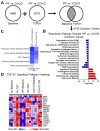
The comparison of IPF and post-COVID fibrosis gene signatures. (A) Schematic showing the determination of common DEGs from the IPF vs. post-COVID fibrosis comparison. The transcriptomic signature was found by comparing the DEGs between IPF lungs and post-COVID fibrosis samples in both TGF-β1 treated and untreated culture conditions. The common genes were then used for downstream analyses. (B) Reactome pathway analysis results for IPF vs. COVID common genes; results reflect pathway activation/inhibition in the IPF cell cultures. (C) IPA canonical pathway results comparing the DEG lists for the IPF vs. COVID comparison under control and TGF-β1 treatment conditions; results reflect the inhibition of pathways observed in the IPF cell cultures compared to the post-COVID fibrosis samples. (D) Heatmap displaying gene expression levels for genes involved in the TGF-β1 signaling pathway, a key driver of fibrosis. (NL = “normal lung”/non-IPF control, IPF = idiopathic pulmonary fibrosis, COVID = post-COVID fibrosis).
Figure 6
Figure 6
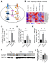
Evaluation of BMP signaling in patient-derived small airway cell cultures. (A) Mechanism of inhibition of TGFβ signaling by BMP. (B) Heatmap displaying the expression levels of genes involved in the BMP signaling pathway (NL = “normal lung”/non-IPF control, IPF = idiopathic pulmonary fibrosis, COVID = post-COVID fibrosis). (CE) RT-PCR quantification of fold changes for genes associated with the BMP signaling pathway in small airway cell cultures (** indicates p-value < 0.001, * indicates p-value < 0.05, N = 3). (F) Western blot showing the detection of the SMAD7 protein in human normal (NL), IPF, and post-COVID fibrosis (COVID) lung tissue. (G) Quantification of SMAD7 protein levels in normal lung (N = 3), IPF (N = 4), and post-COVID fibrosis (N = 3) lung tissue.
Figure 7
Figure 7
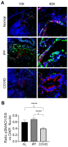
Localization of pSMAD1/5/8 to the nucleus in the patient sample tissue. (A) IHC images of patient samples (IPF = idiopathic pulmonary fibrosis, COVID = post-COVID fibrosis) stained for DAPI (blue), pSMAD1/5/8 (red), and the epithelial cell marker KRT5 (green). The scale bar was equal to 50 μM. Images are shown at two different magnifications; the red square in the 10× images indicates the region of interest pictured in the 60× image. (B) Quantification of the nuclear localization of pSMAD1/5/8 across different tissue types (**** indicates p-value < 0.0001).

References

    1. Stancil I.T., Michalski J.E., Schwartz D.A. An Airway-Centric View of Idiopathic Pulmonary Fibrosis. Am. J. Respir. Crit. Care Med. 2022;206:410–416. doi: 10.1164/rccm.202109-2219PP. - DOI - PMC - PubMed
    1. Blackwell T.S., Tager A.M., Borok Z., Moore B.B., Schwartz D.A., Anstrom K.J., Bar-Joseph Z., Bitterman P., Blackburn M.R., Bradford W., et al. Future Directions in Idiopathic Pulmonary Fibrosis Research. An NHLBI Workshop Report. Am. J. Respir. Crit. Care Med. 2014;189:214–222. doi: 10.1164/rccm.201306-1141WS. - DOI - PMC - PubMed
    1. Stancil I.T., Michalski J.E., Davis-Hall D., Chu H.W., Park J.A., Magin C.M., Yang I.V., Smith B.J., Dobrinskikh E., Schwartz D.A. Pulmonary fibrosis distal airway epithelia are dynamically and structurally dysfunctional. Nat. Commun. 2021;12:4566. doi: 10.1038/s41467-021-24853-8. - DOI - PMC - PubMed
    1. Verleden S.E., Tanabe N., McDonough J.E., Vasilescu D.M., Xu F., Wuyts W.A., Piloni D., De Sadeleer L., Willems S., Mai C., et al. Small airways pathology in idiopathic pulmonary fibrosis: A retrospective cohort study. Lancet Respir. Med. 2020;8:573–584. doi: 10.1016/S2213-2600(19)30356-X. - DOI - PMC - PubMed
    1. Abdelgied M., Uhl K., Chen O.G., Schultz C., Tripp K., Peraino A.M., Paithankar S., Chen B., Tamae Kakazu M., Castillo Bahena A., et al. Targeting ATP12A, a Non-Gastric Proton Pump Alpha Subunit, for Idiopathic Pulmonary Fibrosis Treatment. Am. J. Respir. Cell Mol. Biol. 2023;68:638–650. doi: 10.1165/rcmb.2022-0264OC. - DOI - PMC - PubMed

Publication types