. 2023 Oct 23;12(20):2509.
doi: 10.3390/cells12202509.

SLC38A5 Modulates Ferroptosis to Overcome Gemcitabine Resistance in Pancreatic Cancer

Affiliations

SLC38A5 Modulates Ferroptosis to Overcome Gemcitabine Resistance in Pancreatic Cancer

Myeong Jin Kim et al. Cells. .

Abstract

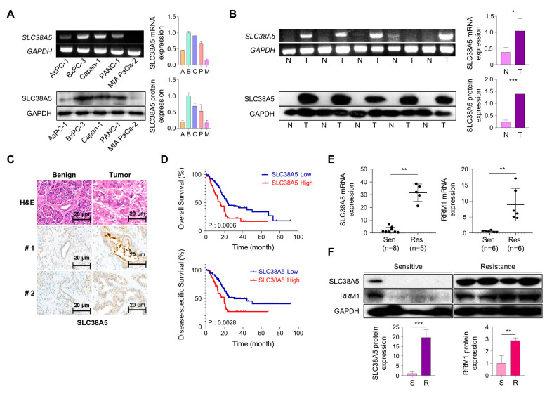
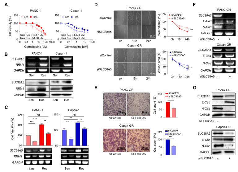
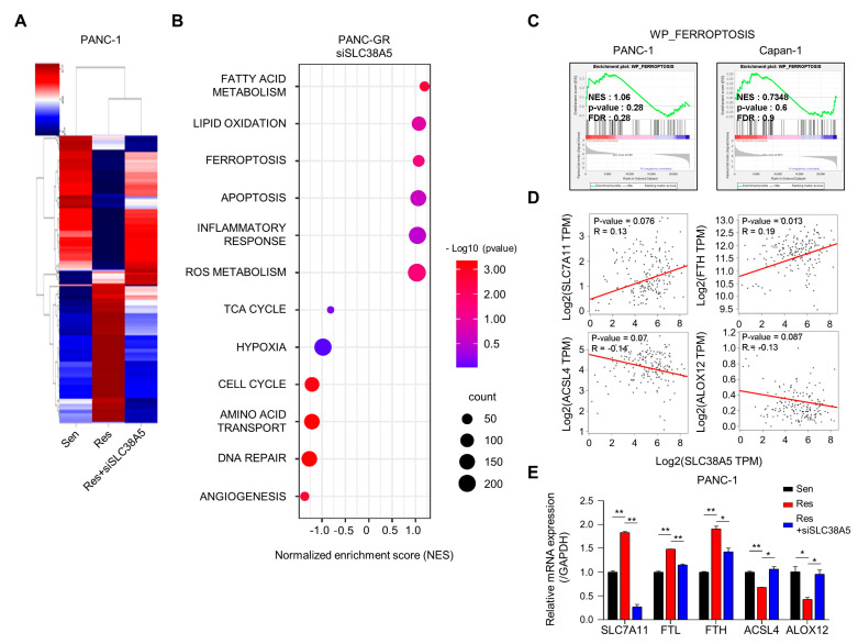
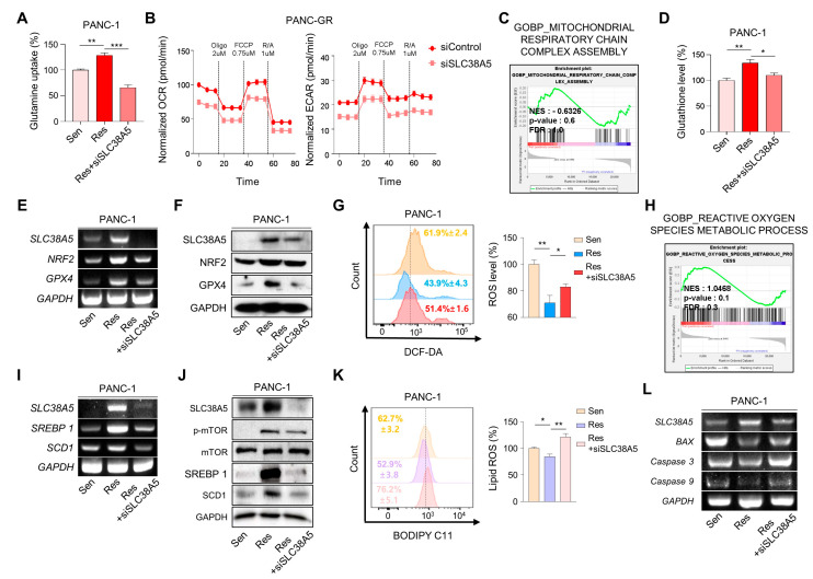
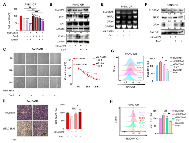
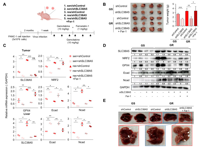
Pancreatic cancer is characterized by a poor prognosis, with its five-year survival rate lower than that of any other cancer type. Gemcitabine, a standard treatment for pancreatic cancer, often has poor outcomes for patients as a result of chemoresistance. Therefore, novel therapeutic targets must be identified to overcome gemcitabine resistance. Here, we found that SLC38A5, a glutamine transporter, is more highly overexpressed in gemcitabine-resistant patients than in gemcitabine-sensitive patients. Furthermore, the deletion of SLC38A5 decreased the proliferation and migration of gemcitabine-resistant PDAC cells. We also found that the inhibition of SLC38A5 triggered the ferroptosis signaling pathway via RNA sequencing. Also, silencing SLC38A5 induced mitochondrial dysfunction and reduced glutamine uptake and glutathione (GSH) levels, and downregulated the expressions of GSH-related genes NRF2 and GPX4. The blockade of glutamine uptake negatively modulated the mTOR-SREBP1-SCD1 signaling pathway. Therefore, suppression of SLC38A5 triggers ferroptosis via two pathways that regulate lipid ROS levels. Similarly, we observed that knockdown of SLC38A5 restored gemcitabine sensitivity by hindering tumor growth and metastasis in the orthotopic mouse model. Altogether, our results demonstrate that SLC38A5 could be a novel target to overcome gemcitabine resistance in PDAC therapy.

Keywords: PDAC; SLC38A5; ferroptosis; gemcitabine resistance; lipid ROS.


Conflict of interest statement

The authors declare no conflict of interest.

Figures

Figure 1
Figure 1
SLC38A5 correlates with gemcitabine-resistant pancreatic cancer patients. (A) mRNA and protein expression of SLC38A5 in pancreatic cancer cell lines. (B) mRNA and protein expression of SLC38A5 in normal tissues and pancreatic cancer tissues. (C) H&E staining of the SLC38A5 in tumor tissue. (D) Kaplan–Meier analysis for overall survival, and disease-specific survival between the SLC38A5 high group and the low group. (E) Dot plot of SLC38A5 and RRM1 gene expression level in purified mRNA from gemcitabine-sensitive and gemcitabine-resistant pancreatic cancer patient tissue. (F) Protein expression of SLC38A5 and RRM1 between gemcitabine-sensitive and gemcitabine-resistant pancreatic cancer patient tissue. All experiments were performed at least three times. Bars represent means ± SD. *, p < 0.05; **, p < 0.01; ***, p < 0.001.
Figure 2
Figure 2
Deletion of SLC38A5 downregulates cell viability and migration in gemcitabine-resistant pancreatic cancer cells. (A) Median inhibitory concentration (IC50) values of gemcitabine for each cell line (PANC-1 and Capan-1). (B) mRNA and protein expression of SLC38A5 and RRM1 in gemcitabine-sensitive PDAC cell lines (PANC-1 and Capan-1) and gemcitabine-resistant PDAC cell lines (PANC-GR and Capan-GR). (C) Cell viability of PANC-1, PANC-GR, Capan-1, and Capan-GR was determined using the WST assay. Each cell line was transfected with siSLC38A5. Transfection was confirmed via RT-PCR. (D) Migration of cells was measured via wound healing assay. The statistical analysis of wound closure percentage was measured using Image J software (version 1.8.0). (E) Representative photographs of the number of invaded cells that pass through the Matrigel-coated Transwell invasion assay. (F,G) The EMT marker expression of siRNA transfected PANC-GR and Capan-GR was analyzed via RT-PCR (F) and Western blot (G). All experiments were performed at least three times. Bars represent means ± SD. ns = not significant; **, p < 0.01; ***, p < 0.001.
Figure 3
Figure 3
Inhibition of SLC38A5 induces changes in ferroptosis-regulating genes. (A) Heat map analysis of the distribution of genes from PANC-1, PANC-GR, and PANC-GR with siSLC38A5 cells. (B) DAVID-based gene ontology bubble plot analysis of mRNA sequencing results from PANC-GR and PANC-GR with siSLC38A5 cells. (C) GSEA for significant enrichment for ferroptosis. (D) Correlation analysis of SLC38A5 and ferroptosis-related genes in pancreatic cancer. (E) mRNA levels of the ferroptosis-related genes were quantified via qPCR in PANC-1, PANC-GR, and PANC-GR with siSLC38A5. All experiments were performed at least three times. Bars represent means ± SD. *, p < 0.05; **, p < 0.01.
Figure 4
Figure 4
SLC38A5 modulates lipid ROS through GSH-mediated ROS and mTOR-SREBP1 signaling in gemcitabine-resistant pancreatic cancer cells. (A) Comparison of glutamine uptake of PANC-1, PANC-GR, and PANC-GR with siSLC38A5. (B) OCR and ECAR of PANC-GR cells (siControl and siSLC38A5) were determined and calculated. (C) GSEA for significant enrichment for mitochondrial activity. (D) Comparison of glutathione levels of PANC-1, PANC-GR, and PANC-GR with siSLC38A5. (E,F) Expression of glutathione-related genes in PANC-1, PANC-GR, and PANC-GR with siSLC38A5 was analyzed via RT-PCR (E) and Western blot (F). (G) Relative ROS production in PANC-1, PANC-GR, and PANC-GR with siSLC38A5 was measured using flow cytometry after dichlorofluorescein (DCF) staining. The bar graph was analyzed using Image J software. (H) GSEA for significant enrichment for ROS level. (I,J) Expression of mTOR-SREBP1 signaling genes was analyzed via RT-PCR (I) and Western blot (J). (K) Flow cytometry analysis of lipid ROS in PANC-1, PANC-GR, and PANC-GR with siSLC38A5. The bar graph was analyzed using Image J software. (L) mRNA levels of apoptosis-related genes in PANC-1, PANC-GR, and PANC-GR with siSLC38A5. All experiments were performed at least three times. Bars represent means ± SD. *, p < 0.05; **, p < 0.01; ***, p < 0.001.
Figure 5
Figure 5
Inhibition of SLC38A5 induces ferroptosis in gemcitabine-resistant pancreatic cancer cells. (A) A WST assay was performed to determine cell viability using indicated PANC-GR groups. (B) The cell proliferation marker was confirmed via Western blot. (C,D) Migration of cells was measured through wound healing assay (C) and Matrigel-coated transwell invasion assay (D). (E,F) Expression of glutathione-related genes was measured via RT-PCR (E) and Western blot (F). (G) Relative ROS production in indicated PANC-GR groups was measured via FACS analysis. (H) FACS analysis of lipid ROS in indicated PANC-GR groups. The bar graph was analyzed using Image J software. An amount of 20 μM of Ferrostatin-1 and 10 μM of Erastin were treated in the control and knockdown groups. All experiments were performed at least three times. Bars represent means ± SD. ns = not significant; *, p < 0.05; **, p < 0.01; ***, p < 0.001, #, p < 0.05, ##, p < 0.01, ###, p < 0.001.
Figure 6
Figure 6
Knockdown of SLC38A5 suppresses tumor growth and metastasis in vivo. (A) Schematic of mouse experimental grouping and processing of the orthotopic model. (B) Images and weights of the tumors harvested from mice. (C) mRNA levels of the EMT and ferroptosis-related genes in tumors and livers were quantified via qPCR. (D) Protein expression of EMT and ferroptosis-related genes in tumors. (E) Liver metastasis images. The white arrows: liver metastasis. All experiments were performed at least three times. Bars represent means ± SD. ns = not significant; *, p < 0.05; **, p < 0.01; ***, p < 0.001.

References

    1. Halbrook C.J., Lyssiotis C.A., Pasca di Magliano M., Maitra A. Pancreatic cancer: Advances and challenges. Cell. 2023;186:1729–1754. doi: 10.1016/j.cell.2023.02.014. - DOI - PMC - PubMed
    1. Chu L.C., Goggins M.G., Fishman E.K. Diagnosis and detection of pancreatic cancer. Cancer J. 2017;23:333–342. doi: 10.1097/PPO.0000000000000290. - DOI - PubMed
    1. Ansari D., Tingstedt B., Andersson B., Holmquist F., Sturesson C., Williamsson C., Sasor A., Borg D., Bauden M., Andersson R. Pancreatic cancer: Yesterday, today and tomorrow. Future Oncol. 2016;12:1929–1946. doi: 10.2217/fon-2016-0010. - DOI - PubMed
    1. Samanta K., Setua S., Kumari S., Jaggi M., Yallapu M.M., Chauhan S.C. Gemcitabine combination nano therapies for pancreatic cancer. Pharmaceutics. 2019;11:574. doi: 10.3390/pharmaceutics11110574. - DOI - PMC - PubMed
    1. Amrutkar M., Gladhaug I.P. Pancreatic cancer chemoresistance to gemcitabine. Cancers. 2017;9:157. doi: 10.3390/cancers9110157. - DOI - PMC - PubMed

Publication types