Review
. 2023 Oct 21;30(10):9335-9345.
doi: 10.3390/curroncol30100675.

Conversion in a Resectable Tumor after Denosumab Neoadjuvant in a Large Dorsal Giant Cells Tumor: A Case Report and a Literature Review

Affiliations
Review

Conversion in a Resectable Tumor after Denosumab Neoadjuvant in a Large Dorsal Giant Cells Tumor: A Case Report and a Literature Review

María Sereno et al. Curr Oncol. .

Abstract

Giant cell tumors of bone are a rare entity, usually occurring in young patients and characteristically arising in the long bones. The spinal location is rare and usually presents with pain and/or neurological symptoms. The treatment of choice is surgery. Treatment with Denosumab, a bisphosphonate inhibitor of RANK-L, which is highly expressed in these tumors, has shown extensive activity in unresectable patients or those undergoing incomplete surgery. Preoperative treatment with this drug is gaining increasing interest, as its high potency in tumor reduction in this subtype of neoplasm has allowed resectability in selected patients. We present the case of a young patient with a large spinal tumor who, after neoadjuvant Denosumab, underwent complete en bloc surgery with clean margins and a great pathological response.

Keywords: bone giant cell tumor; denosumab; neoadjuvant.


Conflict of interest statement

All authors have completed the ICMJE uniform disclosure form. The authors declare no conflict of interest.

Figures

Figure 1
Figure 1
Images of the mass (July 2022) by CT with contrast (A) and by MRI (B), showing infiltration in the ipsilateral posterior third costal arch and in the right T2 and T3 vertebral hemibody, as well as occupation of the conjunctival foramina and right lateral epidural grase in T2–T3 and T3–T4, without invading thecal sac or medullary cord (B).
Figure 2
Figure 2
Pathological study of BAG: Haematoxylin-eosin staining 20×, different magnifications. (A,B). Multitude of multinucleated giant cells, without atypical features. Positivity for CD 163, CD 68 (C) and CD 45 (×40) (D). Negative pattern for pancytokeratin marker (E), TTF-1, CD34, S100, CEA, Ki 67, p40, and AML.
Figure 3
Figure 3
A/B. Re-evaluation studies after neoadjuvant treatment (January 2023). (A) Thoracic CT scan showing response to treatment, with a significant decrease in the size of the right posterior costovertebral mediastinal mass, with diameters currently approximately 3.5 × 5.9 × 4.3 cm (AP × T × CC) and similar invasion/affectation of the right side of the T2 and T3 vertebrae, respecting the contiguous T1 and T4 vertebrae. (B) MRI of the dorsal spine also showing a marked decrease in the soft tissue mass associated with lytic lesion in the right lateral mass of the T3 vertebral body and a decrease in the dimensions of the spinal canal on the right side without obliteration of the exit foramina of the T2 and T3 roots.
Figure 4
Figure 4
(A,B) Intraoperative images of the result of T2–T3 vertebrectomies with expandable PEEK radiolucent vertebral body replacement (XRL®, Johnson & Johnson) filled with a synthetic bone graft substitute (Vitoss®, Stryker) was implanted.
Figure 5
Figure 5
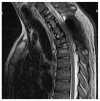
T1-weighted MR image two months after surgery showing post-surgical changes with no evidence of recurrence.
Figure 6
Figure 6
Pathophysiology of RANKL. We know that tumor giant cells are activated osteoclasts through an indirect effect on osteoblasts and stromal cells by the presence of a stimulatory factor: RANKL. Denosumab specifically binds to RANKL, extinguishing the RANK positivity of GCT cells, preventing their osteolytic functionality [6].

References

    1. Del Baena-Ocampo L.C., Ramirez-Perez E., Linares-Gonzalez L.M., Delgado-Chavez R. Epidemiology of bone tumors in Mexico City: Retrospective clinicopathologic study of 566 patients at a referral institution. Ann. Diagn. Pathol. 2009;13:16–21. doi: 10.1016/j.anndiagpath.2008.07.005. - DOI - PubMed
    1. Amelio J.M., Rockberg J., Hernandez R.K., Sobocki P., Stryker S., Bach B.A., Engellau J., Liede A. Population-based study of giant cell tumor of bone in Sweden (1983–2011) Cancer Epidemiol. 2016;42:82–89. doi: 10.1016/j.canep.2016.03.014. - DOI - PubMed
    1. Rendina D., Mossetti G., Soscia E., Sirignano C., Insabato L., Viceconti R., Ignarra R., Salvatore M., Nunziata V. Giant cell tumor and Paget’s disease of bone in one family: Geographic clustering. Clin. Orthop. Relat. Res. 2004;421:212–218. doi: 10.1097/00000118702.46373.e3. - DOI - PubMed
    1. Li H., Gao J., Gao Y., Lin N., Zheng M., Ye Z. Denosumab in Giant Cell Tumor of Bone: Current Status and Pitfalls. Front. Oncol. 2020;10:580605. doi: 10.3389/fonc.2020.580605. - DOI - PMC - PubMed
    1. Campanacci M., Baldini N., Boriani S., Sudanese A. Giant-cell tumor of bone. J. Bone Joint Surg. Am. 1987;69:106–114. doi: 10.2106/00004623-198769010-00018. - DOI - PubMed

MeSH terms