Review
. 2023 Oct 2;10(10):417.
doi: 10.3390/jcdd10100417.

Statins and Cardiomyocyte Metabolism, Friend or Foe?

Affiliations
Review

Statins and Cardiomyocyte Metabolism, Friend or Foe?

Tim Somers et al. J Cardiovasc Dev Dis. .

Abstract

Statins inhibit HMG-CoA reductase, the rate-limiting enzyme in cholesterol synthesis, and are the cornerstone of lipid-lowering treatment. They significantly reduce cardiovascular morbidity and mortality. However, musculoskeletal symptoms are observed in 7 to 29 percent of all users. The mechanism underlying these complaints has become increasingly clear, but less is known about the effect on cardiac muscle function. Here we discuss both adverse and beneficial effects of statins on the heart. Statins exert pleiotropic protective effects in the diseased heart that are independent of their cholesterol-lowering activity, including reduction in hypertrophy, fibrosis and infarct size. Adverse effects of statins seem to be associated with altered cardiomyocyte metabolism. In this review we explore the differences in the mechanism of action and potential side effects of statins in cardiac and skeletal muscle and how they present clinically. These insights may contribute to a more personalized treatment strategy.

Keywords: beneficial and adverse effects; cardiomyocytes; clinical translation; metabolism; statins.


Conflict of interest statement

The authors declare no conflict of interest.

Figures
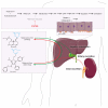
Figure 1
Figure 1
Statins and the cholesterol pathway. The conversion of acetyl-CoA by HMG-CoA reductase leading to the production of cholesterol. Statins (in red) inhibit this enzyme, not only leading to less cholesterol, but also isoprenoids, sterols and steroids. The reduction in cholesterol leads to upregulation of the LDL receptor in hepatocytes (displayed in the panel above the liver). Statins are bidirectionally converted in the liver from the prodrug (lactone) into the active form (acid) or back. The acid form is primarily excreted in the urine (dotted red line) and the lactone form into bile (dotted green line). Abbreviations: LDL: low-density lipoprotein; IPP: isopentenyl pyrophosphate; GPP: geranyl diphosphate; FPP: farnesyl diphosphate.
Figure 2
Figure 2
Beneficial effects of statins on the heart. Factors that stimulate cardiac hypertrophy and fibrosis are indicated in green and in red for factors that reduce cardiac hypertrophy and fibrosis (top). Statins inhibit hypertrophic and fibrotic pathways, as they have a stimulatory effect on factors that reduce both pathologies (red factors). The molecular details of these pathways are shown. In green, factors that stimulate or deteriorate hypertrophy, apoptosis, fibrosis and calcium homeostasis are shown. In red, factors that inhibit hypertrophy or fibrosis (bottom) are shown. The panel most to the left shows part of the mevalonate pathway and the way statins inhibit the important Ras, Rho and Rac proteins. Abbreviations: Cyt C: cytochrome c; EGFR: epidermal growth factor; FPP: farnesyl pyrophosphate; GGPP: geranylgeranyl-pyrophosphate; eNOS: endothelial nitric oxide synthase; ERK: extracellular signal-regulated protein kinase; GSKβ: glycogen synthase kinase 3 β; JNK: c-Jun N-terminal kinase; NADPH: nicotinamide adenine dinucleotide phosphate; NO: nitric oxide; PKA: protein kinase A; PKB: protein kinase B; ROS: reactive oxygen species; TGFβ: transforming growth factor-β; SERCA: sarcoplasmic reticulum Ca2+ ATPase.
Figure 3
Figure 3
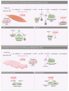
Adverse effects of statins on skeletal and cardiac muscle cells. Molecular mechanisms underlying the adverse effects of statins on skeletal muscle cells (top) and cardiomyocytes (bottom). In green are factors that stimulate apoptosis and mitochondrial dysfunction. On the right are small boxes with details of the pathways seen in the larger boxes. Abbreviations: Akt: PKB/protein kinase B; AMPK: adenosine monophosphate-activated protein kinase; ATP: adenosine triphosphate; ERK: extracellular signal-regulated protein kinase; FPP: farnesyl pyrophosphate; GGPP: geranylgeranyl-pyrophosphate; GPP: geranyl pyrophosphate; GSK3β: glycogen synthase kinase 3 β; IPP: isopentenyl pyrophosphate; MPTP: mitochondrial permeability transition pore; mTOR: mechanistic target of rapamycin; OXPHOS: oxidative phosphorylation; PGC1α: peroxisome proliferator-activated receptor gamma coactivator 1-α; PKC: protein kinase C.
Figure 4
Figure 4
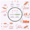
Mitohormesis 2.0. The proposed mechanisms behind the metabolic differences between cardiac and skeletal muscle cells. Central to the figure is the previously described mitohormesis theory. Besides this difference, other proposed mechanisms include differences in calcium-dependent ROS, mitochondrial content, transporter expression resulting in different rates of influx and efflux of statins, cholesterol homeostasis and MCT expression. Together, these variations in mechanisms are proposed as a new mitohormesis (i.e., mitohormesis 2.0) theory. Numbers on mRNA protein expression were obtained from the Human Protein Atlas and displayed on a logarithmic scale. Abbreviations: BCRP: breast cancer resistance protein; LDL: low-density lipoprotein; MCT: monocarboxylate transporter; MRP: multidrug resistance-associated protein; NO: nitric oxide; OATP: organic-anion-transporting polypeptides; PGC-1: peroxisome proliferator-activated receptor gamma coactivator 1-α; P-gp: P-glycoprotein; ROS: reactive oxygen species; RyR: ryanodine receptor. An increase is displayed as upward arrow and a decrease as downward arrow.

References

    1. Ridker P.M., Danielson E., Fonseca F.A.H., Genest J., Gotto A.M., Kastelein J.J.P., Koenig W., Libby P., Lorenzatti A.J., MacFadyen J.G., et al. Rosuvastatin to Prevent Vascular Events in Men and Women with Elevated C-Reactive Protein. N. Engl. J. Med. 2008;359:2195–2207. doi: 10.1056/NEJMoa0807646. - DOI - PubMed
    1. Rogers J.K., Jhund P.S., Perez A.C., Bohm M., Cleland J.G., Gullestad L., Kjekshus J., van Veldhuisen D.J., Wikstrand J., Wedel H., et al. Effect of rosuvastatin on repeat heart failure hospitalizations: The CORONA Trial (Controlled Rosuvastatin Multinational Trial in Heart Failure) JACC Heart Fail. 2014;2:289–297. doi: 10.1016/j.jchf.2013.12.007. - DOI - PubMed
    1. Yoshimura S., Uchida K., Daimon T., Takashima R., Kimura K., Morimoto T., Tanada S., Iida T., Kuroda J., Nose A., et al. Randomized Controlled Trial of Early Versus Delayed Statin Therapy in Patients With Acute Ischemic Stroke. Stroke. 2017;48:3057–3063. doi: 10.1161/STROKEAHA.117.017623. - DOI - PubMed
    1. Pisaniello A.D., Scherer D.J., Kataoka Y., Nicholls S.J. Ongoing challenges for pharmacotherapy for dyslipidemia. Expert Opin. Pharmacother. 2015;16:347–356. doi: 10.1517/14656566.2014.986094. - DOI - PubMed
    1. Hennessy E., Adams C., Reen F.J., O’Gara F. Is There Potential for Repurposing Statins as Novel Antimicrobials? Antimicrob. Agents Chemother. 2016;60:5111–5121. doi: 10.1128/AAC.00192-16. - DOI - PMC - PubMed

LinkOut - more resources