Review
. 2023 Oct 20;14(10):531.
doi: 10.3390/jfb14100531.

The Preparation and Clinical Efficacy of Amnion-Derived Membranes: A Review

Affiliations
Review

The Preparation and Clinical Efficacy of Amnion-Derived Membranes: A Review

Alison L Ingraldi et al. J Funct Biomater. .

Abstract

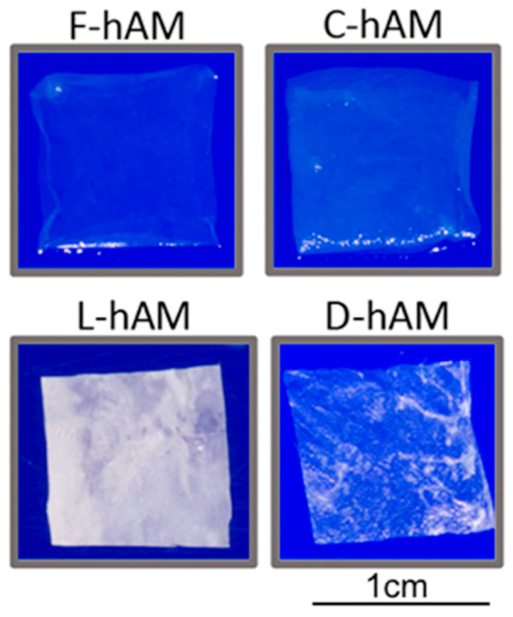
Biological tissues from various anatomical sources have been utilized for tissue transplantation and have developed into an important source of extracellular scaffolding material for regenerative medicine applications. Tissue scaffolds ideally integrate with host tissue and provide a homeostatic environment for cellular infiltration, growth, differentiation, and tissue resolution. The human amniotic membrane is considered an important source of scaffolding material due to its 3D structural architecture and function and as a source of growth factors and cytokines. This tissue source has been widely studied and used in various areas of tissue repair including intraoral reconstruction, corneal repair, tendon repair, microvascular reconstruction, nerve procedures, burns, and chronic wound treatment. The production of amniotic membrane allografts has not been standardized, resulting in a wide array of amniotic membrane products, including single, dual, and tri-layered products, such as amnion, chorion, amnion-chorion, amnion-amnion, and amnion-chorion-amnion allografts. Since these allografts are not processed using the same methods, they do not necessarily produce the same clinical responses. The aim of this review is to highlight the properties of different human allograft membranes, present the different processing and preservation methods, and discuss their use in tissue engineering and regenerative applications.

Keywords: amnion; amnion–chorion; anti-inflammatory; chorion; chronic wound; clinical application; human amniotic membrane; processing; regenerative medicine; skin substitute.


Conflict of interest statement

A.L.I., R.G.A. and A.J.T. are employees of the Carmell Corporation, formerly Axolotl Biologix.

Figures

Figure 1
Figure 1
Structure and components of the amnion and chorion tissue layers. Created with www.biorender.com (accessed on 10 October 2023).
Figure 2
Figure 2
After isolation of amniotic tissue from human placenta, the membrane is dissected to separate the amnion membrane (hAM) from the chorion membrane (hCM), possibly folded or kept together as amnion–chorion membrane (hACM), for further processing. There exists a variety of ways for the membrane to be prepared, preserved, sterilized, or cross-linked for research or clinical use. The process is determined by the company and its specified membrane produced. Modified from Ref. [34] Burns, Vol 46(6), Gholipourmalekabadi M, et al., How preparation and preservation procedures affect the properties of amniotic membrane? How safe are the procedures? 1254–1271, Copyright (2020), with permission from Elsevier.
Figure 3
Figure 3
Comparison of the impact of preservation methods on amniotic membrane properties for tissue engineering applications. Human amniotic membrane formats: F-hAM, fresh, C-hAM, cryopreserved, L-hAM, lyophilized, and D-hAM, decellularized and lyophilized. Note the varying visual appearance based on preservation method. Reprinted from Ref. [65] Materials Science and Engineering: C, Vol 104, Fénelon M et al., Comparison of the impact of preservation methods on amniotic membrane properties for tissue engineering applications, 109903, Copyright (2019), with permission from Elsevier.
Figure 4
Figure 4
Example of gingival recession treatment using the coronally advanced flap technique to expose the root. (A) Baseline photograph; (B) flap reflection and recipient bed preparation; (C) amnion membrane placement; (D) six months postoperative. Reprinted from Ref. [126] the Journal of Clinical and Diagnostic Research, 11(8), Jain A, et. al., Comparative Evaluation of Platelet Rich Fibrin and Dehydrated Amniotic Membrane for the Treatment of Gingival Recession- A Clinical Study, ZC24-ZC28, 2017. Copyright Creative Commons License, https://creativecommons.org/licenses/by-nc-nd/4.0/ (accessed on 13 October 2023).
Figure 5
Figure 5
hAM patch and graft ocular application: when used as a patch (overlay), hAM is sized to cover the defect and sutured into place with the stromal or epithelial in contact with the defect. When used as a graft (inlay), hAM is trimmed to the size of the defect and is placed epithelial side up (stromal side in contact with the cornea) to facilitate epithelialization. Created with www.biorender.com (accessed on 4 October 2023).
Figure 6
Figure 6
Example of a left forehead Mohs micrographic surgery defect treated with dhAAM. The image on the far (left) shows the patient’s initial wound exam before treatment. The (middle) image displays 3-week follow-up post treatment, with the last image (far (right)) depicting complete wound closure by week five. Reprinted from Ref. [177] Journal of Medical Case Reports and Case Series 4(17): Ingraldi AL, Lee D, Tabor AJ (2023) Post-Mohs Surgical Defect Repair with Dehydrated Human Amnion-Amnion Membrane: A Retrospective Clinical Case Study.
Figure 7
Figure 7
Long-term photographic results—amnion. (a) Fifteen-year-old female patient with partial-thickness facial burns after a house fire. (b) Follow-up pictures at intermediate timepoint. (c) 1 year follow-up shows complete healing and re-pigmentation. Reprinted from Ref. [171] Burns, Vol 34(3), Branski LK et al., Amnion in the treatment of pediatric partial-thickness facial burns, 393–399, Copyright (2008), with permission from Elsevier.

References

    1. Hu Z., Luo Y., Ni R., Hu Y., Yang F., Du T., Zhu Y. Biological importance of human amniotic membrane in tissue engineer-ing and regenerative medicine. Mater. Today Bio. 2023;22:100790. doi: 10.1016/j.mtbio.2023.100790. - DOI - PMC - PubMed
    1. Jay R.M., Huish J.P., Wray J.H. Amniotic membrane in clinical medicine: History, current status, and future use. In: Mooradian D.L., editor. Extracellu-lar Matrix-Derived Implants in Clinical Medicine. 1st ed. Elsevier Inc.; Amsterdam, The Netherlands: 2016. pp. 151–176. - DOI
    1. Fénelon M., Catros S., Meyer C., Fricain J.C., Obert L., Auber F., Louvrier A., Gindraux F. Applications of human amni-otic membrane for tissue engineering. Membranes. 2021;11:387. doi: 10.3390/membranes11060387. - DOI - PMC - PubMed
    1. Tehrani F.D., Firouzeh A., Shabani I., Shabani A. A review on modifications of amniotic membrane for biomedical applica-tions. Front. Bioeng. Biotechnol. 2021;8:606982. doi: 10.3389/fbioe.2020.606982. - DOI - PMC - PubMed
    1. Elkhenany H., El-Derby A., Elkodous M.A., Salah R.A., Lotfy A., El-Badri N. Applications of the amniotic membrane in tissue engineering and regeneration: The hundred-year challenge. Stem Cell Res. Ther. 2022;13:8. doi: 10.1186/s13287-021-02684-0. - DOI - PMC - PubMed

LinkOut - more resources