. 2023 Sep 29;9(10):980.
doi: 10.3390/jof9100980.

Polypeptides Targeting Paracoccidioides brasiliensis Drk1

Affiliations

Polypeptides Targeting Paracoccidioides brasiliensis Drk1

Caroline Maria Marcos et al. J Fungi (Basel). .

Abstract

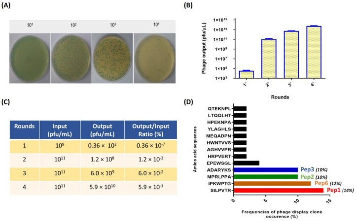
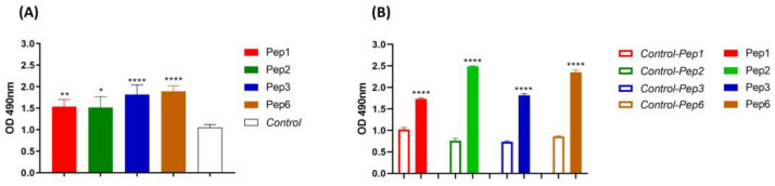
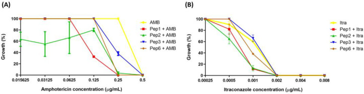
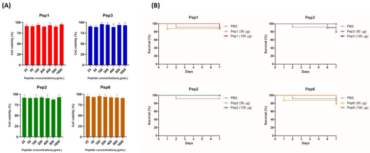
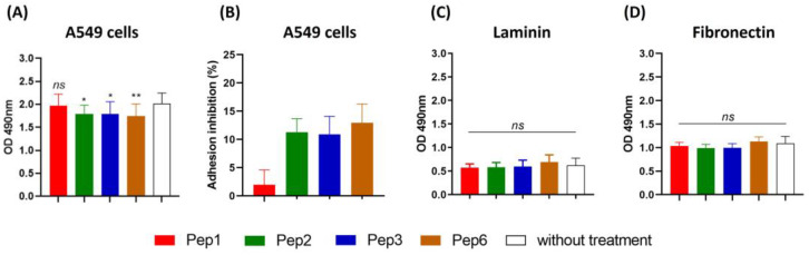
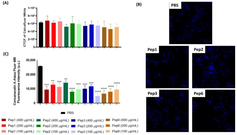
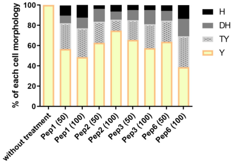
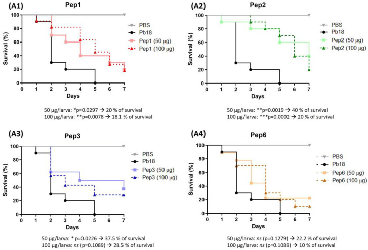
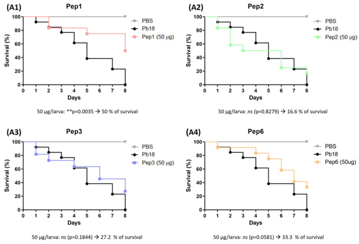
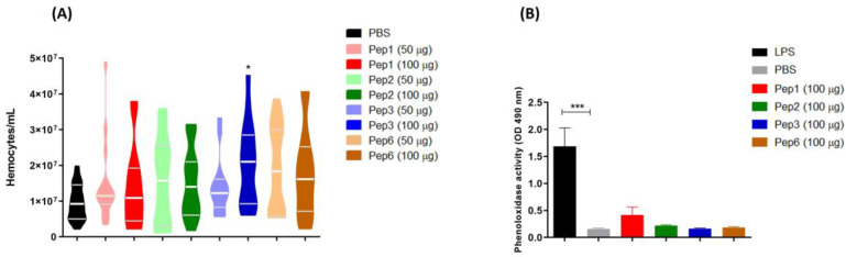
Considering the toxicity of conventional therapeutic approaches and the importance of precise mechanistic targets, it is important to explore signaling pathways implicated in fungal pathobiology. Moreover, treatment of paracoccidioidomycosis, a systemic mycosis caused by a dimorphic fungus, requires prolonged therapeutic regimens. Among the numerous factors underpinning the establishment of Paracoccidioides spp. infection, the capacity to transition from the mycelial to the yeast form is of pivotal importance. The Drk1 protein of Paracoccidioides brasiliensis likely plays a decisive role in this morphological shift and subsequent virulence. We identified peptides with affinity for the PbDrk1 protein using the phage-display method and assessed the effects of these peptides on P. brasiliensis. The peptides were found to inhibit the phase transition of P. brasiliensis. Furthermore, a substantial proportion of these peptides prevented adhesion to pneumocytes. Although these peptides may not possess inherent antifungal properties, they can augment the effects of certain antifungal agents. Notably, the cell wall architecture of P. brasiliensis appears to be modulated by peptide intervention, resulting in a reduced abundance of glycosylated proteins and lipids. These peptides were also evaluated for their efficacy in a Galleria mellonella model and shown to contribute to enhanced larval survival rates. The role of PbDrk1, which is notably absent in mammals, should be further investigated to improve the understanding of its functional role in P. brasiliensis, which may be helpful for designing novel therapeutic modalities.

Keywords: Drk1; Paracoccidioides brasiliensis; peptides; phage-display.


Conflict of interest statement

The authors declare no conflict of interest.

Figures

Figure 1
Figure 1
Enrichment of positive phage clones specifically binding to the recombinant PbDrk1 and most abundant peptides identified. (A) Example of counting plaques on LB plates with IPTG/X-gal media with serial dilution used to phage titering; recombinant phage from the phage library that carried the lacZα gene are shown as blue plaques on the plates. (B) Quantification of blue phage plaques recovered after each round of biopanning via M13KE used in Ph.D.TM—7 Phage Display Peptide Library on immobilized PbDrk1. (C) Enrichment of phages for each selection round from the phage-displayed heptapeptide library. (D) Amino acid sequences and frequencies of occurrence from 78 phage plaques randomly selected for sequencing after three affinity-selection rounds against PbDrk1. To simplify the naming of the peptides identified, the terms Pep1 (SILPVTR), Pep2 (MPRLPPA), Pep3 (ADARYKS), and Pep6 (IPKWPTG) were adopted. Pfu: plaque-forming unit.
Figure 2
Figure 2
Peptides affinity to P. brasiliensis and PbDrk1 protein determined using ELISA. The figures show the ELISA absorbance values of peptides binding to (A) P. brasiliensis using anti-cell free and (B) PbDrk1 using an anti-Drk1 antibody as primary antibodies, respectively. In Control: peptides were omitted. In Control-Pep1, Control-Pep2, Control-Pep3, and Control-Pep6: PbDrk1 protein was omitted. **** p < 0.0001; ** p = 0.0061, and * p = 0.0158 compared to the respective controls using one-way ANOVA followed by Tukey’s multiple comparisons tests.
Figure 3
Figure 3
Potentiation effect of selected peptides on antifungal activity. The peptide concentrations were fixed at 200 µg/mL, and antifungal concentrations ranged from (A) 0.015625 to 0.5 µg/mL to amphotericin (AMB) and (B) 0.00025 to 0.008 to itraconazole (Itra). p < 0.0001 for all peptides with 0.25 µg/mL of AMB; p = 0.0001 (to Pep1), p = 0.0138 (to Pep2) and p = 0.0022 (to Pep6) with 0.001 µg/mL of Itra analyzed using two-one way ANOVA with Dunnett’s comparison test.
Figure 4
Figure 4
Toxicity of selected peptides for pneumocytes A549 cells and Galleria mellonella. (A) Cytotoxicity in A549 cells determined using the resazurin method. Monolayer cells were treated with increasing concentrations of indicated peptides ranging from 25 to 1.600 µg/mL; after 24 h, metabolic activity was quantified using the resazurin assay, and the absorbance values were normalized to those of the untreated control. The result was expressed as cell viability (%) and analyzed using one-way ANOVA with Tukey’s multiple comparison test. (B) Toxicity in G. mellonella invertebrate model; groups of larvae (n = 10) were injected with 50 and 100 µg/larva of indicated peptides; survival was monitored for 7 days post-injection and analyzed using the log-rank (Mantel–Cox) test. p = 0.9447 (to Pep1), p = 0.4724 (to Pep2), p = 0.3683 (to Pep3), and p = 0.7828 (to Pep6).
Figure 5
Figure 5
Adhesion inhibition to A549 cells and extracellular matrix mediated by peptides. Paracoccidioides brasiliensis yeast cells were treated for 1 h with 200 µg/mL of each peptide and then used in a monolayer of A549 cells and 96-well coated plate with 10 µg/mL of each laminin and fibronectin for 15 h of incubation and evaluated using ELISA. (A) Absorbance at 490 nm following ELISA for A549 cells, and (B) adhesion inhibition to A549 cells as a percentage for each peptide. (C) ELISA for laminin. (D) ELISA for fibronectin. ns: not significant, * p < 0.05 and ** p < 0.01. p = 0.0156 (to Pep2; 11.2% reduction), p = 0.0178 (to Pep3, 10.9% reduction), p = 0.0063 (to Pep6, 12.9% reduction) compared to the control without treatment. One-way ANOVA followed by Dunnett’s multiple comparison test.
Figure 6
Figure 6
Alterations of cell-wall components of P. brasiliensis after peptide treatment. Yeast cells of P. brasiliensis were treated for 3 h with 100, 200, and 400 µg/mL of each peptide. The cells were labeled with (A,B) calcofluor white (CFW) to measure chitin levels using fluorescence microscopy and (C) concanavalin-Alexa Fluor conjugated to glycosylated proteins levels using cytometry. ** p < 0.01, *** p < 0.001, **** p < 0.0001 compared to the PBS control group using one-way ANOVA followed by Dunnett’s multiple comparisons tests. CTCF: corrected total cell fluorescence. Scale bars are included = 10 µM.
Figure 7
Figure 7
Morphological transition in P. brasiliensis after peptide treatment. The mycelium phase of P. brasiliensis was treated with different concentrations (50 and 100 µg/mL) of each peptide and incubated at 37 °C until the control (without treatment) reached complete conversion to the yeast phase (9 days). Aliquots of each group were analyzed using optical microscopy to discriminate the cell morphology as described by Nunes et al. [38]; H: hyphae, DH: differentiate hyphae, TY: transforming yeasts, and Y: yeasts.
Figure 8
Figure 8
Use of peptides to protect G. mellonella larvae against P. brasiliensis infection. The survival curve of G. mellonella larvae (n = 10 per group) was treated with the selected peptides (A1) to Pep1, (A2) to Pep2, (A3) to Pep3 and (A4) to Pep6 at both concentrations (50 and 100 µg/larva) 3 h before infection with P. brasiliensis. Statistical analyses were performed using the log-rank (Mantel–Cox) test of three independent experiments. * p < 0.05, ** p < 0.01, *** p < 0.001 and ns: not significant. The p values for each peptide and concentration as well as the % of larvae survival are described in the figure.
Figure 9
Figure 9
Use of peptides to combat G. mellonella infection with P. brasiliensis. Survival curve of G. mellonella larvae (n = 10 per group) infected with P. brasiliensis and treated with the selected peptides (A1) to Pep1, (A2) to Pep2, (A3) to Pep3 and (A4) to Pep6 at 50 µg/larva for 3 h. Statistical analyses were performed using the log-rank (Mantel–Cox) test of three independent experiments. ** p < 0.01, and ns: not significant. The p values for each peptide and concentration as well as the % of larvae survival are described in the figure.
Figure 10
Figure 10
Galleria mellonella immunomodulation following peptide treatment. Larvae (n = 10) of G. mellonella were treated with each peptide at different concentrations (50 and 100 µg/larva for hemocyte assay and 100 µg/larva for phenoloxidase assay) for 3 h, and the (A) hemocytes density and (B) phenoloxidase activity were evaluated. * p = 0.0331 compared to PBS control, analyzed using the Kruskal–Wallis test followed by Dunn’s multiple comparison test. *** p < 0.0001 compared to PBS control with one-way ANOVA with Tukey’s multiple comparison test.

References

    1. Kim J.H., Cheng L.W., Land K.M. Advances in Antifungal Development: Discovery of New Drugs and Drug Repurposing. Pharmaceuticals. 2022;15:787. doi: 10.3390/ph15070787. - DOI - PMC - PubMed
    1. De Oliveira H.C., Bezerra B.T., Rodrigues M.L. Antifungal Development and the Urgency of Minimizing the Impact of Fungal Diseases on Public Health. ACS Bio Med. Chem. Au. 2022;3:137–146. doi: 10.1021/acsbiomedchemau.2c00055. - DOI - PMC - PubMed
    1. Yu Y., Hube B., Kämper J., Meyer V., Krappmann S. When green and red mycology meet: Impressions from an interdisciplinary forum on virulence mechanisms of phyto- and human-pathogenic fungi. Virulence. 2017;8:1435–1444. doi: 10.1080/21505594.2017.1356502. - DOI - PMC - PubMed
    1. Borges-Walmsley M.I., Chen D., Shu X., Walmsley A.R. The pathobiology of Paracoccidioides brasiliensis. Trends Microbiol. 2002;10:80–87. doi: 10.1016/S0966-842X(01)02292-2. - DOI - PubMed
    1. San-Blas G., Niño-Vega G., Iturriaga T. Paracoccidioides brasiliensis and paracoccidioidomycosis: Molecular approaches to morphogenesis, diagnosis, epidemiology, taxonomy and genetics. Med. Mycol. 2002;40:225–242. doi: 10.1080/mmy.40.3.225.242. - DOI - PubMed

LinkOut - more resources