. 2023 Nov 28;42(11):113316.
doi: 10.1016/j.celrep.2023.113316. Epub 2023 Oct 26.

Pain and itch coding mechanisms of polymodal sensory neurons

Affiliations

Pain and itch coding mechanisms of polymodal sensory neurons

Changxiong Guo et al. Cell Rep. .

Abstract

Pain and itch coding mechanisms in polymodal sensory neurons remain elusive. MrgprD+ neurons represent a major polymodal population and mediate both mechanical pain and nonhistaminergic itch. Here, we show that chemogenetic activation of MrgprD+ neurons elicited both pain- and itch-related behavior in a dose-dependent manner, revealing an unanticipated compatibility between pain and itch in polymodal neurons. While VGlut2-dependent glutamate release is required for both pain and itch transmission from MrgprD+ neurons, the neuropeptide neuromedin B (NMB) is selectively required for itch signaling. Electrophysiological recordings further demonstrated that glutamate synergizes with NMB to excite NMB-sensitive postsynaptic neurons. Ablation of these spinal neurons selectively abolished itch signals from MrgprD+ neurons, without affecting pain signals, suggesting a dedicated itch-processing central circuit. These findings reveal distinct neurotransmitters and neural circuit requirements for pain and itch signaling from MrgprD+ polymodal sensory neurons, providing new insights on coding and processing of pain and itch.

Keywords: CP: Neuroscience; MrgprD; glutamate; itch; neural coding; neuromedin B; neurotransmitters; pain; polymodal sensory neurons.


Conflict of interest statement

Declaration of interests The authors declare no competing interests.

Figures
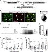
Figure 1.
Figure 1.. Chemogenetic activation of MrgprD+ sensory neurons produces both pain and itch behavioral responses
(A) Breeding strategy for the generation of MrgprdhM3Dq/+ mice. MrgprdCreERT2/+ are crossed with TgCAG-hM3Dq/+ mice to produce MrgprdCreERT2/+; TgCAG-hM3Dq/+ (MrgprdhM3Dq/+) mice and control littermates. After tamoxifen-induced cre recombination, the floxed stop codon between the hM3Dq allele and CAG promoter is excised in MrgprD-expressing cells. (B) Immunofluorescence staining showed that 63.5% of IB4+ sensory neurons express mCitrine. Arrows indicate mCitrine+ neurons (n = 3 mice). Findings were confirmed by RNAscope. See also Figure S1. (C and D) Whole-cell current-clamp recordings of cultured IB4 (C) and IB4+ (D) DRG neurons from MrgprdhM3Dq/+ mice. Traces are representative of neuronal responses to CNO stimulation (0.1 mM, bath applied). (E) Quantification of CNO responses in cultured DRG from MrgprdhM3Dq/+ mice (n ≥ 12 neurons per group, from 4 mice). (F and G) Intradermal cheek injections of CNO produced simultaneous pain and itch responses in MrgprdhM3Dq/+ mice. When injected into the cheek (20 μL, in 1% DMSO in saline vehicle), CNO robustly induced scratching (F) and wiping (G) in a dose-dependent manner (n ≥ 5 mice per genotype per dose). Please note that the vehicle control induced mild pain-related wiping behavior in both control and MrgprdhM3Dq/+ mice, which may mask pain responses at the lowest tested CNO dose. Data are presented as mean ± SEM. Nonparametric Mann-Whitney U tests were used to determine statistical significance for (F) and (G). n.s., no significance. *p ≤ 0.05, **p ≤ 0.01, ***p ≤ 0.001. Scale bars: 50 μm.
Figure 2.
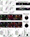
Figure 2.. Glutamate and neuromedin B (NMB) are released by MrgprD+ neurons for synaptic transmission
(A) Fluorescence-activated cell sorting (FACS) analysis of GFP+ neurons from wild-type and MrgprdEGFP/+ mice. Gated GFP+ fraction accounted for ~15% of all events. (B) RT-PCR analysis of solute vesicle and neurotransmitter precursor expression in FACS-purified MrgprD-GFP neurons. The glutamate transporters Vglut1 and Vglut3 and the neuropeptides Cgrp, Grp, and Nppb were not detected in MrgprD-GFP neurons, while Vglut2 and Nmb were robustly expressed by this population. BSA-purified, unsorted DRG neurons were used as a control. (C) Immunofluorescence staining of DRGs from MrgprdEGFP/+ mice showed broad co-expression of VGlut2 in GFP+ neurons, indicating that most MrgprD+ neurons expressed VGlut2 (n = 3 mice). (D) In situ analysis showed that all Mrgprd+ neurons expressed Nmb (n = 3 mice). (E) Extracellular glutamate level of cultured DRGneurons from control and MrgprdhM3Dq/+ mice after vehicle or CNO treatment (500 μM), as determined by enzymatic assay. Data are normalized to vehicle treatment (n = 3 biological pairs). (F) Extracellular NMB level of cultured DRG neuronsfrom control and MrgprdhM3Dq/+ mice after vehicle or CNO treatment, as determined by ELISA (n = 3 biological pairs). (G) Extracellular glutamate level of cultured DRG neurons from wild-type mice after β-alanine (1 or 2 mM, as indicated) or KCl (30 mM) treatment. Data are normalized to vehicle treatment. Mean value is indicated above each column (n ≥ 5 biological replicates). (H) Extracellular NMB level of cultured DRG neurons from wild-type mice after vehicle, β-alanine (1 mM), or KCl (30 mM) treatment. Data are normalized to vehicle treatment. (n = 7 biological replicates). All data are presented as mean ± SEM. Shapiro-Wilk tests were used to determine data normality for (E)–(H). Unpaired Student’s t tests was used to determine statistical significance for (E) and (F). Paired (pre/post) Student’s t tests were used for (G) and (H). *p %≤ 0.05, **p ≤ 0.01. Scale bars: 50 μm.
Figure 3.
Figure 3.. Glutamate is required for signal coding and transmission of both pain and itch in MrgprD+ sensory neurons
(A) Breeding strategy for the generation of Mrgprd-Vglut2 cKO mice. After tamoxifen-induced cre recombination, the floxed exon 2 of Vglut2 is excised in Mrgprd-expressing cells, resulting in the conditional deletion of VGlut2 in MrgprD+ cells. Note, the deletion of exon 2 produces a reading frameshift; no subsequent exon is translated in Vglut2. (B and C) Immunofluorescence staining of VGlut2 in DRGs from control and MrgprD-Vglut2 cKO mice. After tamoxifen-induced recombination, VGlut2 expression was reduced from 96.9% IB4+ trigeminal ganglia (TG) and DRG neurons in control mice (B) to only 27.5% in MrgprD-Vglut2 cKO mice (C) (n ≥ 3 mice per genotype). See also Figure S2. (D–J) MrgprD-Vglut2 cKO mice showed defects in mechanical pain and nonhistaminergic itch. (D) MrgprD-Vglut2 cKO mice exhibited significantly lower sensitivity to noxious mechanical stimuli (n = 10 per group). (E and F) Mrgprd-Vglut2 cKO mice did not show defects in thermal nociception, as measured by (E) Hargreaves assay (n ≥ 5 per group) and (F) tail flick assay (50°C, n ≥ 5 per group). (G and H) Mrgprd-Vglut2 cKO mice did not develop (G) spontaneous itch (n = 8 per group) or (H) enhanced itch responses to chloroquine (0.16 μmol in 20 μL, n = 6 per group). In contrast, (I) β-alanine- (1 μmol) and (J) diethylstilbestrol (DES; 1 μmol in 50 μL ethanol vehicle for topical nape application)-induced scratching were significantly reduced (n ≥ 12 and n = 6 per genotype, respectively). Unless otherwise indicated, all injections were 20 μL at the cheek using saline vehicle. Itch behavior was recorded and analyzed for 15 min, except for the chloroquine assay (30 min). Data are presented as mean ± SEM. Nonparametric Mann-Whitney U tests were used to determine statistical significance for (D)–(J). n.s., no significance. *p ≤ 0.05, **p ≤ 0.01, ***p ≤ 0.001. Scale bars: 50 μm.
Figure 4.
Figure 4.. The neuropeptide NMB is selectively required for itch sensation in MrgprD+ neurons
(A) Breeding strategy for the generation of Mrgprd-Nmb cKO mice. After tamoxifen-induced cre recombination, the floxed exon 2 of Nmb, which encodes the NMB signaling peptide, is excised in Mrgprd-expressing cells, resulting in the conditional deletion of NMB in MrgprD+ cells. (B and C) Single-cell RT-PCR of IB4+ DRG neurons from control and Mrgprd-Nmb cKO mice. After tamoxifen treatment, (B) Nmb expression was not affected in DRG neurons from control mice but (C) was undetectable in Mrgprd+ neurons from Mrgprd-Nmb cKO mice. Note that IB4+/MrpgrD neurons from both genotypes express Nmb (n = 3 mice per genotype). (D) Mrgprd-Nmb cKO mice showed similar response frequencies to von Frey stimulation as control littermates (n ≥ 6 per group). (E and F) Mrgprd-Nmb cKO mice did not develop defects in thermal nociception, as measured by (E) Hargreaves assay (n ≥ 5 per group) and (F) tail flick assay (50C, n ≥ 5 per group). (G and H) β-alanine- (1 μmol, saline vehicle) (G) and DES (H) (1 μmol in 50 μL ethanol vehicle for topical nape application)-induced scratching (bouts/15 min) were significantly reduced (n ≥ 6 per genotype for both experiments). (I) CNO-induced itch is selectively diminished in MrgprdhM3Dq/+;Mrgprd-Nmb cKO mice. While there was no significant difference in CNO (20 nmol, 1% DMSO saline vehicle)-induced wiping behavior between control and cKO mice, itch-related scratching responses were significantly attenuated in the Mrgprd-Nmb cKO mice (n ≥ 5 per genotype). Pain and itch behavior was recorded and analyzed for 30 min. Unless otherwise indicated, all injections were 20 μL at the cheek. Data are presented as mean ± SEM. Nonparametric Mann-Whitney U tests were used to determine statistical significance for (D)–(I). n.s., no significance. *p ≤ 0.05, ***p ≤ 0.001.
Figure 5.
Figure 5.. NMB-sensitive medullary dorsal horn (MDH) neurons receive itch input from MrgprD+ afferents
(A–F) Ex vivo slice recording of brainstems from NmbrEGFP+ mice. (A) Representative whole-cell current-clamp recording of NMB (4 μM, bath perfusion) response in NmbrEGFP+ MDH neurons. (B) Quantification of NmbrEGFP+ MDH neuron responses to NMB. NMB dose-dependently induced membrane potential increase in NmbrEGFP+ neurons (n ≥ 6 neurons from 3 mice for each group). (C) β-alanine (1 mM, bath perfusion)-induced increase in membrane potential in a subset of NmbrEGFP+ MDH neurons (13/25 neurons, from 10 mice). Synaptic connections were also confirmed by immunostaining; see also Figure S3. (D) Representative whole-cell current-clamp recording of AMPA (1 μM, bath perfusion). (E) Representative whole-cell current-clamp recording of AMPA and NMB (1 μM each, bath perfusion). (F) Quantification of NmbrEGFP+ MDH neuron responses to AMPA alone and AMPA with NMB. AMPA-enhanced NMB induced membrane potential increase and action potential firing in NmbrEGFP+ neurons (n ≥ 5 neurons from 3 mice for each group). Please note that for (D)–(F), recordings were performed in the presence of 100 mM picrotoxin and 1 μM strychnine to block GABA and glycine receptors. (G) Validation of NmbrEGFP+ MDH neuron ablation after intracisternal NMB-saporin injection. GFP signal was found in the MDH of blank-saporin-injected NmbrEGFP+ mice. In contrast, no GFP signal was found in the MDH of NMB-saporin-injected NmbrEGFP+ mice (n ≥ 3 mice per group). (H) Intracisternal NMB-saporin injection did not affect response to von Frey stimulation (n = 6 mice per group). (I and J) Thermal nociception, as measured by (I) Hargreaves assay (n ≥ 6 per group) and (J) tail flick assay (n ≥ 6 per group), was not significantly changed by NMB-saporin injection. (K) Intracisternal NMB-saporin injection attenuated β-alanine (1 μmol, 20 μL cheek, saline vehicle)-induced itch-related scratching behavior (bouts/15 min, n ≥ 5 mice per group). (L) CNO (20 nmol in 20 μL cheek, 1% DMSO saline vehicle)-induced wiping responses were not different between blank- or NMB-saporin-injected MrgprdhM3Dq/+ mice, but itch-related scratching was nearly abolished by NMB-saporin injections (n ≥ 5 mice per group). Pain and itch behavior was recorded and analyzed for 30 min. Data are presented as mean ± SEM. Shapiro-Wilk tests were used to determine data normality, and paired (pre/post) Student’s t tests were used to determine statistical significance for (A), (C), and (F). Nonparametric Mann-Whitney U tests were used to determine statistical significance for (H)–(L). *p ≤ 0.05, **p ≤ 0.01, ***p ≤ 0.001. Scale bars: 50 μm.
Figure 6.
Figure 6.. The combinatory model for pain and itch coding by MrgprD+ polymodal neurons
MrgprD+ polymodal afferents detect both noxious mechanical forces and pruritic chemical stimuli including β-alanine. Mechanical stimuli induce brief action potential firings in these neurons and glutamate release at their central terminals. In contrast, chemical pruritogens like β-alanine induce burst firings in MrgprD+ neurons, which triggers the release of neuropeptide NMB as well as a low level of glutamate release at central terminals. Itch signals are received and processed by NMBR+ spinal neurons and related itch pathways.

References

    1. Basbaum AI, Bautista DM, Scherrer G, and Julius D (2009). Cellular and molecular mechanisms of pain. Cell 139, 267–284. - PMC - PubMed
    1. Bautista DM, Wilson SR, and Hoon MA (2014). Why we scratch an itch: the molecules, cells and circuits of itch. Nat. Neurosci. 17, 175–182. 10.1038/nn.3619. - DOI - PMC - PubMed
    1. Patel KN, and Dong X (2011). Itch: Cells, Molecules, and Circuits. ACS Chem. Neurosci. 2, 17–25. 10.1021/cn100085g. - DOI - PMC - PubMed
    1. Han L, Ma C, Liu Q, Weng HJ, Cui Y, Tang Z, Kim Y, Nie H, Qu L, Patel KN, et al. (2013). A subpopulation of nociceptors specifically linked to itch. Nat. Neurosci. 16, 174–182. 10.1038/nn.3289. - DOI - PMC - PubMed
    1. Knowlton WM, Palkar R, Lippoldt EK, McCoy DD, Baluch F, Chen J, and McKemy DD (2013). A sensory-labeled line for cold: TRPM8-expressing sensory neurons define the cellular basis for cold, cold pain, and cooling-mediated analgesia. J. Neurosci. 33, 2837–2848. 10.1523/JNEUROSCI.1943-12.2013. - DOI - PMC - PubMed

Publication types