. 2023 Oct 27;9(43):eadi8986.
doi: 10.1126/sciadv.adi8986. Epub 2023 Oct 27.

Deciphering sources of PET signals in the tumor microenvironment of glioblastoma at cellular resolution

Affiliations

Deciphering sources of PET signals in the tumor microenvironment of glioblastoma at cellular resolution

Laura M Bartos et al. Sci Adv. .

Abstract

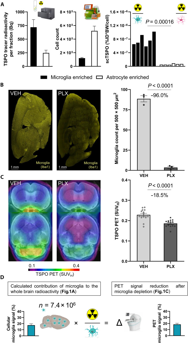
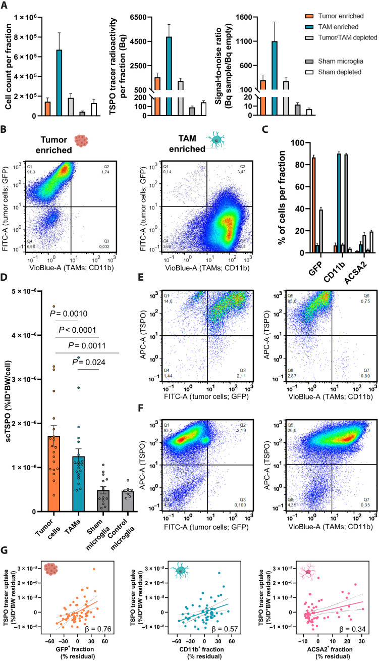
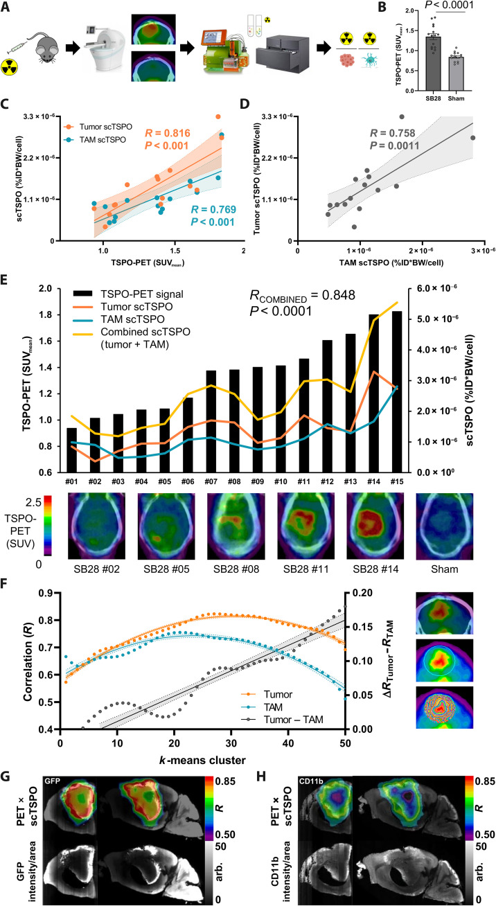
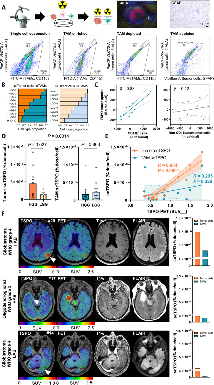
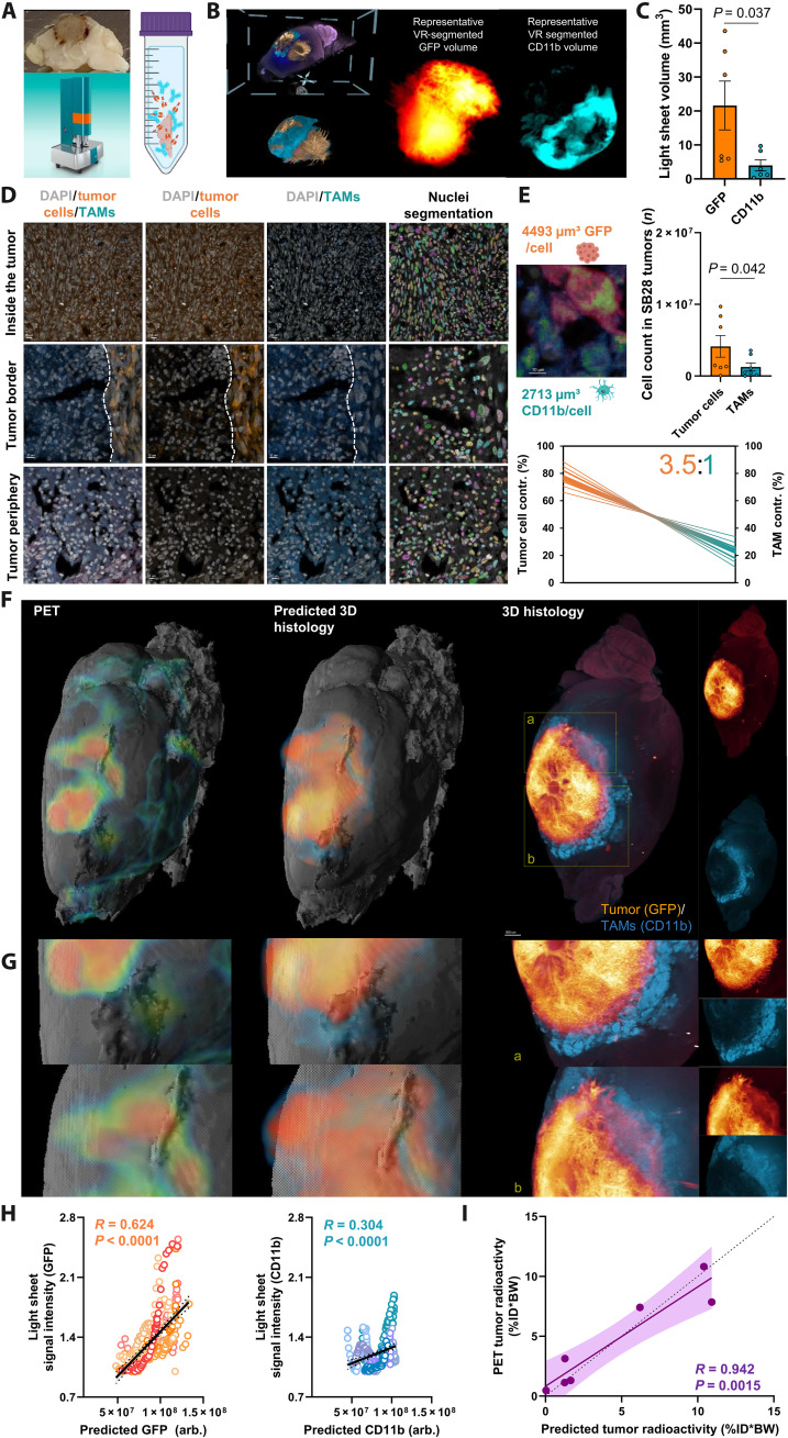
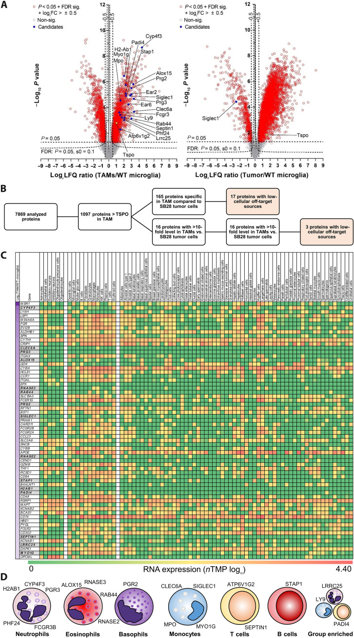
Various cellular sources hamper interpretation of positron emission tomography (PET) biomarkers in the tumor microenvironment (TME). We developed an approach of immunomagnetic cell sorting after in vivo radiotracer injection (scRadiotracing) with three-dimensional (3D) histology to dissect the cellular allocation of PET signals in the TME. In mice with implanted glioblastoma, translocator protein (TSPO) radiotracer uptake per tumor cell was higher compared to tumor-associated microglia/macrophages (TAMs), validated by protein levels. Translation of in vitro scRadiotracing to patients with glioma immediately after tumor resection confirmed higher single-cell TSPO tracer uptake of tumor cells compared to immune cells. Across species, cellular radiotracer uptake explained the heterogeneity of individual TSPO-PET signals. In consideration of cellular tracer uptake and cell type abundance, tumor cells were the main contributor to TSPO enrichment in glioblastoma; however, proteomics identified potential PET targets highly specific for TAMs. Combining cellular tracer uptake measures with 3D histology facilitates precise allocation of PET signals and serves to validate emerging novel TAM-specific radioligands.


Figures

Fig. 1.
Fig. 1.. TSPO-PET signal reduction in the whole brain after microglia depletion corresponds to the whole-brain signal attributable to tracer uptake in microglia.
(A) Components of scRadiotracing after in vivo tracer injection. Upon in vivo tracer injection and magnetic cell separation, cell pellets were analyzed by a high-sensitive gamma counter to measure the radioactivity (in becquerels) in the sample (left) (n = 6, means ± SEM) and by flow cytometry to determine the cell count (middle) (n = 6, means ± SEM). After calculation of radioactivity per single cell [single-cell TSPO tracer (scTSPO), normalized to ID and BW] in each sample, untreated mice showed significantly higher TSPO tracer uptake in microglia when compared to astrocytes (right) (each bar represents a single animal; n = 6, paired t test). (B) Representative sagittal Iba1 immunohistochemistry sections of vehicle treatment (VEH; n = 3) and PLX5622 (PLX)–treated (n = 3) mice and quantification of stained Iba1 area (%). Unpaired t test, means ± SEM. (C) Group average TSPO-PET images of healthy mice upon a magnetic resonance imaging (MRI) template after VEH (n = 14) or CSF1R inhibition (PLX5622; microglia depletion, n = 15) indicated a distinct signal reduction in the whole brain of PLX5622-treated animals (−18.5%). Unpaired t test, means ± SEM. (D) Extrapolation of the signal attributable to microglia using published cell numbers (76) and tracer uptake per single microglia cell as calculated by scRadiotracing. The calculated 17.5% contribution of microglia to the whole-brain TSPO-PET signal corresponded to the observed TSPO-PET signal reduction after microglia depletion (18.5%). Means ± SEM.
Fig. 2.
Fig. 2.. scRadiotracing in the SB28 glioblastoma mouse model allows differential assessment of TSPO tracer uptake in tumor cells and TAMs.
(A) Acquired components of scRadiotracing after in vivo tracer injection in SB28 glioblastoma (n = 20) and sham (n = 14) mice. Using MACS, tumor cells (GFP+) and CD11b+ TAMs from SB28-bearing mice and CD11b+ microglia from sham-injected mice were enriched, thus leaving the respective residual–depleted cell fractions. Absolute cell numbers (left) and measured radioactivity (middle) resulted in satisfying signal-to-noise ratios (right) for all enriched and depleted fractions investigated. Means ± SEM. (B) MACS of tumor cells and TAMs led to >90% purity in enriched fractions as confirmed by flow cytometry. (C) Distribution of tumor cells (GFP), TAMs (CD11b), and astrocytes (ACSA2) in enriched and depleted fractions of SB28 glioblastoma (n = 20) and sham (n = 14) mice. Means ± SEM. (D) Comparison of the scTSPO uptake of isolated tumor cells (n = 20) and TAMs (n = 20) in SB28 glioblastoma mice as well as microglia of sham (n = 14) and untreated control (n = 8) mice. Paired t test for tumor cells versus TAMs, one-way ANOVA for all other comparisons, means ± SEM. (E and F) TSPO costaining in flow cytometry shows that nearly all tumor cells (GFP) (E) and TAMs (CD11b) (F) were also positive for TSPO. Notably, the minor population of CD11b cells in the TAM-enriched fraction did not show positivity for TSPO (F, lower left quadrant), confirming the specificity of the TSPO costaining. Pooled data from n = 3 tumors. (G) Regression model including the enriched and depleted fractions of SB28 mice indicated highest contribution of tumor cells (left) to the radioactivity in the sample, followed by TAMs (middle) and astrocytes (right). Linear regression, β = standardized regression coefficient. n = 60 samples. Error bands represent 95% confidence interval.
Fig. 3.
Fig. 3.. Correlation of single-cell TSPO tracer uptake of tumor cells and TAMs with TSPO-PET indicates association of TSPO-PET heterogeneity with single-cell tracer enrichment.
(A) Schematic illustration of the scRadiotracing workflow. (B) TSPO-PET imaging indicated significantly higher lesion site signals in SB28 tumor mice (n = 15) than in sham (n = 14) animals at day 18 after inoculation. Unpaired t test, means ± SEM. (C) Correlation between scRadiotracing and TSPO-PET showed a strong dependence of PET signals from both tumor and TAM scTSPO uptake (normalized to ID and BW) in SB28 mice. N = 15, R = Pearson’s coefficient of correlation. Error bands represent 95% confidence interval. (D) Intercorrelation of scTSPO of tumor cells and TAMs in the SB28 glioblastoma mouse model. N = 15, R = Pearson’s coefficient of correlation. Error band represents 95% confidence interval. (E) Association between combined scTSPO and heterogeneous TSPO-PET signals. Black bars symbolize the individual TSPO-PET signal for all SB28 mice investigated (n = 15). Curves represent scTSPO of tumor cells (orange) and TAMs (blue) and a combined vector of cellular tracer uptake (yellow) for each individual animal. Axial sections of TSPO-PET images upon the individual contrast-enhanced computed tomography (CT) illustrate interindividual TSPO-PET signal heterogeneity of SB28 tumors. R = Pearson’s coefficient of correlation. (F) Visualization of the regional k-means cluster analysis. A sphere was placed over the entire signal enhancement in TSPO-PET, which was followed by application of k-means clustering. This resulted in 50 volumes of interest (VOIs) defining 50 intratumoral regions of increasing signal intensity (images on the right). (G and H) Strong regional agreement between tumor and TAM cell density (GFP/CD11b, light sheet microscopy) and regions with high scTSPO-to-PET associations. Coronal and sagittal slices show projections of correlation coefficients (R) onto k-means cluster VOIs of an individual mouse. arb., arbitrary units.
Fig. 4.
Fig. 4.. Translation of scRadiotracing to human glioma samples.
(A) Schematic illustration of the workflow and the gating strategy in human glioma samples (n = 20). The single-cell suspension (left) was separated into TAM-enriched (CD11b+; second from left) and tumor-enriched (third from left) fractions. Tumor cells were defined via glial fibrillary acidic protein (GFAP) or 5-aminolevulinic acid (5-ALA) after confirmation of 5-ALA positivity during surgery or after confirmation of GFAP positivity during neuropathological workup. 5-ALA+ cells colocalized with GFAP+ cells (right). (B) Relative distribution of tumor cells and TAMs in the single-cell suspension of human high-grade glioma (HGG; n = 10) and low-grade glioma (LGG; n = 8) samples. The first two patients did not receive an analysis of the single-cell suspension. (C) Strong contribution of TAMs (CD11b+ cells) but not of CD11b/nontumor cells to the measured activity (n = 10 biopsy samples). Linear regression, β = standardized regression coefficient. Error bands represent 95% confidence interval. (D) Comparison of scTSPO uptake of tumor cells and TAMs in samples of human HGG (n = 11) and LGG (n = 9) by a multivariate model including tumor grade, age, and sex. Means ± SEM. (E) Correlation of TSPO-PET signals with scTSPO of tumor cells and TAMs. N = 13, R = Pearson’s coefficient of correlation. Error bands represent 95% confidence interval. (F) Three patient examples with similar signals in amino acid [O-(2-[18F]fluoroethyl)-l-tyrosine (FET)] PET and only little contrast enhancement in MRI. The patient with high tumoral TSPO-PET signal [top row, glioblastoma, World Health Organization (WHO) grade-4, high-affinity binding status (HAB), patient #20] showed distinctly more scTSPO compared to the patients with only faint (middle row, oligodendroglioma, WHO grade-3, HAB, patient #17) or low [bottom row, glioblastoma, WHO grade-4, low-affinity binding status (LAB), patient #14] tumoral signal in TSPO-PET.
Fig. 5.
Fig. 5.. Integrated analysis of regional PET signals, cellular tracer uptake, and 3D histology.
(A) Tissue clearing with a modified version of 3DISCO (65) and light sheet florescent microscope imaging. (B) Representative masks of tumor cells and TAMs in the virtual reality (VR) annotation tool. This process determined the occupied volume of GFP+ tumor cells and CD11b+ TAMs within the whole tumor using segmentation via syGlass (69). (C) Quantitative comparison of GFP+ and CD11b+ volumes within individual SB28 tumors (n = 7, paired t test). (D) Confocal microscopy determined the average GFP+ volume per single tumor cell and the average CD11b+ volume per single TAM. Nuclei segmentation using cellpose (68) determined cell numbers per section (right column). (E) Quantitative comparison of the absolute tumor and TAM cell count (n = 7, paired t test) and contribution of tumor cells and TAMs to the overall TSPO-PET signal (3.5:1). Thick lines indicate the mean of scTSPO × top/bottom standard error of the mean (SEM) band of tumor volumes; thin lines indicate top/bottom SEM band of scTSPO × top/bottom SEM band of tumor volumes. (F) Regional TSPO-PET signals (left) were combined with single-cell tracer uptake values of tumor cells and TAMs to predict cell type abundance within individual SB28 tumors (middle) with validation by light sheet microscopy (right). All mice are illustrated in fig. S7. (G) Representative magnified areas within the SB28 tumor underline the regional agreement between predicted and standard of truth 3D histology. (H) Correlation of predicted and standard of truth 3D histology within k-means clusters (Fig. 3). n = 350 single regions from n = 7 tumors. R = Pearson’s coefficient of correlation. Error bands represent 95% confidence interval. (I) Prediction of radioactivity measured in PET by cellular abundance and cellular tracer uptake. R = Pearson’s coefficient of correlation. Error bands represent 95% confidence interval. Dashed line represents line of identity (y = x).
Fig. 6.
Fig. 6.. Proteome analysis of the SB28 TME identifies TAM-specific radiotracer targets for glioblastoma.
(A) Volcano plot representation of the different protein levels in isolated TAMs (n = 5) and isolated SB28 tumors cells (n = 3) in comparison with control microglia isolated from age-matched mice (WT-microglia; n = 6). TSPO (yellow) showed intermediate elevation of protein levels in TAMs and SB28 tumor cells. A total of 1098 of 7869 analyzed proteins indicated higher protein levels in TAMs than did TSPO. FC, fold change. (B) Selection process of potential radiotracer targets for specific detection of TAMs over SB28 tumor cells. A total of 165 of the identified proteins in TAMs were not detected in SB28 tumor cells, and another 16 proteins had >10-fold higher levels in TAMs compared to SB28 tumor cells. A total of 20 proteins showed low-cellular off-target sources and were identified as potential TAM radiotracer targets. (C) Heatmap screening for TAM specificity of the 181 proteins of interest. The Human Protein Atlas (17) was used to determine RNA expression levels of all proteins of interest in (i) resident off-target cells of the brain (left cell type columns), (ii) resident and infiltrating cells in the presence of glioblastoma (middle cell type columns), and (iii) off-target cells of the organism (right cell type columns). Genes are sorted by protein level differences in TAMs compared to control microglia of untreated mice (top to bottom; log2 label-free quantification ratio; first column). Sixty-four proteins with highest elevation [log2 (TAMs/WT-microglia) > 2; left column] are illustrated in (C) and all proteins are provided in fig. S8. Proteins of interest are highlighted in gray. (D) Immune cell expression cluster allocation of proteins of interest. Single-cell RNA data of the Human Protein Atlas were used to categorize the final set of identified proteins.

References

    1. Jin M.-Z., Jin W.-L., The updated landscape of tumor microenvironment and drug repurposing. Signal Transduct. Target. Ther. 5, 166 (2020). - PMC - PubMed
    1. Pitt J. M., Marabelle A., Eggermont A., Soria J. C., Kroemer G., Zitvogel L., Targeting the tumor microenvironment: Removing obstruction to anticancer immune responses and immunotherapy. Ann. Oncol. 27, 1482–1492 (2016). - PubMed
    1. Fernandes B., Feltes P. K., Luft C., Nazario L. R., Jeckel C. M. M., Antunes I. F., Elsinga P. H., de Vries E. F. J., Potential PET tracers for imaging of tumor-associated macrophages. EJNMMI Radiopharm. Chem. 7, 11 (2022). - PMC - PubMed
    1. Kist de Ruijter L., van de Donk P. P., Hooiveld-Noeken J. S., Giesen D., Elias S. G., Lub-de Hooge M. N., Oosting S. F., Jalving M., Timens W., Brouwers A. H., Kwee T. C., Gietema J. A., Fehrmann R. S. N., Fine B. M., Sanabria Bohórquez S. M., Yadav M., Koeppen H., Jing J., Guelman S., Lin M. T., Mamounas M. J., Eastham J. R., Kimes P. K., Williams S. P., Ungewickell A., de Groot D. J. A., de Vries E. G. E., Whole-body CD8+ T cell visualization before and during cancer immunotherapy: A phase 1/2 trial. Nat. Med. 28, 2601–2610 (2022). - PMC - PubMed
    1. Low J. T., Ostrom Q. T., Cioffi G., Neff C., Waite K. A., Kruchko C., Barnholtz-Sloan J. S., Primary brain and other central nervous system tumors in the United States (2014-2018): A summary of the CBTRUS statistical report for clinicians. Neurooncol. Pract. 9, 165–182 (2022). - PMC - PubMed