. 2024 Feb 15;143(7):619-630.
doi: 10.1182/blood.2023021359.

Acute myeloid leukemias with UBTF tandem duplications are sensitive to menin inhibitors

Affiliations

Acute myeloid leukemias with UBTF tandem duplications are sensitive to menin inhibitors

Juan M Barajas et al. Blood. .

Abstract

UBTF tandem duplications (UBTF-TDs) have recently emerged as a recurrent alteration in pediatric and adult acute myeloid leukemia (AML). UBTF-TD leukemias are characterized by a poor response to conventional chemotherapy and a transcriptional signature that mirrors NUP98-rearranged and NPM1-mutant AMLs, including HOX-gene dysregulation. However, the mechanism by which UBTF-TD drives leukemogenesis remains unknown. In this study, we investigated the genomic occupancy of UBTF-TD in transformed cord blood CD34+ cells and patient-derived xenograft models. We found that UBTF-TD protein maintained genomic occupancy at ribosomal DNA loci while also occupying genomic targets commonly dysregulated in UBTF-TD myeloid malignancies, such as the HOXA/HOXB gene clusters and MEIS1. These data suggest that UBTF-TD is a gain-of-function alteration that results in mislocalization to genomic loci dysregulated in UBTF-TD leukemias. UBTF-TD also co-occupies key genomic loci with KMT2A and menin, which are known to be key partners involved in HOX-dysregulated leukemias. Using a protein degradation system, we showed that stemness, proliferation, and transcriptional signatures are dependent on sustained UBTF-TD localization to chromatin. Finally, we demonstrate that primary cells from UBTF-TD leukemias are sensitive to the menin inhibitor SNDX-5613, resulting in markedly reduced in vitro and in vivo tumor growth, myeloid differentiation, and abrogation of the UBTF-TD leukemic expression signature. These findings provide a viable therapeutic strategy for patients with this high-risk AML subtype.


Conflict of interest statement

Conflict-of-interest disclosure: J.E.R. has been a consultant for Biomea Fusion. The remaining authors declare no competing financial interests.

Figures

None
Graphical abstract
Figure 1.
Figure 1.
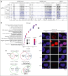
UBTF-TD/menin/KMT2A co-occupy genomic loci of genes dysregulated in UBTF-TD AML. (A) IGV tracks of HA:UBTF-TD (black), demonstrating features from 3 cbCD34+ donors transduced with UBTF-TD–expressing lentiviral vectors and maintained in culture for 40 days. CUT&RUN for UBTF, H3K4me3, H3K27ac, POLR2A, menin, and KMT2A in blue were performed in donor A. (B) GO-enrichment pathway analysis of significant targets occupied by UBTF-TD in all 3 donors (n = 226). (C) Overlap of genomic regions occupied by UBTF-TD (HA), KMT2A, and menin. Significance of overlap was calculated using hypergeometric distribution. (D) In situ PLA of endogenous UBTF/menin and UBTF/KMT2A in a UBTF-TD patient sample. Single targets (menin, KMT2A, and UBTF) are shown as controls.
Figure 2.
Figure 2.
UBTF is localized to HOXA/HOXB regions in UBTF-TD AML but not in normal hematopoietic progenitors. (A) Genomic occupancy of UBTF-WT and UBTF-TD in cbCD34+ cells. UBTF occupancy was assessed via HA:CUT&RUN in cbCD34+ cells expressing HA-UBTF-WT (blue) or HA:UBTF-TD at days 10 (D10) for UBTF-WT and UBTF-TD, as well as day 32 (D32) and day 45 (D45) for UBTF-TD. (B) IGV tracks of UBTF at the rDNA, HOXB, and HOXA genomic loci of normal cbCD34+ cells from 3 biological replicates (blue), 2 different UBTF-TD PDX (colored in red), a KMT2A-r PDX (KMT2A::MLLT3, colored in black), and an MPAL PDX (black). (C) Rank ordered peak scores of UBTF peaks subset to promoters or rDNA in normal cbCD34+ cells, UBTF-TD PDX from B (UBTF-TD PDX–1), and the second UBTF-TD PDX (UBTF-TD PDX–2). Peaks at HOXA/HOXB (red) or rDNA (blue) are annotated.
Figure 3.
Figure 3.
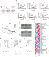
Leukemic transformation is dependent on UBTF-TD expression. (A) Schematic of FKBP12F36V-HA-UBTF-TD lentiviral construct and generation of cbCD34+ UBTF-TD degradation model. (B) Time-course degradation of FKBP12F36V-HA-UBTF-TD after treatment with dTAG-13 (1 μM) in cbCD34+ cells. Immunoblot with HA- and β-actin–specific antibodies. (C) Growth rate after dTAG-13 treatment. cbCD34+ cells expressing FKBP12F36V-HA-UBTF-TD or HA-UBTF-TD were counted over time while treated with DMSO or 1μM dTAG-13. Log of fold change was calculated from starting cell number of 200K cells. (∗∗∗ indicates P value < .001; ∗∗ indicates P value < .01, after adjustment by 2-stage step-up method (Benjamini, Krieger, and Yekutieli). (D) Immunophenotyping of cells from panel C after 10 days of treatment. (E) Wright-Giemsa staining to assess cellular morphology of cells from panels A and B after 10 days of treatment. DMSO, dimethyl sulfoxide.
Figure 4.
Figure 4.
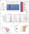
UBTF-TD genomic localization is required for gene activation. (A) Experimental design of FKBP12F36V-HA-UBTF-TD genomic occupancy experiment. (B) Tornado plots depicting the merged genomic occupancy (n = 3 replicates) at significantly depleted HA: FKBP12F36V-HA-UBTF-TD target regions (n = 266 regions, FDR < 0.5) after treatment with dTAG-13 for 3 days. Occupancy for KMT2A and menin are also shown. (C) Heatmap of the top depleted regions and their closest genes (n = 29). Heatmap colors depict row normalized enrichment. (D) Genomic tracks of merged coverage (n = 3) of HA: FKBP12F36V-HA-UBTF-TD cells treated with DMSO (black) or dTAG-13 (red) for HA, KMT2A, menin, H3K27ac (27ac), or H3K4me3 (me3). (E) Volcano plot of differentially expressed genes in FKBP12F36V-HA-UBTF-TD cells treated with DMSO or dTAG-13. Top targets (29 genes from panel C) are annotated in red. (F) mRNA levels of top target genes from panel C as compared with nontargets. (G) Upstream regulator analysis from ingenuity pathway analysis. Top targets from panel B were used for the prediction. FDR, false discovery rate; DMSO, dimethyl sulfoxide.
Figure 5.
Figure 5.
UBTF-TD leukemias are sensitive to menin inhibitors. (A) Schema for in vitro culture of primary UBTF-TD AMLs. (B) Primary tumors from 5 individual patients (Pt1-5) harboring a UBTF-TD alteration treated with menin inhibitor SNDX-5613 in vitro. Primary sample from a single patient with a RUNX1::RUNX1T1 alteration was used as a control. (C) MEIS1 mRNA steady-state levels in patient cells treated with SNDX-5613. (D) CFU capacity of UBTF-TD leukemias after exposure to SNDX-5613. Cells from Pt4 and Pt5 were treated with SNDX-5613 (250nM, SFEMII media) for 12 days and then plated in methylcellulose (#H4435, STEMCELL Technologies). (E) Heatmap depicting mRNA levels of the top 30 differentially expressed genes after 7 days of treatment with SNDX-5613. The divergent color map depicts row normalized expression. (F) CFU of UBTF-TD PDX cells plated in methylcellulose (H4435, STEMCELL) and treated with DMSO or SNDX-5613 (100nM, 500nM, 1μM). (G) Immunophenotyping of cells from panel F for stem-cell marker CD117 and CD11b. (H) Annexin V+ staining of cells from panel F. For panels F and G, statistical significances were calculated using a 1-way analysis of variance test with Dunnett’s multiple comparison adjustment using DMSO as control. CFU, colony forming unit; DMSO, dimethyl sulfoxide.
Figure 6.
Figure 6.
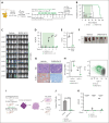
Preclinical model of UBTF-TD leukemia shows in vivo sensitivity to menin inhibitor SNDX-5613. (A) Schematic of in vivo SNDX-5613 treatment of UBTF-TD PDX. (B) Kaplan-Meier curves of UBTF-TD PDX model treated with vehicle or SNDX-5613 (n = 6 per group). Treatment period is shaded in green. (C) IVIS images of mice treated with vehicle or SNDX-5613 over time. Treatment period is shaded in green. Luminescence is row normalized to each time point. (D) Human CD45 chimerism (% of live) in the peripheral blood of mice from panel B. Treatment period is shaded in green (∗∗∗∗ indicates P value < .0001, 2-stage step-up Benjamini, Krieger, and Yekutieli test). (E) Spleen weight of mice harvested after 5 weeks of treatment with SNDX-5613 or vehicle (n = 3 per group) (2-tailed unpaired t test). (F) Spleen size of mice from panel E. (G) H&E and human NuMA1 IHC staining of spleens from panel F. (H) Flow cytometry analysis of bone marrow isolated from mice from panel D (P values are calculated with 2-tailed unpaired t test). MFI of CD11b-APC/Cy7 and CD117-PE/Cy7 (left panel) and representative flow plot (right panel) are shown (P values are calculated with 2-tailed unpaired t test). (I) Schematic of serial transplant experiment. Cells from each group in panel E were plated onto methylcellulose (#H4435, STEMCELL Technologies) (n = 3) or serially transplant into NSG-SGM3 mice (n = 3). (J) CFU capacity of cells from panel I (P value was calculated using an unpaired, 2-tailed t test). (K) Peripheral blood chimerism of mice from panel I. P values were calculated using 2-stage step-up Benjamini, Krieger, and Yekutieli test. CFU, colony-forming unit; IVIS, in vivo imaging system; H&E, hematoxylin and eosin; IHC, immunohistochemistry; MFI, mean fluorescent intensity; P.O, per os.

Comment in

References

    1. Sanij E, Hannan RD. The role of UBF in regulating the structure and dynamics of transcriptionally active rDNA chromatin. Epigenetics. 2009;4(6):374–382. - PubMed
    1. Moss T, Mars JC, Tremblay MG, Sabourin-Felix M. The chromatin landscape of the ribosomal RNA genes in mouse and human. Chromosome Res. 2019;27(1-2):31–40. - PubMed
    1. Hamdane N, Stefanovsky VY, Tremblay MG, et al. Conditional inactivation of upstream binding factor reveals its epigenetic functions and the existence of a somatic nucleolar precursor body. PLoS Genet. 2014;10(8) - PMC - PubMed
    1. Umeda M, Ma J, Huang BJ, et al. Integrated genomic analysis identifies UBTF tandem duplications as a recurrent lesion in pediatric acute myeloid leukemia. Blood Cancer Discov. 2022;3(3):194–207. - PMC - PubMed
    1. Kaburagi T, Shiba N, Yamato G, et al. UBTF-Internal tandem duplication as a novel poor prognostic factor in pediatric acute myeloid leukemia. Genes Chromosomes Cancer. 2023;62(4):202–209. - PubMed

Publication types

MeSH terms

Substances