. 2023 Dec;299(12):105396.
doi: 10.1016/j.jbc.2023.105396. Epub 2023 Oct 27.

A conserved core region of the scaffold NEMO is essential for signal-induced conformational change and liquid-liquid phase separation

Affiliations

A conserved core region of the scaffold NEMO is essential for signal-induced conformational change and liquid-liquid phase separation

Christopher J DiRusso et al. J Biol Chem. 2023 Dec.

Abstract

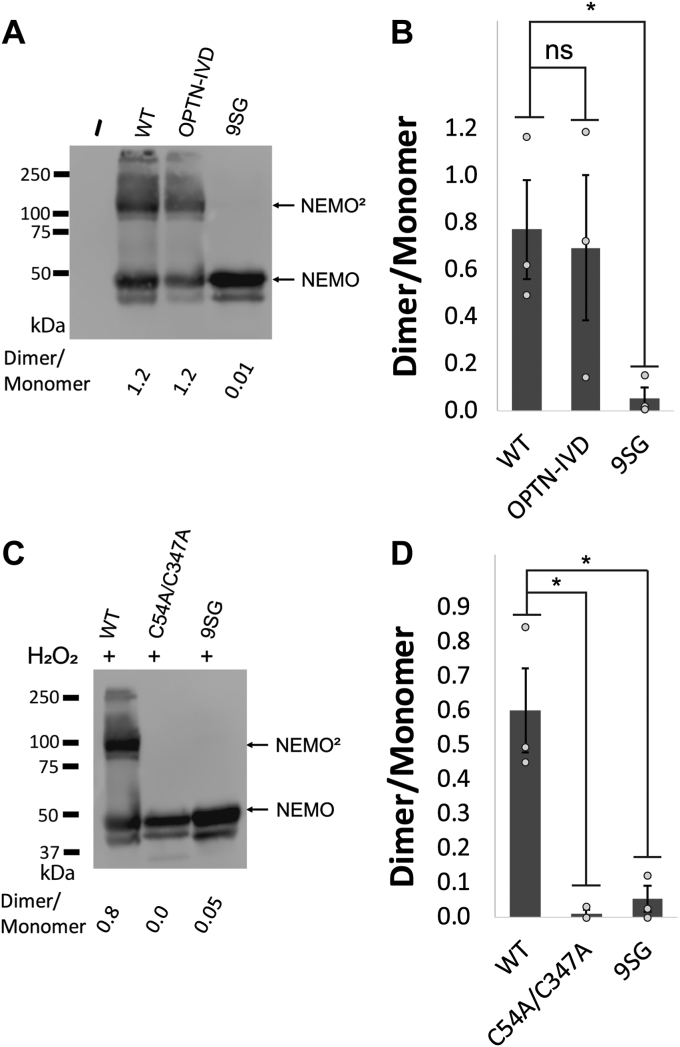
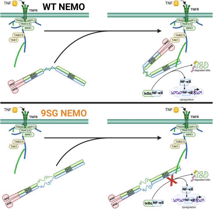
Scaffold proteins help mediate interactions between protein partners, often to optimize intracellular signaling. Herein, we use comparative, biochemical, biophysical, molecular, and cellular approaches to investigate how the scaffold protein NEMO contributes to signaling in the NF-κB pathway. Comparison of NEMO and the related protein optineurin from a variety of evolutionarily distant organisms revealed that a central region of NEMO, called the Intervening Domain (IVD), is conserved between NEMO and optineurin. Previous studies have shown that this central core region of the IVD is required for cytokine-induced activation of IκB kinase (IKK). We show that the analogous region of optineurin can functionally replace the core region of the NEMO IVD. We also show that an intact IVD is required for the formation of disulfide-bonded dimers of NEMO. Moreover, inactivating mutations in this core region abrogate the ability of NEMO to form ubiquitin-induced liquid-liquid phase separation droplets in vitro and signal-induced puncta in vivo. Thermal and chemical denaturation studies of truncated NEMO variants indicate that the IVD, while not intrinsically destabilizing, can reduce the stability of surrounding regions of NEMO due to the conflicting structural demands imparted on this region by flanking upstream and downstream domains. This conformational strain in the IVD mediates allosteric communication between the N- and C-terminal regions of NEMO. Overall, these results support a model in which the IVD of NEMO participates in signal-induced activation of the IKK/NF-κB pathway by acting as a mediator of conformational changes in NEMO.

Keywords: IKK; NEMO; NF-kappaB; conformational change; liquid-liquid phase separation; mutant; optineurin; scaffold protein; signal transduction.


Conflict of interest statement

Conflict of interest The authors declare that they have no conflicts of interest with the contents of this article.

Figures

Figure 1
Figure 1
Evolutionary analysis and comparison of NEMO and OPTN proteins.A, NEMO domain structure, with colored areas corresponding to the functional domains of NEMO and proposed unstructured regions represented by black lines. HLX1/KBD, Helix 1/Kinase Binding Domain; CC1/IVD, Coiled-Coil 1/Intervening Domain; HLX2, Helix 2; CC2, Coiled-Coil 2; LZ, Leucine Zipper; UBAN, Ubiquitin Binding in ABIN and NEMO domain; ZF, Zinc Finger. B, plot of the sequence conservation obtained using Rate4Site and the residue hydropathy calculated using the Kyte & Doolittle scale for human NEMO, represented by a moving average. The red bars denote the location of the hot spot regions of the NEMO-IKKβ binding interaction (Fig. S2). C, structural comparison of NEMO and OPTN protein domain structures, with the black dotted line indicating a gap in the alignment. LIR, LC3 Interacting Region. D, multiple sequence alignment for the IVD core region of NEMO and OPTN, from a broad spread of vertebrate proteins. Sequences were aligned with Clustal Omega (EMBL-EBI) using WT human NEMO as a reference sequence. The sequence alignment was visualized and colored according to residue conservation using MView (EMBL-EBI) (60).
Figure 2
Figure 2
Effect of mutations in the core region of the IVD on the in vivo function and structural integrity of NEMO.A, IVD mutants were used in this study. Light blue residues indicate conservative amino acid substitutions, while orange residues indicate non-conservative substitutions. B, anti-NEMO (top) and anti-phospho-IκBα (bottom) Western blots of 293T NEMO KO cells transfected with the indicated FLAG-tagged NEMO expression vectors or an empty vector plasmid (−). Cells were treated with TNFα (final concentration 20 ng/ml) for 10 min prior to lysis (+) or left untreated (−) as indicated. Molecular weight markers (in kDa) are indicated to the left. The dotted line separates two sections of the same blot that were probed with the antibody indicated on the right. C, quantitation of IκBα phosphorylation from at least three experiments per transfection condition. The amount of phosphorylation of IκBα is expressed as the ratio of phospho-IκBα to NEMO relative to the average ratio of wild-type NEMO transfected cells (100). Shown are the means ± SEM; ns, not significant; ∗p < 0.05; ∗∗p < 0.01 by t test assuming equal or unequal variance as appropriate; n = ≥ three individual experiments. Dots indicate the values of individual experiments. D, The CD spectra of indicated NEMO constructs determined at 25 °C at 10 μM, with and without equimolar equivalents of a 45-mer peptide of IKKβ, incubated for 1 h at room temperature. E, CD-monitored thermal denaturation of NEMO constructs by measuring the loss in secondary structure as an increase in the 222 nm signal, ± equimolar equivalents of a 45-mer peptide of IKKβ, incubated for 1 h at room temperature. Insets show the peak of the first derivative as the melting temperature (Tm). F, table of key CD results. The ellipticity ratios of the constructs is indicated as the ratio of the signal at 222 nm to 208 nm with and without the IKKβ peptide in (B), Tm with and without IKKβ peptide in (D), and KD values for the binding affinity of the constructs for a FITC-labeled 45-mer peptide of IKKβ determined using a fluorescence anisotropy assay (and see Fig. S6).
Figure 3
Figure 3
Mutations in a core region of the NEMO IVD affect its conformation and ability to form dimers.A, anti-NEMO Western blot of 293T NEMO KO cells transfected with FLAG-tagged expression vectors containing the indicated NEMO proteins or an empty vector (−). Samples were prepared in SDS sample buffer lacking β-mercaptoethanol and were not boiled prior to SDS-PAGE separation. NEMO, monomer, NEMO2, NEMO dimer. Molecular weight markers (in kDa) are indicated to the left. B, quantitation of the dimer/momomer ratio for three experiments performed as in (A). Shown are the means ± SEM; ns, not significant; ∗p < 0.05 by t test assuming equal or unequal variance as appropriate. Dots indicate values of individual experiments. C, anti-NEMO Western blot of 293T NEMO KO cells transfected as in (A). Cells were treated with 50 μM hydrogen peroxide for 10 min prior to lysis. Samples were boiled in SDS-sample buffer lacking β-mercaptoethanol prior to separation by SDS-PAGE. Molecular weight markers (in kDa) are indicated to the left. D, quantitation of the dimer/monomer ratio for three experiments performed as in (C). Shown are the means ± SEM; ns, not significant; ∗p < 0.05 by t test assuming equal or unequal variance as appropriate. Dots indicate values of individual experiments.
Figure 4
Figure 4
Mutation of the core region of the NEMO IVD abolishes its ability to undergo liquid-liquid phase separation.A, fluorescent images of liquid droplets of the indicated NEMO proteins induced by M1-Ub8 at different concentrations. Liquid droplets formed after mixing 6 μM of the indicated NEMO proteins with the indicated concentrations of M1-Ub8 for 45 min at 37 °C. Scale bar, 50 μm. B, quantitation of fluorescence intensity of the liquid droplets in (A). Shown are the means ± SEM, n = 10 areas, dots are the fluorescence intensities of individual areas. C, comparison of fluorescence at 1.1 μM Ub-8 for the indicated proteins. Values were normalized to the average intensity of WT NEMO (1.0). Shown are the means ± SEM, n = 10 areas, dots are the fluorescence intensities of individual areas. D, coomassie blue-stained gel of the bacterially expressed proteins used in (A). Samples were prepared in SDS sample buffer containing β-mercaptoethanol and were boiled prior to SDS-PAGE. E, Pull-down assay with GST and GST-Ub2 of the indicated NEMO proteins. The top panel shows an anti-FLAG Western blot of GST or GST-Ub2 pull-downs of lysates from 293T NEMO KO cells transfected with FLAG-tagged expression vectors containing the indicated NEMO proteins or an empty vector (−). Input lanes (I) contain 0.5% of the cell lysate used in each pulldown. The bottom panel is a Coomassie blue-stained gel of 1% of the GST and GST-Ub2 proteins used in the pulldowns. For (D) and (E), molecular weight markers (in kDa) are indicated to the left. Dotted lines separate different sections of the same blot.
Figure 5
Figure 5
Inactivating mutations in the core region of the NEMO IVD affect puncta formation in vivo.A, anti-p65 Western blot of cytoplasmic (c) and nuclear (n) extracts from DF-1 cells treated for the indicated times with Pam2CSK at a final concentration of 10 μg/ml. Molecular weight markers (in kDa) are indicated to the left. B, representative fluorescent images of puncta formation in DF-1 cells transfected with the expression vectors for the indicated FLAG-tagged NEMO proteins. Cells were treated with 10 μg/ml Pam2CSK for 4 h (+) or were left untreated (−) prior to saponin extraction and fixation. C, quantitation of puncta per cell of transfected cells in (B). Shown are the means ± SEM; n = ≥20 cells per condition in at least two experiments. ns, not significant; ∗∗p < 0.001 by t test assuming equal or unequal variance as appropriate. Dots are the number of puncta in individual cells. D, NEMO KO 293T cells were co-transfected with expression vectors for HA-TRAF6 and for the indicated FLAG-tagged NEMO proteins or an empty vector (−). Extracts from cells were immunoprecipitated with anti-FLAG beads (IP) and then subjected to Western blotting for either HA-TRAF6 or FLAG-NEMO. The lanes marked Input include ∼5% of the extract used in the given immunoprecipitations, also subjected to Western blotting.
Figure 6
Figure 6
Impact of the IVD on NEMO structure.A, protein domain map showing the residues present in each truncated NEMO construct. B, CD-monitored thermal denaturation of NEMO constructs measured by the loss in secondary structure monitored at 222 nm; inset showing the peak of the first derivative as the melting temperature (Tm). C, CD-monitored urea denaturation of NEMO constructs measuring the loss in secondary structure monitored at 222 nm (25 °C and 10 μM), fitted to an equation for two-state denaturation by nonlinear regression. D, tabulated values of truncated NEMO constructs for the following: Tm values determined via CD from (B) or via nanoDSF. The [urea]50 indicates the concentration of urea at which half of the protein is unfolded from (C). The ΔGu values were calculated from linearized data in (C). The % helix was calculated from the CD spectra using the BeStSel server, and the ellipticity ratios were calculated from the signal at 222 nm to 208 nm. E, CD experiments conducted with 1 to 419 9SG, ± equimolar equivalents of a 45-mer peptide of IKKβ, incubated for 1 h at room temperature. The top panel shows the CD spectra (25 °C; 10 μM) and the bottom panel shows the CD-monitored thermal denaturation with the inset showing Tm as in (B). F, CD-monitored urea denaturation of NEMO measured as in (C). G, tabulated values for 1 to 419 7XAla NEMO and 1 to 419 9SG NEMO of the following: Tm determined by CD in (E); the [urea]50 in (F); the ΔGu values calculated from linearized data in (F); the % alpha-helix calculated by comparing the 222 nm protein signal in TFE to that in buffer; and the % helix and ellipticity ratios calculated as in (D). H, bar plots comparing the Δhelicity ratio and ΔTm for the indicated constructs ± IKKβ 45-mer peptide.
Figure 7
Figure 7
Models for role of the NEMO IVD in ligand-induced conformational change.Top Panel, in WT NEMO the IVD exists largely as an α-helical coiled coil and differing upstream and downstream constraints cause an asymmetric twist of angles between IVD helices that drive the helices towards diverging positions. In the presence of an activating signal for the NF-κB pathway, such as binding of TNFα to its receptor, the IKK complex binds to polyubiquitin chains localized to protein complexes at surface receptors. IKKβ binding to NEMO is proposed to lock the IVD into a strained geometry, causing an IVD-centered “kink” to act as a hinge for the observed conformational change in NEMO. Polyubiquitin binding to NEMO causing additional conformational effects may similarly be mediated by the IVD, already structurally primed from IKKβ binding, and leading to larger organizational changes of these protein complexes. This initiates the phosphorylation of IκBα, a signal for its K48 polyubiquitination and subsequent proteasomal degradation, freeing the NF-κB dimer to enter the nuclease, bind DNA, and upregulate its target genes. Bottom Panel, in 9SG NEMO, absent the directing of the native IVD core, ligand binding causes rigidification and a symmetrical tightening of the coiled-coils, of the upstream and downstream regions leading to an overall lengthening of the NEMO dimer. Without the intact IVD, NEMO is unable to undergo the ligand-induced conformational changes upon IKKβ and ubiquitin binding, and is unable to activate the NF-κB pathway.

Update of

References

    1. DiRusso C.J., Dashtiahangar M., Gilmore T.D. Scaffold proteins as dynamic integrators of biological processes. J. Biol. Chem. 2022;298 - PMC - PubMed
    1. Ghosh S. CRC Press; Boca Rotan, FL: 2006. Handbook of NF-kappaB; p. 232.
    1. Israël A. The IKK complex, a central regulator of NF-κB activation. Cold Spring Harb. Perspect. Biol. 2010;2 - PMC - PubMed
    1. Trares K., Ackermann J., Koch I. The canonical and non-canonical NF-κB pathways and their crosstalk: a comparative study based on petri nets. Biosystems. 2022;211 - PubMed
    1. Maubach G., Schmädicke A.-C., Naumann M. NEMO links nuclear factor-κB to human diseases. Trends Mol. Med. 2017;23:1138–1155. - PubMed

Publication types

LinkOut - more resources