. 2023 Oct 27;13(1):18443.
doi: 10.1038/s41598-023-45304-y.

Regional and interhemispheric differences of neuronal representations in dentate gyrus and CA3 inferred from expression of zif268

Affiliations

Regional and interhemispheric differences of neuronal representations in dentate gyrus and CA3 inferred from expression of zif268

Lars-Patrick Schmill et al. Sci Rep. .

Abstract

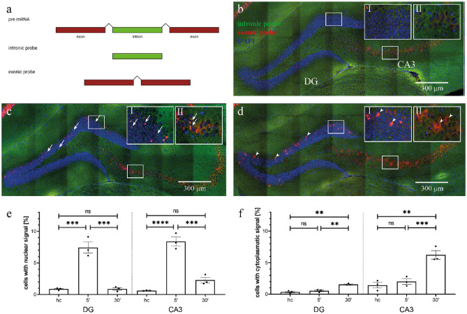
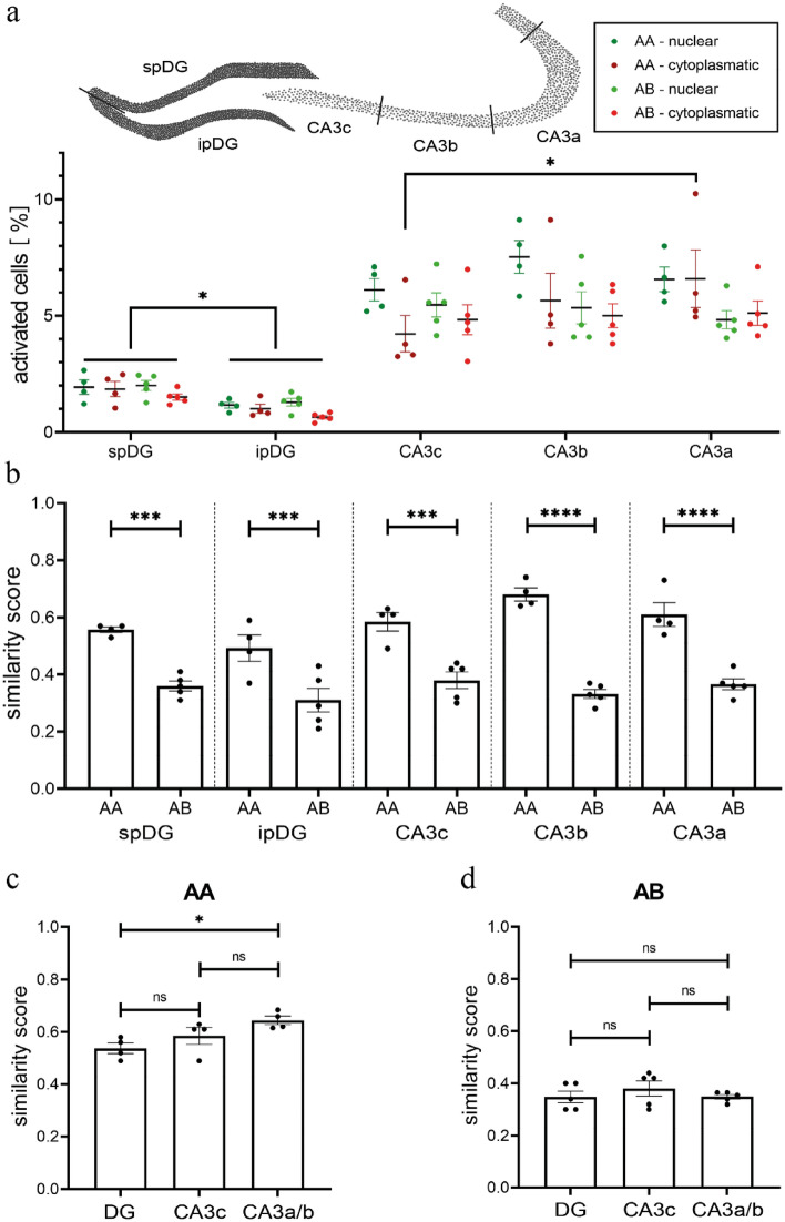
The hippocampal formation is one of the best studied brain regions for spatial and mnemonic representations. These representations have been reported to differ in their properties for individual hippocampal subregions. One approach that allows the detection of neuronal representations is immediate early gene imaging, which relies on the visualization of genomic responses of activated neuronal populations, so called engrams. This method permits the within-animal comparison of neuronal representations across different subregions. In this work, we have used compartmental analysis of temporal activity by fluorescence in-situ hybridisation (catFISH) of the immediate early gene zif268/erg1 to compare neuronal representations between subdivisions of the dentate gyrus and CA3 upon exploration of different contexts. Our findings give an account of subregion-specific ensemble sizes. We confirm previous results regarding disambiguation abilities in dentate gyrus and CA3 but in addition report novel findings: Although ensemble sizes in the lower blade of the dentate gyrus are significantly smaller than in the upper blade both blades are responsive to environmental change. Beyond this, we show significant differences in the representation of familiar and novel environments along the longitudinal axis of dorsal CA3 and most interestingly between CA3 regions of both hemispheres.


Conflict of interest statement

The authors declare no competing interests.

Figures

Figure 1
Figure 1
Double fluorescent in situ hybridization for zif268 reliably distinguishes sequential exposures separated by 30 min. (a) Intron containing pre-mRNA is localized in the nucleus. After removal of introns mRNA is shuttled to the cytosol. Probes are directed against intronic or exonic sequences, respectively. (b–d) Example images of coronal hippocampal sections from (b) home cage, (c) 5’ and (d) 30’ hybridized with exonic (red) and intronic (green) probes and stained for nuclei with DAPI (blue). Insets show magnifications of boxed areas. Arrows mark nuclear signals, arrowheads indicate cytoplasmatic signals. Scale bars are 300 µm. (e) Nuclear signals were strongly increased 5 min after exposure to a new environment in both DG and CA3 but returned to home cage (hc) levels 30 min after the exposure. (f) Cytoplasmatic signals were at hc levels 5 min after exposure to a new environment but significantly increased 30 min after the exposure. Stars indicate the level of significance of the results of Bonferroni’s multiple comparisons test as post-hoc test for two-way ANOVA. ns = not significant; **p < 0.005; ***p < 0.0005; ****p < 0.0001.
Figure 2
Figure 2
Both environments are explored equally and activate a similar percentage of neurons in DG and CA3. (a) and (b) show examples of movement tracks in context A (a) and context B (b). Black dots indicate the position of objects. (c) We found no differences in the total distance moved between the different exposures of the AA and AB groups. (d) All mice showed similar exploration of the environments during individual exposures as indicated by the distance moved in individual zones. (e, f) Neither the percentage of neurons with nuclear (e) nor with cytoplasmic (f) zif268 catFISH signals in DG and CA3 was significantly different between the individual exposures of the AA or AB groups. ns = not significant.
Figure 3
Figure 3
Ensemble sizes and similarity scores in sub-regions of DG and CA3. (a) Top: Schematic illustrating the division of DG and CA3 into subregions. Bottom: A significantly higher percentage of neurons shows nuclear or cytoplasmic signals in the spDG than in the ipDG in both the AA and the AB group. Within CA3, percentages of activated neurons do not show consistent differences. Only in group AA a higher number of cytoplasmic signals was detected in CA3a than in CA3c. (b) For all subregions of the DG and CA3, the similarity score was significantly higher for repeated exploration of context A (AA) than for exploration of two different environments (AB). (c) The similarity score in the AA group is significantly higher in CA3a/b than in the DG. (d) For group AB, no differences in similarity score were obvious between the subregions of CA3 and the DG or within CA3. ns = not significant; *p < 0.05; ***p < 0.0005; ****p < 0.0001.
Figure 4
Figure 4
Ensemble sizes and similarity scores differ along the rostro-caudal axis and between hemispheres. (a) Along the rostro-caudal axis of the dorsal hippocampus the percentage of activated neurons does not change in the DG but is significantly larger in rostral than caudal parts of CA3. (b) The similarity scores for both groups were significantly higher in rostral than caudal parts of CA3. (c) The percentage of activated neurons was not significantly different between hemispheres in either the AA or the AB group. (d) The AA group showed significantly higher similarity scores in CA3 of the right than the left hemisphere, whereas no such differences were present for the AB group. ns = not significant; *p < 0.05; **p < 0.005; ***p < 0.0005.

Similar articles

References

    1. Scoville WB, Milner B. Loss of recent memory after bilateral hippocampal lesions. J. Neurol. Neurosurg. Psychiatry. 1957;20:11–21. doi: 10.1136/jnnp.20.1.11. - DOI - PMC - PubMed
    1. Guzowski JF, McNaughton BL, Barnes CA, Worley PF. Environment-specific expression of the immediate-early gene Arc in hippocampal neuronal ensembles. Nat. Neurosci. 1999;2:1120–1124. doi: 10.1038/16046. - DOI - PubMed
    1. Liu X, et al. Optogenetic stimulation of a hippocampal engram activates fear memory recall. Nature. 2012;484:381–385. doi: 10.1038/nature11028. - DOI - PMC - PubMed
    1. Hainmueller T, Bartos M. Parallel emergence of stable and dynamic memory engrams in the hippocampus. Nature. 2018;558:292–296. doi: 10.1038/s41586-018-0191-2. - DOI - PMC - PubMed
    1. Marr D. Simple memory: A theory for archicortex. Philos. Trans. R. Soc. Lond. B Biol. Sci. 1971;262:23–81. doi: 10.1098/rstb.1971.0078. - DOI - PubMed

Publication types