. 2023 Oct 27;14(1):6853.
doi: 10.1038/s41467-023-42005-y.

Gut microbiota impacts bone via Bacteroides vulgatus-valeric acid-related pathways

Affiliations

Gut microbiota impacts bone via Bacteroides vulgatus-valeric acid-related pathways

Xu Lin et al. Nat Commun. .

Abstract

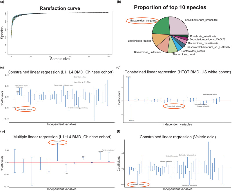
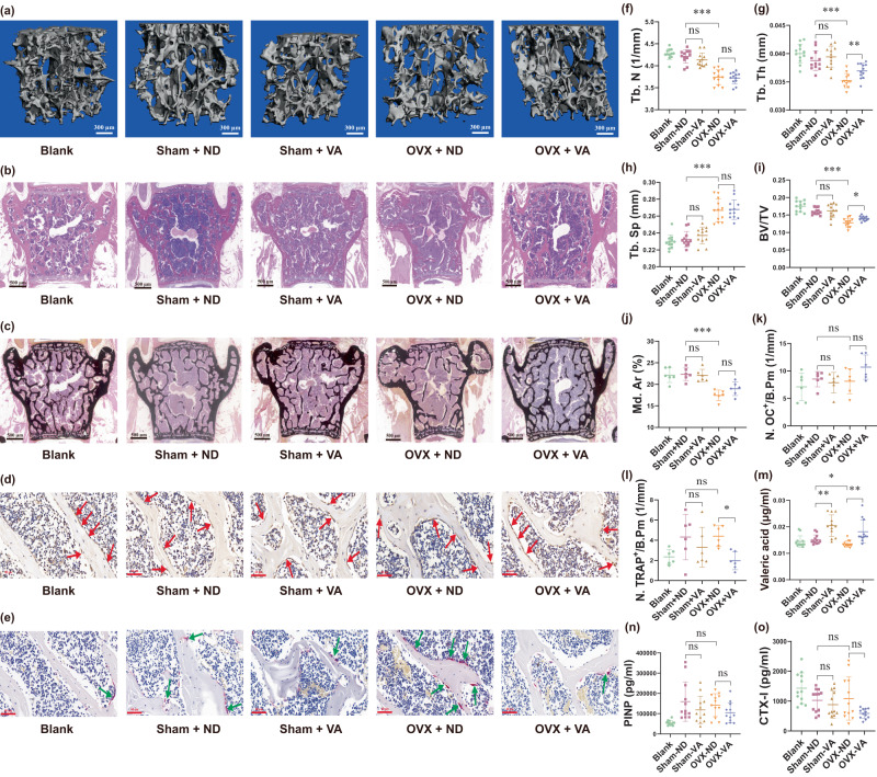
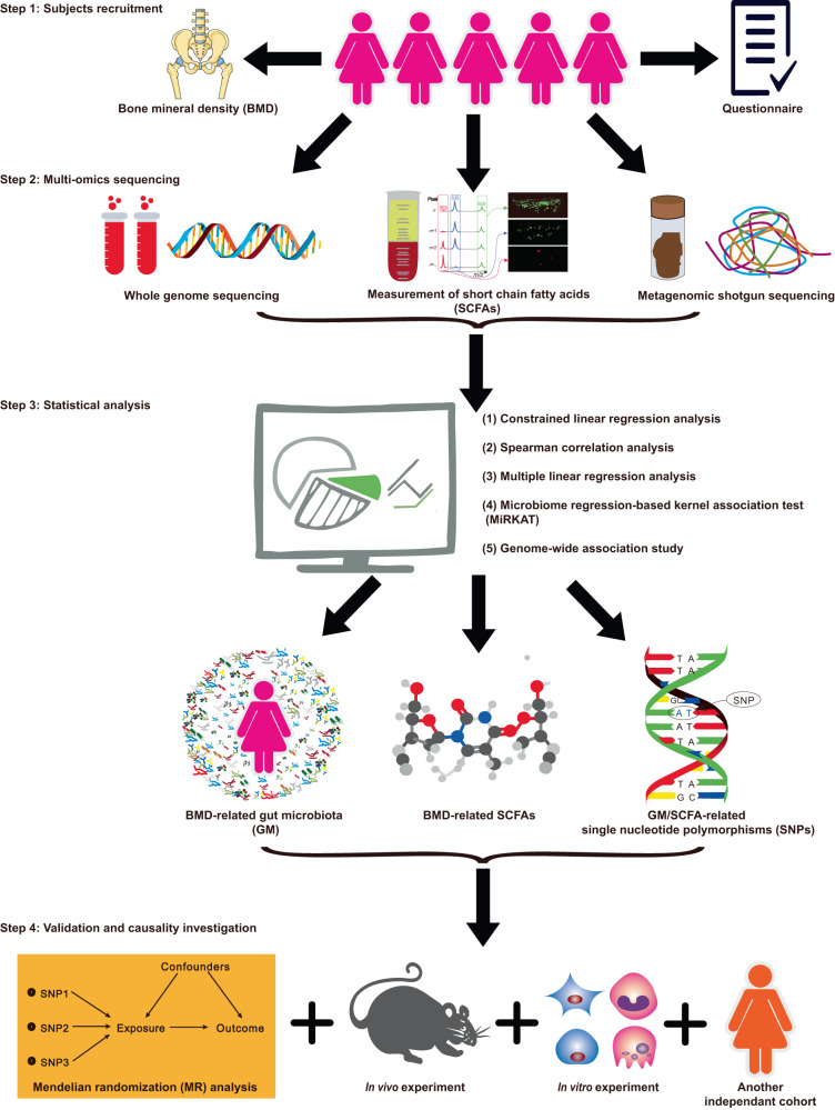
Although the gut microbiota has been reported to influence osteoporosis risk, the individual species involved, and underlying mechanisms, remain largely unknown. We performed integrative analyses in a Chinese cohort of peri-/post-menopausal women with metagenomics/targeted metabolomics/whole-genome sequencing to identify novel microbiome-related biomarkers for bone health. Bacteroides vulgatus was found to be negatively associated with bone mineral density (BMD), which was validated in US white people. Serum valeric acid (VA), a microbiota derived metabolite, was positively associated with BMD and causally downregulated by B. vulgatus. Ovariectomized mice fed B. vulgatus demonstrated increased bone resorption and poorer bone micro-structure, while those fed VA demonstrated reduced bone resorption and better bone micro-structure. VA suppressed RELA protein production (pro-inflammatory), and enhanced IL10 mRNA expression (anti-inflammatory), leading to suppressed maturation of osteoclast-like cells and enhanced maturation of osteoblasts in vitro. The findings suggest that B. vulgatus and VA may represent promising targets for osteoporosis prevention/treatment.


Conflict of interest statement

The authors declare no competing interests.

Figures

Fig. 1
Fig. 1. Bacterial species composition and associations among bacterial speices, SCFAs and BMD.
a Rarefaction curve: the number of bacterial species (Y-axis) was plotted against the number of samples (X-axis, n = 499). b Bacterial species composition in the study cohort composed of top 10 bacterial species. cf Bacterial species/SCFAs related to BMD/VA in the Chinese cohort and US cohort by constrained linear regression analysis or multiple linear regression analysis. Regression coefficients (Y-axis) were plotted against independent variables (X-axis). Species/SCFAs and covariates were considered as independent variables and represented by the blue lines. The length of the blue line indicates the 95% confidence interval (CI). c L1-L4 BMD-related species of Chinese cohort (n = 499); d HTOT BMD-related speices of the US white cohort (n = 59); e L1-L4 BMD-related SCFAs of of Chinese cohort (n = 500); f Valeric acid-related species of Chinese cohort (n = 499). Source data are provided as a Source Data file. SCFAs short chain fatty acids, BMD bone mineral density, L1-L4 lumbar spine, HTOT left total hip, YSM years since menopause, BMI body mass index.
Fig. 2
Fig. 2. Bacteroides vulgatus regulates bone-associated phenotypes of mice in vivo.
Changes in various bone-associated phenotypes in mice after gavage with B. vulgatus or NS for 8 weeks after OVX, compared with sham (non-OVX) and blank (non-OVX and no oral gavage) female mice (n_mice = 12/group): ae representative microCT/HE staining/Von Kossa staining/IHC-OC staining/TRAP staining images of the 5th lumbar vertebral body. Arrows point to osteocalcin positive cells (d) and osteoclasts positive cells (e). fi Quantitative indices of trabecular bone volume and structure, including Tb.N, Tb.Th, Tb.Sp, and BV/TV (n_mice = 12/gourp). j Mineralized volumes of mice bone are quantified by percentages of mineralized area in Von Kossa staining (n_mice = 6/group). k, l Quantitative data of osteocalcin positive cells and osteoclasts, respectively (n_mice = 6/group). mo Relative abundance of B. vulgatus and valeric acid-producing species (Megasphaera elsdenii and Oscillibacter valericigenes) (n_mice = 12/group). pr serum levels of valeric acid, PINP concentrations, and CTX-I concentrations (n_mice = 12/group). microCT - micro-computed tomography, B.V. Bacteroides vulgatus, BV/TV bone volume/tissue volume, CTX-I C-telopeptide of type I collagen, HE hematoxylin-eosin, IHC-OC immunohistochemistry-osteocalcin, Md. Ar mineralized area, NS normal saline, N.OC+/B.Pm osteocalcin positive cells number per analyzed bone perimeter, N.TRAP+/B.Pm TRAP-stained osteoclast number per analyzed bone perimeter, OVX ovariectomized, PINP procollagen I N-terminal propeptide. Tb.N trabecular number, Tb.Sp trabecular separation, Tb.Th trabecular thickness. Data are presented as mean values with SD. ns indicates non-significant, * indicates q-value < 0.05, ** indicates q-value < 0.01, *** indicates q-value < 0.001. Source data are provided as a Source Data file.
Fig. 3
Fig. 3. Valeric acid influences bone-associated phenotypes of mice in vivo.
Changes in various bone-associated phenotypes in mice after free-drinking water with/without valeric acid for 8 weeks after OVX, compared with sham (non-OVX) and blank (non-OVX and no oral gavage) female mice (n_mice = 12/group): ae representative microCT/HE staining/Von Kossa staining/IHC-OC staining/TRAP staining images of the 5th lumbar vertebral body. Arrows point to osteocalcin positive cells (d) and osteoclasts positive cells (e). fi Quantitative indices of trabecular bone volume and structure, including Tb.N, Tb.Th, Tb.Sp, and BV/TV (n_mice = 12/group). j mineralized volumes of mice bone are quantified by percentages of mineralized area in Von Kossa staining (n_mice = 6/group). k, l Quantitative data of osteocalcin positive cells and osteoclasts, respectively (n_mice = 6/group). mo Serum levels of valeric acid, PINP concentrations, and CTX-I concentrations (n_mice = 12/group). Data are presented as mean values with SD. ns indicates non-significant, * indicates q-value < 0.05, ** indicates q-value < 0.01, *** indicates q-value < 0.001. Source data are provided as a Source Data file. VA valeric acid, ND normal drinking water without valeric acid treatment.
Fig. 4
Fig. 4. Valeric acid influences osteoclast-like cell and osteoblast differentiation in vitro.
Effects of valeric acid (VA) on osteoclast-like cell differentiation of RAW264.7 cells (induced by receptor activator of nuclear factor-κB ligand [RANKL]), osteoblast differentiation of MC3T3-E1 cells. Two-sample t-test was performed to identify the difference between groups. False discovery rate (q-value) was calculated for multiple testing correction on the p-values (two-sided): a Microscopic images of tartrate-resistant acid phosphatase (TRAP) staining of osteoclast-like cells induced from RAW264.7 cells after 5 days of osteoclastogenesis with/without VA treatment (n = 6). VA significantly decreased the number of mature osteoclast-like cells (two-sample two-sided t-test p-value < 0.001). Arrows indicate TRAP-positive multinucleated cells (TRAP+MNCs). b Alkaline phosphatase (ALP) staining for osteoblast differentiation and alizarin red S (ARS) staining for extracellular matrix mineralization by MC3T3-E1 cells after 14 days of osteoblastogenesis with/without VA treatment (n = 6). VA significantly increased ALP activity (q-value < 0.001) and mineralization of the extracellular matrix (q-value = 0.007); dot plots show ALP activity (top panel) and quantification of ARS staining (bottom panel). c Western blot of p-RELA, p-NFKBIA, p-CHUK/IKBKB proteins in osteoclast-like cells with/without VA treatment (n = 3). VA significantly increased expression of p-CHUK/IKBKB (q-value = 0.009) and p-NFKBIA (q-value = 0.031), and decreased expression of p-RELA (q-value = 0.017). d Western blot of p-RELA and p-NFKBIA proteins in osteoblasts with/without VA treatment (n = 3). VA significantly increased expression of p-NFKBIA (q-value = 0.005) and caused a trend of decreased expression of p-RELA (p-value = 0.038, q-value = 0.057). e IL10 and TNF mRNA expressions in osteoclast-like cells with/without VA treatment (n = 6). VA significantly increased mRNA levels of IL10 and TNF (q-values < 0.01). f IL10 and TNF mRNA expressions in osteoblasts with/without VA treatment (n = 6). VA caused a trend of increased mRNA levels of IL10 (p-value = 0.036, q-value = 0.053). The “induce” means the MC3T3-E1 cells were induced into osteoblasts by osteoblastogenic medium without VA treatment. Data are presented as mean values with SD. * indicates q-value < 0.05, ** means q-value < 0.01, *** indicates q-value < 0.001. Source data are provided as a Source Data file.
Fig. 5
Fig. 5. Mechanisms of B. vulgatus on bone via VA production and NF-κB signaling pathway.
B. vulgatus inhibits valeric acid (VA)-producing species to reduce VA production within the gut. The VA suppresses pro-inflammatory RELA protein production in the osteoclast-like cells and osteoblasts to suppress maturation of osteoclast-like cells and promote maturation of osteoblasts. Thus, B. vulgatus decreases VA levels to enhance RELA protein production, which promotes inflammation and osteoclast activity, and suppresses osteoblast activity, then decreases bone mineral density (BMD).
Fig. 6
Fig. 6. Workflow of this study.
517 peri-/post-menopausal Chinese women were randomly recruited from Guangzhou City in China. Their stool and blood samples were collected for metagenomics/targeted metabolomics/whole-genome sequencing. By performing various statistical association analyses for dual energy X-ray absorptiometry-derived BMD, several BMD-related bacterial species/SCFAs/GM functional capacity were identified. An independent cohort of US white people and MR analysis were used for validation/causality investigation. Finally, in vitro and in vivo functional experiments were performed to validate the findings.

References

    1. Singer A, et al. Burden of illness for osteoporotic fractures compared with other serious diseases among postmenopausal women in the United States. Mayo Clin. Proc. 2015;90:53–62. doi: 10.1016/j.mayocp.2014.09.011. - DOI - PubMed
    1. Khosla S, et al. Addressing the crisis in the treatment of osteoporosis: a path forward. J. Bone Min. Res. 2017;32:424–430. doi: 10.1002/jbmr.3074. - DOI - PubMed
    1. Hendrickx G, Boudin E, Hul WV. A look behind the scenes: the risk and pathogenesis of primary osteoporosis. Nat. Rev. Rheumatol. 2015;11:462–474. doi: 10.1038/nrrheum.2015.48. - DOI - PubMed
    1. Sjögren K, et al. The gut microbiota regulates bone mass in mice. J. Bone Min. Res. 2012;27:1357–1367. doi: 10.1002/jbmr.1588. - DOI - PMC - PubMed
    1. Li C, et al. Gut microbiota composition and bone mineral loss-epidemiologic evidence from individuals in Wuhan, China. Osteoporos. Int. 2019;30:1003–1013. doi: 10.1007/s00198-019-04855-5. - DOI - PubMed

Publication types

Substances

Supplementary concepts