. 2023 Oct 28;64(1):31.
doi: 10.1186/s40529-023-00397-6.

Five new Camillea (Xylariales) species described from French Guiana

Affiliations

Five new Camillea (Xylariales) species described from French Guiana

Jacques Fournier et al. Bot Stud. .

Abstract

Background: The genus Camillea was created in 1849 from collections made in French Guiana with eight species included. Numerous species assigned to Camillea were subsequently discovered, especially in the forests of the Amazon basin, but new discoveries have not been reported from French Guiana since 1849. Recent fieldwork in French Guiana has begun to fill this gap by identifying five new species, most of which were collected in the vicinity of Saül village.

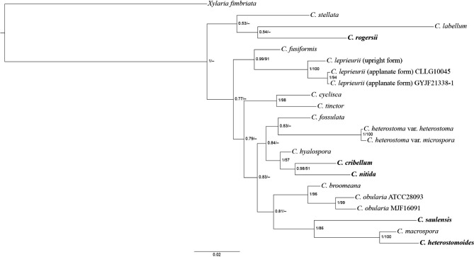
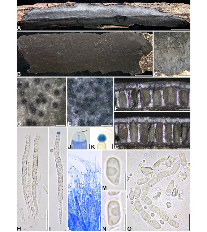
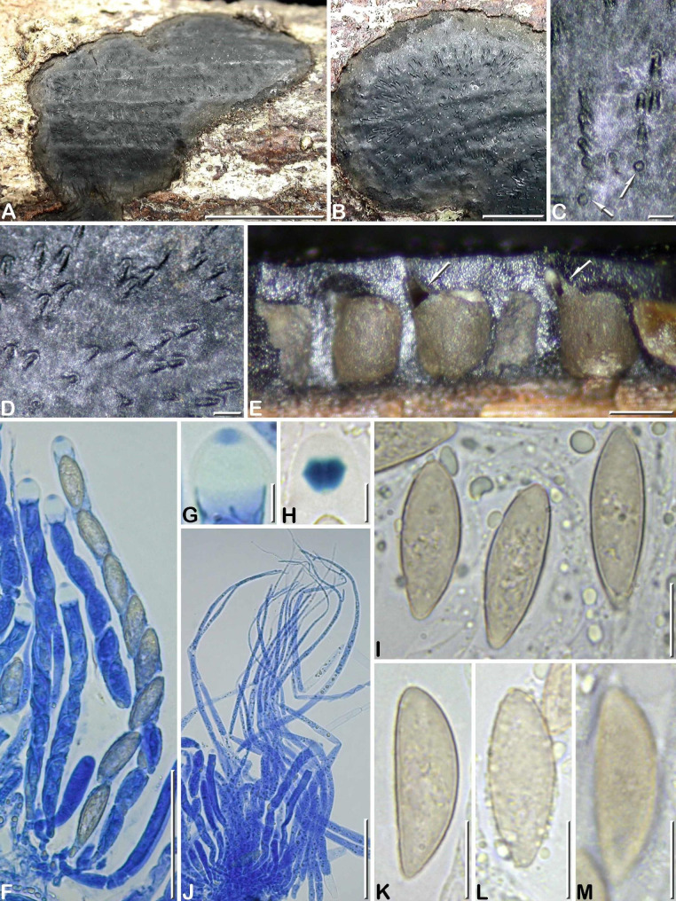
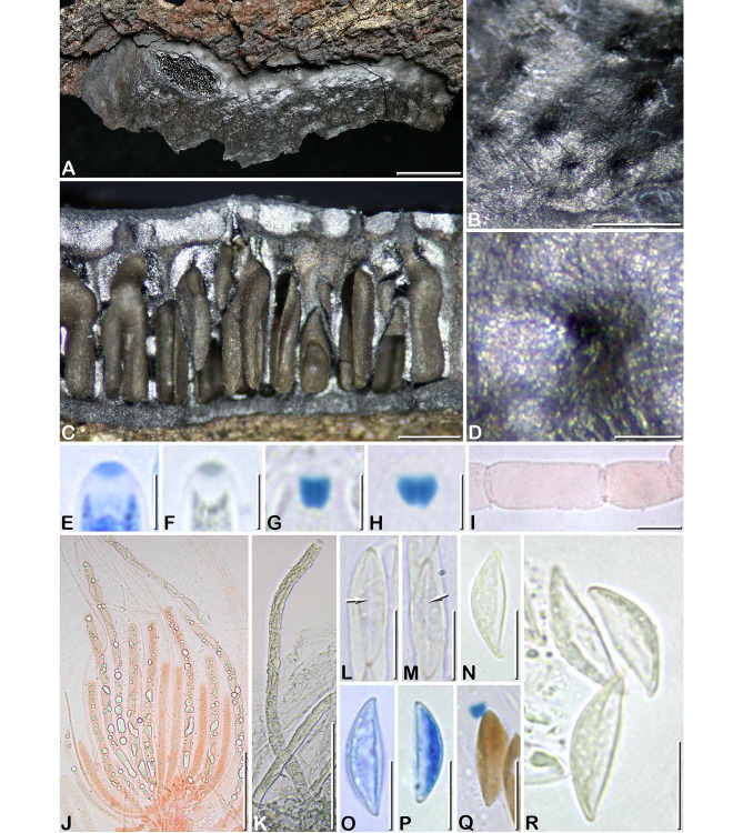
Results: Based on macro- and micromorphological study of their stromata, including SEM images of ascospore wall ornamentation, five new species were recognized, including C. cribellum, C. heterostomoides, C. nitida, C. rogersii and C. saulensis. Cultures could be obtained for C. heterostomoides and C. rogersii, and ITS and LSU sequences were obtained for all of the five new species. Camillea heterostoma and its variety microspora were shown to be conspecific. Provisional molecular phylogenetic analyses support the possible reinstatement of Hypoxylon melanaspis, currently regarded as merely an applanate form of C. leprieurii.

Conclusion: The current study is based on a relatively limited fieldwork in its duration and sampling area but was able to substantially increase the number of Camillea species known from French Guiana. This augurs an exceptional and still unknown diversity of the genus in this area and by extension in the adjacent neotropical forests.

Keywords: Ascomycota; Graphostromataceae; Neotropics; Phylogeny; SEM; Saül; Taxonomy.


Conflict of interest statement

The authors declare that they have no competing interests.

Figures

Fig. 1
Fig. 1
Phylogenetic tree generated by BI analysis from the ITS-LSU dataset. The newly described species are in boldface. Numbers at internodes represent posterior probability values and are immediately followed by bootstrap values greater than 50 generated by ML analysis
Fig. 2
Fig. 2
Camillea cribellum. (A, E, F, J–O. GYJF 18137 from holotype; B–D, G–I from paratype CLL 0820). A Habit of a narrowly elongate stroma showing a slightly shiny surface; B Fragmentary widely spread stroma with dull black surface; C Grey margin of a stroma and fertile part finely dotted with black; D, E Surface of two different stromata in close-up, showing blackish halos around ostiolar openings; F, G Vertical sections of two different stromata; H, I Asci showing oily content around ascospores, in 1% SDS and Melzer’s reagent respectively; J Ascus apical apparatus showing a minute apical pulvillus slightly stained by blue Pelikan ink (arrow); K Rhomboid subapical apparatus stained by Melzer’s reagent; L Hymenium stained by blue Pelikan ink showing paraphyses tapering above asci and containing large refractive guttules; M, N Ascospores in side view; O Variously shaped ascospores interspersed with oily guttules and amorphous refractive bodies (M-O in 1% SDS). Scale bars: A, B = 10 mm; C = 1 mm; D-G = 0.2 mm; H, I, L = 20 μm; J, K = 2 μm; M, N = 5 μm; O = 10 μm
Fig. 3
Fig. 3
Ascospores of Camillea species by SEM. A C. cribellum (from holotype). B C. heterostomoides (from holotype). C C. nitida (from holotype). D, E C. rogersii (from holotype). F, G C. saulensis (from holotype). Scale bars: B, D, F = 5 μm; A, C = 2.5 μm; E, G = 1 μm
Fig. 4
Fig. 4
Camillea heterostomoides (from holotype). A, B Habit of stromata on host surface; C, D Stromatal surface in close-up showing rounded (arrows) to elongate ostiolar depressions; E Stroma in vertical section showing carbonaceous tissue surrounding the perithecia opening through eccentric ostiolar canals (arrows); F Immature and mature asci, in blue Pelikan ink; G Ascus apex in blue Pelikan ink, showing an apical pulvillus stained blue; H Ascus apex showing the rhomboid subapical apparatus bluing in Melzer’ reagent; I Variously shaped ascospores; J Bundle of paraphyses, in blue Pelikan ink; K Ascospore in side view; L Freshly released ascospores coated with oily guttules; M Ascospore in side view with focus on epispore showing an obscure reticulate ornamentation (I, K-M in 1% SDS). Scale bars: A = 5 mm; B = 2 mm; C, D, E = 0.2 mm; F, J = 50 μm; G, H = 5 μm; I, K–M = 10 μm.
Fig. 5
Fig. 5
Cultures of Camillea species. A–C C. heterostomoides (from holotype). A Colony at 1 wk; B Colony at 3 wk; C Reverse at 3 wk; D–F C. rogersii (from holotype). D Colony at 1 wk; E Colony at 3 wk; F Reverse at 3 wk. Scale bars: A–F = 1 cm.
Fig. 6
Fig. 6
Camillea nitida. (A–D, I–P from holotype; E–H from paratype GYJF 12119). A, B Habit of two narrowly elongate stromata on host surface, showing a shiny surface; C Ellipsoid stroma in top view showing a shiny surface mottled with grey deposits of ascospores around the ostioles; D Stroma in vertical section showing clusters of perithecia with convergent ostiolar necks, separated by regions composed of brownish black sterile tissue (arrows); E Carbonaceous crust removed from the stromatal surface to show a rosette of empty carbonaceous perithecial cells arranged around two ostiolar canals (arrows); F, H Keyhole-shaped and rounded ostioles (arrows) in an outwardly radiating pattern; G Keyhole-shaped ostiole showing a slightly prominent rim (arrow); I Immature and mature asci and paraphyses, in blue Pelikan ink; J Ascus apical apparatus showing an apical plug stained by blue Pelikan ink; K Diamond-shaped subapical apparatus stained by Melzer’s reagent; L Freshly released ascospores with remnants of oily globules attached; M, N Ascospores respectively in side and dorsal view, in Melzer’s reagent; O Paraphyses in blue Pelikan ink; P Ascospores and apical apparati, in Melzer’s reagent. Scale bars: A, B = 10 mm; C = 5 mm; D, E = 0.5 mm; F = 0.2 mm; G, H = 0.1 mm; I, O = 20 μm; J, K = 2 μm; L, P = 10 μm; M, N = 5 μm
Fig. 7
Fig. 7
Camillea rogersii (from holotype). A, C Habit of stromata on host surface with wide sterile margins; B Close-up on the fertile part of a stroma showing an outwardly radiating arrangement pattern of ostiolar depressions; D, I Stroma in vertical section showing variously shaped perithecia with pale olivaceous content; E, F Asci in blue Pelikan ink, with paraphyses (arrow); G Ascus apex in blue Pelikan ink, showing a darker apical pulvillus (arrow); H Ascus apex showing the subapical apparatus bluing in Melzer’ reagent; J Ostiolar depression in close-up showing a minute apical ostiole (arrow) surrounded by a low rim; K Ascospore in side view in 1% SDS; L Ascospore in side view in Waterman blue ink diluted in lactic acid; M, N Ascospores in side view in Melzer’s reagent. Scale bars: A = 10 mm; B, D, I = 0.5 mm; C = 5 mm; E, F = 20 μm; G, H = 1 μm; J = 100 μm; K-N = 5 μm.
Fig. 8
Fig. 8
Camillea saulensis (from holotype). A Habit of a fragmentary stroma erumpent from bark; B Close-up on the stromatal surface showing scattered papillate ostioles; C Stroma in vertical section showing tubular to flask-shaped perithecia sharing common ostioles; D Close-up on a papillate ostiole; E Ascus apex in blue Pelikan ink, showing an apical pulvillus stained blue; F Ascus apex in blue black Waterman ink, showing an apical pulvillus stained dark grey; G, H Ascus apices showing the subapical apparatus bluing in Melzer’ reagent; I Basal part of a paraphysis, in Congo red; J Bundle of immature and mature asci showing refractive content, in Congo red; K Mature ascus in blue black Waterman ink; L, M Two barely mature ascospore in ventral view showing a narrow straight white line (arrows), in Melzer’s reagent; N Ascospore in side view, in 1% SDS; O, P Ascospores in side view showing a ventral paler area of variable width, in blue Pelikan ink; Q Atypical ascospores with pigmented wall, in Melzer’s reagent; R Discharged ascospores showing a wide, unpigmented ventral zone, in 1% SDS. Scale bars: A = 10 mm; B, C = 0.5 mm; D = 0.2 mm; E-H = 5 μm; I, L-R = 10 μm; J, K = 50 μm

References

    1. Dennis RWG. Further notes on tropical american Xylariaceae. Kew Bull. 1957;12:297–332. doi: 10.2307/4114428. - DOI
    1. Dennis RWG (1970) Fungus flora of Venezuela and adjacent countries. Kew Bulletin Additional Series 3:344–531
    1. Fournier J. Camillea lechatii (Graphostromataceae, Xylariales), a new species from Martinique (French West Indies) Ascomycete org. 2022;14:129–132.
    1. Fries EM (1849) Summa vegetabilium Scandinaviae, sectio posterior. Holmiae & Lipsiae, pp 261–572
    1. Hastrup ACS, Læssøe T. Camillea (Xylariaceae, Ascomycota), including two new species, along a trans-andean altitude gradient in Ecuador. Mycological Progress. 2009;8:305–316. doi: 10.1007/s11557-009-0601-9. - DOI

LinkOut - more resources