Review
. 2023 Dec;22(12):996-1017.
doi: 10.1038/s41573-023-00809-z. Epub 2023 Oct 27.

T cell receptor therapeutics: immunological targeting of the intracellular cancer proteome

Affiliations
Review

T cell receptor therapeutics: immunological targeting of the intracellular cancer proteome

Christopher A Klebanoff et al. Nat Rev Drug Discov. 2023 Dec.

Abstract

The T cell receptor (TCR) complex is a naturally occurring antigen sensor that detects, amplifies and coordinates cellular immune responses to epitopes derived from cell surface and intracellular proteins. Thus, TCRs enable the targeting of proteins selectively expressed by cancer cells, including neoantigens, cancer germline antigens and viral oncoproteins. As such, TCRs have provided the basis for an emerging class of oncology therapeutics. Herein, we review the current cancer treatment landscape using TCRs and TCR-like molecules. This includes adoptive cell transfer of T cells expressing endogenous or engineered TCRs, TCR bispecific engagers and antibodies specific for human leukocyte antigen (HLA)-bound peptides (TCR mimics). We discuss the unique complexities associated with the clinical development of these therapeutics, such as HLA restriction, TCR retrieval, potency assessment and the potential for cross-reactivity. In addition, we highlight emerging clinical data that establish the antitumour potential of TCR-based therapies, including tumour-infiltrating lymphocytes, for the treatment of diverse human malignancies. Finally, we explore the future of TCR therapeutics, including emerging genome editing methods to safely enhance potency and strategies to streamline patient identification.


Conflict of interest statement

Competing interests:

C.A.K. and S.S.C. are inventors on patents related to TCR discovery and public neoantigen-specific TCRs and are recipients of licensing revenue shared according to MSKCC institutional policies. C.A.K. has consulted for or is on the scientific advisory boards for Achilles Therapeutics, Affini-T Therapeutics, Aleta BioTherapeutics, Bellicum Pharmaceuticals, Bristol Myers Squibb, Catamaran Bio, Cell Design Labs, Decheng Capital, G1 Therapeutics, Klus Pharma, Obsidian Therapeutics, PACT Pharma, Roche/Genentech and T-knife. C.A.K. is a scientific co-founder and equity holder in Affini-T Therapeutics. B.M.B. is an inventor on patents related to TCR engineering and neoantigen discovery, has consulted for Eureka Therapeutics and EnaraBio, and is on the scientific advisory board of T-cure Bioscience. S.A.Q is an inventor in patents related to the use of T cell therapies targeting tumour clonal neoantigens in cancer. S.A.Q. is also a founder, CSO, and equity holder of Achilles Therapeutics. A.R. reports personal fees from Amgen, Chugai, Genentech, Merck, Novartis, Roche, Sanofi, Vedanta, 4C Biomed, Appia, Apricity, Arcus, Highlight, Compugen, ImaginAb, Kalthera/ImmPACT Bio, MapKure, Merus, Rgenix, Lutris, Nextech, PACT Pharma, Synthekine, Tango, Advaxis, CytomX, Five Prime, RAPT, Isoplexis, and Kite/Gilead and has received grants from Agilent and Bristol Myers Squibb outside the submitted work.

Figures
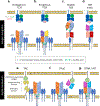
Fig. 1:
Fig. 1:. The molecular architecture of TCR-based therapeutics.
(a-d) Comparison of the structural features of TCR and TCR-like molecules that bind specific peptide/human leukocyte antigen (p/HLA) complexes. (a) The endogenous TCR is comprised of an octameric complex composed of six proteins: the clonotypic TCRα (red) / TCRβ (blue) membrane-anchored heterodimer and the invariant CD3γ, δ, ε, and ζ chains. These proteins assemble with a 1:1:1:1 stoichiometry comprised of the dimeric subunits TCRαβ:CD3δε:CD3γε:CD3ζζ. Each TCR hemichain is composed of an antigen-binding variable (V) domain, a constant (C) domain, a transmembrane domain, and a short non-signaling cytoplasmic tail. The endogenous TCRα/TCRβ hemichains are covalently linked though a single interchain disulfide bond (grey spheres). Non-covalent interactions with the CD3 molecules facilitates intracellular signaling. HLA class I-restricted TCRs bind to a pHLA complex comprised of three alpha subunits (blue), beta-2-microglobulin (light grey) and a short polypeptide sequence typically 8–10 amino acids in length (red). (b) T-cell specificity can be genetically redirected to recognize p/HLA complexes displayed by tumour cells through expression of an exogenous TCRα (light grey) and TCRβ (dark grey) hemichain. Mispairing with the endogenous TCR hemichains can be minimized by introduction of a second interchain disulfide bond. (c) Soluble TCRs are recombinant bispecific proteins that contain a TCR’s α/β variable domains linked in a single-chain format on one end and an antibody-derived antigen binding variable heavy (VH, rouge) and variable light (VL, pink) chains specific for CD3ε on the other. (d) TCR-mimics are an alternative class of recombinant bispecific proteins that use an antibody’s VH/VL domains (yellow and purple) in place of a TCRα/TCRβ to engage a specific p/HLA complex. Shown is a diabody format TCR-mimic. (e-g) Comparison of the structural features of next-generation antigen receptors that repurpose one or several components of the TCR’s CD3 signaling complex. (e) The T cell antigen coupler (TAC) is a bispecific transmembrane protein expressed as a transgene in polyclonal T cells. One domain of a TAC uses an antibody-derived variable sequence to engage a membrane-associated tumor antigen (dark grey) while the other binds CD3ε. (f) The T cell receptor fusion construct (TRuC) is a transgene expressed in polyclonal T cells that covalently links an antibody variable sequence with specificity for a tumour antigen to an exogenous CD3ε molecule. (g) The synthetic TCR antigen receptor (STAR)/HLA-independent TCR (HIT) is a non-HLA restricted receptor that replaces the TCR variable domains with the tumor antigen-binding variable domains of an antibody. By retaining the TCR constant domains, the STAR/HIT receptor can recruit the full complex of CD3 signaling molecules upon ligand binding. V = TCR variable domain, C = TCR constant domain, VH = immunoglobulin variable heavy chain, VL = immunoglobulin variable light chain.
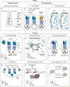
Fig. 2:
Fig. 2:. Discovery of TCR therapeutic candidates.
(a) Fully human TCR gene sequences that confer recognition to tumour-derived peptide/HLA (p/HLA) complexes can be retrieved from healthy donors and cancer patients. Healthy donors have a broad circulating TCR repertoire that has not been subjected to the negative influence of immune-depleting cancer treatments, thymic involution, and peripheral tolerance (left). However, because the frequency of tumour-reactive TCR clonotypes is exceedingly rare within the naïve repertoire, healthy donor T cells must undergo in vitro stimulation (IVS) to enable detection. Tumour antigen-reactive T cells can be detected within the peripheral blood and tumour infiltrating lymphocytes (TILs) of patients with cancer (right). Although TCR diversity (represented as a grey bar) is typically lower in patients compared with healthy donors, the TCR repertoire often is enriched in tumour-reactive T cells that have undergone in vivo clonal expansion (represented as a purple bar). This feature may enable the retrieval of a larger number of tumour-reactive TCR clonotypes than is possible using a comparable sample volume obtained from healthy donors. (b) Tumour-reactive T cells can be generated through antigen-specific vaccination of HLA transgenic mice. HLA transgenic mice possess a diverse TCR repertoire that has not been subjected to central thymic tolerance against human proteins that differ in sequence from their murine counterparts. However, TCRs retrieved from HLA transgenic mice possess immunogenic murine variable sequences capable of triggering host-versus-graft rejection when infused into humans. To overcome this limitation, “humanized” mice have been generated in which the genetic sequences encoding the human TCR variable chains have been knocked into the genetic loci encoding the murine TCR chains (light blue, right). TCRs generated in these mice therefore possess fully human TCR variable sequences. α12 = highly polymorphic domains of HLA class I; α3 = HLA class I constant domain; β2M = beta-2 microglobulin (HLA light chain); V = TCR variable domain; C = TCR constant domain; Mhc = genetic locus encoding the major histocompatibility complex proteins (the murine ortholog of HLA).
Fig. 3:
Fig. 3:. Strategies to resolve the safety profile and potency and of TCR therapeutic candidates.
(a-c) Methods to quantify the safety profile of a TCR candidate. (a) Normal tissue expression assesses the presence of a target antigen in healthy tissues to assess the risk for on-target/off-tumour toxicities from a TCR candidate. Ideally, the risk profile for a novel antigen will be determined using multiple assays, including bulk tissue RNA sequencing, single-cell RNA sequencing, and measurement of protein level expression. (b) Allogeneic (allo) reactivity measures the capacity of a TCR to respond to a mismatched HLA molecule irrespective of the bound peptide sequence. Allo-reactivity is assessed by co-culture of T cells expressing a candidate TCR with a panel of target cells, such as EBV-transformed B-lymphoblastic cell lines (B-LCLs), that express diverse HLA alleles. (c) TCR degeneracy is the capacity of a single TCR to respond to unrelated peptide sequences but restricted by the same HLA molecule. The degeneracy potential of a TCR can be measured using sequential amino acid scanning mutagenesis (eg. X-Scan) or large combinatorial libraries. In the example shown, the TCR is capable of recognizing both the cognate peptide sequence (highlighted boxes) and unrelated peptide sequences with similar or even enhanced potency. In the heatmap, dark blue represents an amino acid that results in enhanced TCR-mediated cytokine release or binding relative to other amino acids. In all instances where potential cross-reactive peptide sequences are identified, confirmatory studies are required to establish physiologic significance. (d-i) Methods to quantify TCR potency. (d) Functional avidity measures the capacity of T cells expressing multiple copies of a membrane-associated TCR to functionally respond to progressively lower concentrations of a specific p/HLA complex. Functional avidity results from the summation of all binding interactions between a T cell and target cell, including contributions from the TCR, the CD8 or CD4 co-receptors, and intercellular adhesion molecules. (e) Structural avidity measures the TCR-p/HLA dissociation rate (koff) using fluorophore-conjugated (star) p/HLA multimers that dissociate into monomers following addition of an inert chemical (grey diamond). Unlike functional avidity, measurement of structural avidity is not affected by the differentiation state of T cells. (f) Co-receptor dependency measures the capacity of T cells expressing a TCR candidate to respond to target cells in the absence of the avidity and signaling contributions facilitated by the CD8α/β or CD4 co-receptors. The ability of a TCR to function in a coreceptor-independent manner suggests the TCR has a relatively high binding affinity. (g) TCR affinity is the strength of interaction between a single TCR molecule and a single p/HLA complex. Most commonly, affinity is measured using surface plasmon resonance by flowing a recombinant, soluble, single-chain TCR over a metal surface containing immobilized p/HLA complexes. Under equilibrium conditions, a TCR’s binding affinity is inversely proportional to its dissociation constant (Kd) which in turn is defined by the ratio of the rates of dissociation and association (koff/kon). Plot illustrates time-dependent changes in binding (measured as relative response units) of a single-chain TCR flowed over a sensor containing immobilized p/HLA complexes at different concentrations. (h) In vitro tumour recognition measures the capacity of a TCR to trigger T cell responses to physiologic levels of an endogenously processed peptide displayed in the context of a specific HLA allele by tumour cells. Tumour recognition can be quantified by measuring T cell-mediated cytolysis or cytokine production. Solid red and grey lines represent the time-dependent cytolytic activity of two therapeutic TCR candidates while the grey dashed lines represents the cytolytic activity of a control TCR. (i) In vivo tumour regression assesses the ability of a TCR candidate (solid red line) to penetrate an established tumour mass and cause a sustained antitumour response over time. Grey solid and dashed lines represent a control TCR and no treatment control, respectively.

References

    1. Hedrick SM, Cohen DI, Nielsen EA & Davis MM Isolation of cDNA clones encoding T cell-specific membrane-associated proteins. Nature 308, 149–153 (1984).

      Together with reference , these papers report the genetic sequence for the TCRβ chain in mice and humans for the first time.

    1. Yanagi Y, et al. A human T cell-specific cDNA clone encodes a protein having extensive homology to immunoglobulin chains. Nature 308, 145–149 (1984). - PubMed
    1. Rosenberg SA IL-2: the first effective immunotherapy for human cancer. Journal of immunology 192, 5451–5458 (2014). - PMC - PubMed
    1. Ribas A & Wolchok JD Cancer immunotherapy using checkpoint blockade. Science 359, 1350–1355 (2018). - PMC - PubMed
    1. Guedan S, Ruella M & June CH Emerging Cellular Therapies for Cancer. Annu Rev Immunol 37, 145–171 (2019). - PMC - PubMed

Publication types