. 2023 Oct 28;23(1):317.
doi: 10.1186/s12866-023-03069-4.

The regulatory subunits of CK2 complex mediate DNA damage response and virulence in Candida Glabrata

Affiliations

The regulatory subunits of CK2 complex mediate DNA damage response and virulence in Candida Glabrata

Qi Ni et al. BMC Microbiol. .

Abstract

Background: Candida glabrata which belongs to normal microbiota, has caused significant concern worldwide due to its high prevalence and drug resistance in recent years. C. glabrata has developed many strategies to evade the clearance of the host immune system, thereby causing persistent infection. Although coping with the induced DNA damage is widely acknowledged to be important, the underlying mechanisms remain unclear.

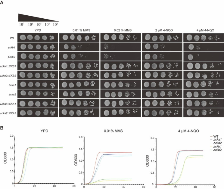
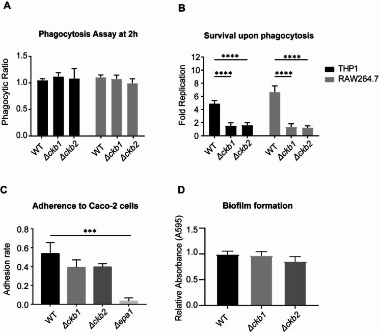
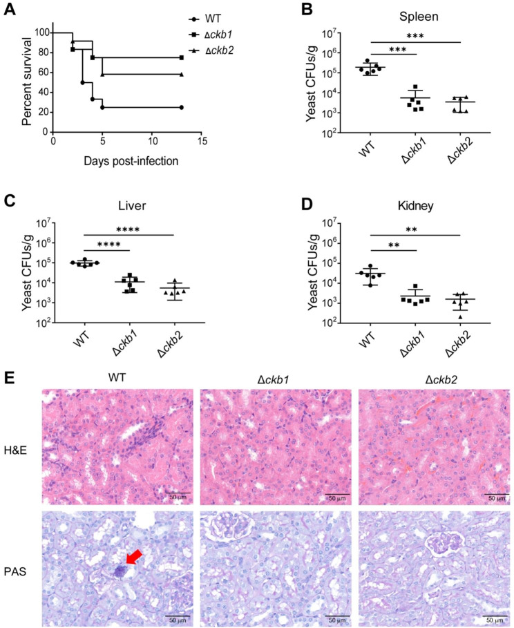
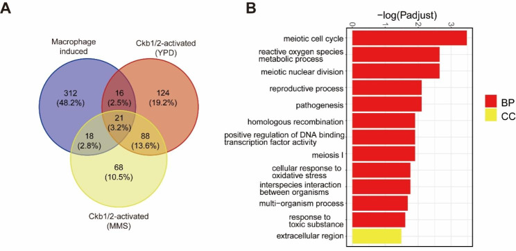
Results: The present study provides hitherto undocumented evidence of the importance of the regulatory subunits of CgCK2 (CgCkb1 and CgCkb2) in response to DNA damage. Deletion of CgCKB1 or CgCKB2 enhanced cellular apoptosis and DNA breaks and led to cell cycle delay. In addition, deficiencies in survival upon phagocytosis were observed in Δckb1 and Δckb2 strains. Consistently, disruption of CgCKB1 and CgCKB2 attenuated the virulence of C. glabrata in mouse models of invasive candidiasis. Furthermore, global transcriptional profiling analysis revealed that CgCkb1 and CgCkb2 participate in cell cycle resumption and genomic stability.

Conclusions: Overall, our findings suggest that the response to DNA damage stress is crucial for C. glabrata to survive in macrophages, leading to full virulence in vivo. The significance of this work lies in providing a better understanding of pathogenicity in C. glabrata-related candidiasis and expanding ideas for clinical therapies.

Keywords: Candida glabrata; Cell cycle; DNA damage; Macrophage; Virulence.


Conflict of interest statement

The authors declare no competing interests.

Figures

Fig. 1
Fig. 1
Phylogenetic trees of fungal Ckb1and Ckb2. (A) and (B) A phylogenetic tree constructed based on fungal Ckb1 or Ckb2 protein sequences to show the sequence homology of Ckb1 or Ckb2 proteins across species. The human Ckb1 or Ckb2 protein sequence was used as the root, and the maximum likelihood method was used for calculation and comparison by MEGA (V11.0.13). (C) Overview of amino acid identity between the C. glabrata and S. cerevisiae Ckb1 and CKb2.
Fig. 2
Fig. 2
CgCKB1 and CgCKB2 genes are essential in response to DNA damage. (A) Strains were diluted serially in 10-fold to being grown on YPD containing different concentrations of DNA damage reagents (MMS, methyl methane sulfonate and 4-NQO, 4-Nitroquinoline-1-oxide) at 30℃ for 2 days. WT, wildtype. (B) Growth curves of each strain after treatment with different concentrations of DNA damage reagents. The OD600 values were obtained by BioTek plate reader every 15 min at 30 °C for 48 h. Data were representative of three independent experiments. All the growth curves were repeated at least three times.
Fig. 3
Fig. 3
Deletion of CgCKB1 or CgCKB2 increases cellular apoptosis and DNA breaks. (A) Cells were treated with 0.01% MMS or 4 µM 4-NQO or left untreated for 2 h and stained with Annexin V-FITC and PI. The Fluorescence intensity was detected by BD Fortessa Flow cytometer. Q1 (Annexin V-/PI+): dead cells; Q2 (Annexin V+/PI+): necrotic cells; Q3 (Annexin V+/PI-): apoptotic cells; Q4 (Annexin V-/PI-): live cells. (B) The percentages of each strain that are apoptotic (black bars) and death (gray bars) were obtained. The relative cell ratio was compared with WT in RPMI 1640 medium (set at 1.0) (C) Representative fluorescence micrographs showing DNA fragmentation and unusual chromosome agglutination. Cells were incubated in RPMI 1640, 0.01% MMS or 4 µM 4-NQO for 12 h at 30 ℃ and then stained with DAPI and FITC. The DNA fragmentation was visualized with FITC (green fluorescence) and unusual chromosome agglutination was visualized with DAPI (blue fluorescence). Data were representative of three independent experiments. (D) The TUNEL-positive cells were quantified. DNase I treatment was used as the positive control. The percentages of TUNEL-positive cells were presented as mean ± SD and assessed for statistical analysis by one-way ANOVA followed by Tukey test; * p < 0.05, ** p < 0.01, *** p < 0.001, **** p < 0.0001.
Fig. 4
Fig. 4
The cell cycle is delayed in CgCKB1 and CgCKB2 null mutants. Yeast cells were synchronized in YP (carbon starvation condition) and released to YPD either in the absence or presence of 0.01% MMS or 4 µM 4-NQO and collected once an hour. Flow cytometry was used to measure DNA content.
Fig. 5
Fig. 5
Effect of CgCKB1 and CgCKB2 on interaction with host cells. (A) After co-incubation of fungi with RAW264.7 macrophages or PMA-treated THP-1 cells for 2 h, the cells were lysed with 0.05% Triton X-100 to release the fungi, and the phagocytosis rate was defined as the number of fungi phagocytosed by macrophages after 2 h to the number of fungi before co-incubation. (B) After cocultured with macrophages for 24 h, survival upon phagocytosis was measured as described above. The ratio of the intracellular yeast cells after 24 h of coculture to that after 2 h coculture was defined as fold replication. (C) Epithelial Caco-2 cells were cocultured with CgCKB1 or CgCKB2 null mutants, respectively. Δepa1 which exhibited deficient adhesion ability was used as a control. (D) Log-phased yeast cells were seeded in a 24-well plate for biofilm formation assay. Crystal violet [0.4% (w/v)] was used for staining mature biofilms. The destaining solution was measured for absorbance at 595 nm. Wells without yeast cells were used as background. CBS138 was the used as the WT and the knockout strains were generated using CBS138 as the parent. Experiments were performed three times. Error bars show standard deviation. Comparisons were performed using one-way ANOVA followed by Tukey test, and asterisks indicate statistically significant differences (*** p < 0.001, **** p < 0.0001).
Fig. 6
Fig. 6
CgCKB1 and CgCKB2 are essential to virulence in vivo. (A) 200 mg/kg cyclophosphamide was intraperitoneally injected per ICR mouse (5–6 weeks old, 24–26 g) on day − 3 and every fourth day later. Then mice were infected with 1 × 108 WT, Δckb1 or Δckb2 cells in 200 µL in 0.9% (w/v) saline (n = 12). Mice in the agonal stage were humanely euthanized by cervical dislocation. Experiments were terminated on day 14. Analysis by the Gehan-Breslow-Wilcoxon test indicated that in contrast to WT, the virulence of CgCKB1 and CgCKB2 null mutants was significantly attenuated (p < 0.05). (B)(C)(D) Fungal burden assays were performed using ICR mice (n = 6). ICR mice were immunosuppressed with 200 mg/kg cyclophosphamide on day − 3 and were further injected with 5 × 107 yeast cells through the tail veil on day 0. Organs were harvested, weighed and mechanically homogenized 3 days post-infection. Tissue homogenates were diluted, plated onto YPD agar, and incubated at 30℃ for 2 days. CFUs were calculated and analyzed. Error bars show standard deviation. Comparisons were performed using one-way ANOVA followed by Tukey test, and asterisks indicate statistically significant differences (**: p < 0.01; ***: p < 0.001, ****: p < 0.0001). CBS138 was the used as the WT and the knockout strains were generated using CBS138 as the parent; (E) Representative HE (Hematoxylin-Eosin) and PAS (Periodic Acid-Schiff) stained sections of kidneys from immunosuppressed ICR mice on day 3 post-infection. Magnification was ×20. PAS-positive yeast cells were indicated by red arrowhead. Data are representative of at least three replicates.
Fig. 7
Fig. 7
Transcriptional analysis in WT, Δckb1, and Δckb2 strains. Log-phase yeast cells were inoculated into fresh YPD medium either with 0.01% MMS added or left untreated, total RNA was extracted and further sequenced. (A) Volcano plot of RNA seq transcriptome data displaying the gene expression pattern. The values of gene expression in each group were compared with those of WT strain under the same conditions. Significantly differentially expressed genes (FDR, p ≤ 0.05) are highlighted in red (upregulated) or blue (downregulated). (B) Heatmap plot of DEGs (Differential expression genes) displaying the pattern of gene expression. The gene expression value is normalized and converted to Z score. (C) Scatterplot where each gene is represented by a dot and its “no MMS/MMS” log2 ratio for Δckb1 is plotted on the x axis and the ratio for Δckb2 is plotted on the y axis. (D and E) Venn plot comparing the upregulated DEGs (URGs) and downregulated DEGs (DRGs) of Δckb1 and Δckb2 under all culture conditions. The differential genes of each group were compared with WT strain under the same conditions. (F) Gene ontology (GO) terms in the biological processes describing the upregulated and downregulated genes in each condition. The differential genes both in Δckb1 and Δckb2 (compared with WT under the same conditions) were clustered to GO analysis. (G) Heatmap of RNA-seq data for meiotic genes in Δckb1 and Δckb2 strains compared with WT under all culture conditions, including programmed formation of DNA double‐strand breaks (DSBs), recombination, and DNA repair coordination of the meiotic cell cycle.
Fig. 8
Fig. 8
Transcriptome data reveal the link between Ckb1/2 and C. glabrata survival in macrophages. (A) Comparison of differential genes (compared with WT) regulated by CgCKB1 and CgCKB2 under control conditions and MMS conditions with the activated genes (compared with RPMI 1640 medium condition) of C. glabrata upon macrophage engulfment. (B) GO analysis demonstrates the biological processes involved in overlapping genes. A schematic diagram of a bar chart for the top enriched GO terms ranked according to the values of − log10 (adjusted p value). BP: Biological Process, CC: Cell Component.

Similar articles

Cited by

References

    1. Kumar K, Askari F, Sahu MS, Kaur R. Candida Glabrata: a Lot more than meets the Eye. Microorganisms. 2019;7(2). - PMC - PubMed
    1. Lamoth F, Lockhart SR, Berkow EL, Calandra T. Changes in the epidemiological landscape of invasive candidiasis. J Antimicrob Chemother. 2018;73(suppl1):i4–i13. doi: 10.1093/jac/dkx444. - DOI - PMC - PubMed
    1. Lotfali E, Fattahi A, Sayyahfar S, Ghasemi R, Rabiei MM, Fathi M, et al. A review on Molecular mechanisms of Antifungal Resistance in Candida Glabrata: Update and recent advances. Microb Drug Resist. 2021;27(10):1371–88. doi: 10.1089/mdr.2020.0235. - DOI - PubMed
    1. Castanheira M, Deshpande LM, Davis AP, Carvalhaes CG, Pfaller MA. Azole resistance in Candida Glabrata clinical isolates from global surveillance is associated with efflux overexpression. J Glob Antimicrob Resist. 2022;29:371–7. doi: 10.1016/j.jgar.2022.05.004. - DOI - PubMed
    1. Timmermans B, De Las Penas A, Castano I, Van Dijck P. Adhesins in Candida Glabrata. J Fungi (Basel). 2018;4(2). - PMC - PubMed

Publication types

Substances