Review
. 2023 Oct 27;19(1):46.
doi: 10.1186/s13005-023-00394-x.

Cost-effective 3D scanning and printing technologies for outer ear reconstruction: current status

Affiliations
Review

Cost-effective 3D scanning and printing technologies for outer ear reconstruction: current status

György Wersényi et al. Head Face Med. .

Abstract

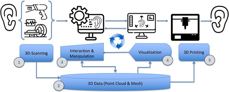
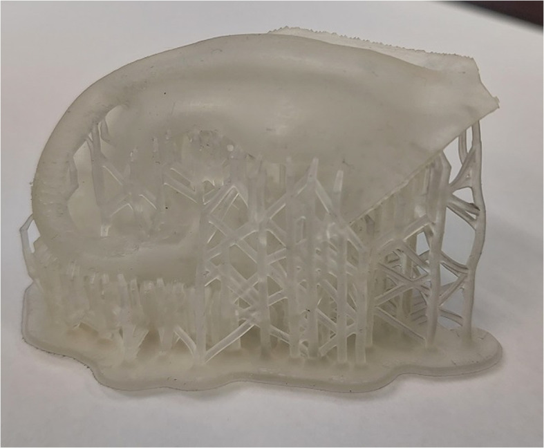
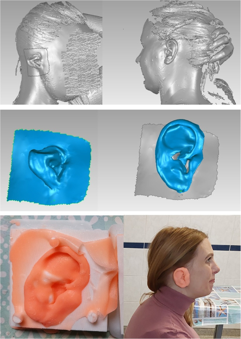
Current 3D scanning and printing technologies offer not only state-of-the-art developments in the field of medical imaging and bio-engineering, but also cost and time effective solutions for surgical reconstruction procedures. Besides tissue engineering, where living cells are used, bio-compatible polymers or synthetic resin can be applied. The combination of 3D handheld scanning devices or volumetric imaging, (open-source) image processing packages, and 3D printers form a complete workflow chain that is capable of effective rapid prototyping of outer ear replicas. This paper reviews current possibilities and latest use cases for 3D-scanning, data processing and printing of outer ear replicas with a focus on low-cost solutions for rehabilitation engineering.

Keywords: 3D printing; 3D scanning and reconstruction; Additive manufacturing; Clinical application; Outer ear; Patient-centered medicine; Patient-individualized therapy; Volumetric scanning.


Conflict of interest statement

The authors declare no competing interests.

Figures

Fig. 1
Fig. 1
Workflow pipeline for rapid prototyping of outer ear replicas. From left to right: (1) 3D-scanning (“3D data acquisition possibilities for the outer ear” section), either volumetric (“Optical 3D-scanning and capturing devices” section) or optical (“Optical 3D-scanning and capturing devices” section); (2) repository of scanned 3D data (local or cloud-based); (3) interactive correction, manipulation and enhancement of the 3D data (“Data processing” section); (4) interactive 3D-visualization and inspection of the ear data; (5) additive 3D manufacturing of the ear replica (“3D printing” section). Steps (2), (3) and (4) are tightly coupled in a closed-loop, as the 3D visual inspection and 3D correction generally go hand-in-hand
Fig. 2
Fig. 2
Anatomy of the outer / external right human ear (of author TW), consisting (from the inside out) of the external end of auditory canal in the center (gray), surrounded by tragus and antitragus (green), the antihelix and helix with their ‘legs’ (crus) on the top (in blue), which enclose the concha and fossa (purple), and the scapa (gray). The bottom part of the ear is the ‘lobe’ (orange)
Fig. 3
Fig. 3
Examples of a 3D reconstruction of a left outer ear from a 3T MRI scan (of author TW) using Slicer 3D software [47]. Left side: posterior-anterior view; center: anterior-posterior view; right side: side-view
Fig. 4
Fig. 4
Examples of facial scans using handheld scanners (Go!SCAN, top row; SIMSCAN 3D, bottom row) for post-processing in MeshLab
Fig. 5
Fig. 5
3D Scanning and reconstruction of the pinna (of author TW) using a digital SLR camera and open-source reconstruction software. The structures of the helix, antihelix, and scapa are reconstructed correctly, while the dimples of the concha and fossa remain as white spots
Fig. 6
Fig. 6
3D Scanning and reconstruction of the pinna (of author TW) using a commercial handheld 3D scanning device (3DSense) (left), and a smartphone (right) in combination with an open-source reconstruction software (PolyCam)
Fig. 7
Fig. 7
Image registration and image fusion of the MRI data (Fig. 3) and optical data (Fig. 6, left) obtained from same person (author TW). Left side: posterior-anterior view; center: anterior-posterior view; right side: side-view. ‘Brown’ indicates data from the optical scanner, while ‘green’ represents data from the MRI. The MRI data has a much higher resolution quality and is much smoother than the data obtained from the optical scanner
Fig. 8
Fig. 8
Image registration fusion of left and right outer ear from the MRI data (Fig. 3). Left side: posterior-anterior view; center: anterior-posterior view; right side: side-view. ‘Green’ represents data from the left ear, ‘purple’ indicates data from the right ear. Some asymmetries can be observed between the ears, which are rarely perceived in daily routine
Fig. 9
Fig. 9
Overview of 3D printing methods, their methodological principles, sub methods, and selected characteristics of the respective method. SLA: stereolithography; DLP: Digital light processing; CDLP: Continuous digital light processing; FDM: fused deposition modeling; SLS: selective laser sintering; MJF: multi jet fusion; EBM: electron beam melting; NPJ: nanoparticle jetting; DOD: Drop on Demand; BJ: Binder jetting. *Layer height varies based on 3D printer and material, so the stated number is a rough guideline value. Green check mark: possible/good performance; red X: not possible/low performance
Fig. 10
Fig. 10
Examples of printed replicas using hard polymer and synthetic resin. The top left is the mirrored version of the top middle replica
Fig. 11
Fig. 11
Printed ear with supporting material
Fig. 12
Fig. 12
Reconstructed right ear replica based on the mirrored image of the left side. Image was scanned with a handheld scanner and post-processed in MeshLab. A series of prototypes were created using a mold and tried on in an iteration process. Fixation methods can be tested after the final model is printed

References

    1. Cooper RA, Ohnabe H, Hobson DA. An introduction to rehabilitation engineering. New York: CRC Press; 2006.
    1. Cooper RA, Cooper R. Rehabilitation Engineering: A perspective on the past 40-years and thoughts for the future. Med Eng Phys. 2019;72:3–12. doi: 10.1016/j.medengphy.2019.08.011. - DOI - PubMed
    1. Lane JP. Rehabilitation Engineering in the Assistive Technology Industry. In: Mihailidis A, Smith R, editors. Rehabilitation Engineering: Principles and Practice. 2022. p. 28. https://www.taylorfrancis.com/chapters/edit/10.1201/b21964-11/rehabilita.... - DOI
    1. Trevelyan J. Reconstructing engineering from practice. Eng Stud. 2010;2(3):175–195. doi: 10.1080/19378629.2010.520135. - DOI
    1. Fuchs JR, Nasseri BA, Vacanti JP. Tissue engineering: a 21st century solution to surgical reconstruction. Ann Thorac Surg. 2001;72(2):577–591. doi: 10.1016/S0003-4975(01)02820-X. - DOI - PubMed