

Double-Outlet Left Ventricle: Case Series and Systematic Review of the Literature
- PMID: 37891996
- PMCID: PMC10605834
- DOI: 10.3390/diagnostics13203175
Double-Outlet Left Ventricle: Case Series and Systematic Review of the Literature
Abstract
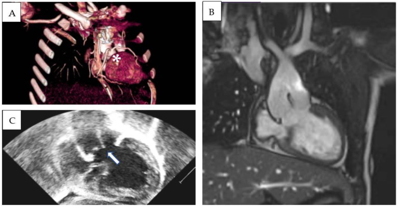
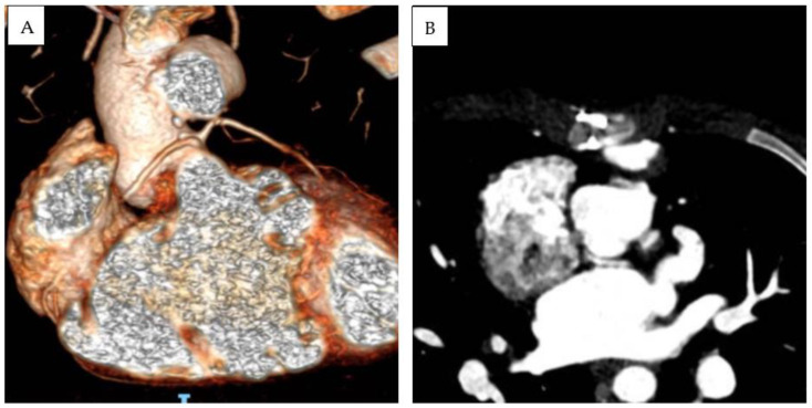
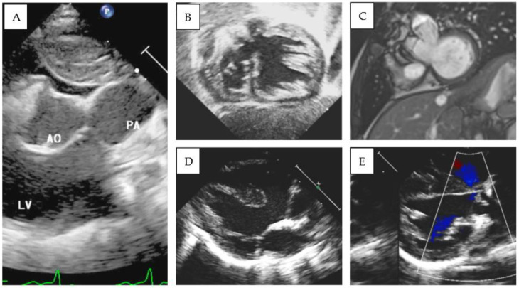
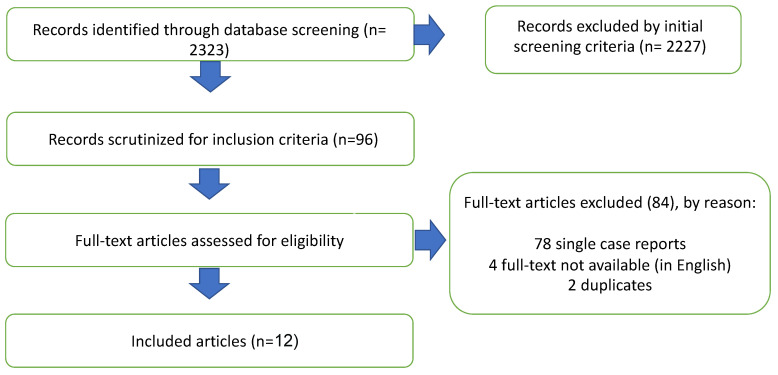
Double-outlet left ventricle (DOLV) is an abnormal ventriculo-arterial connection characterized by the origin of both great arteries from the morphological left ventricle. The aim of our paper is to describe the morphological and imaging features of DOLV and to assess the prevalence of the associated malformations and their surgical outcomes. METHODS From 2011 to 2022, we retrospectively reviewed the electronic case records of patients diagnosed with DOLV at the Bambino Gesu Children's Hospital. A systematic search was developed in MEDLINE, Web of Science, and EMBASE databases to identify reports assessing the morphology and outcomes of DOLV between 1975 and 2023. RESULTS: Over a median follow-up of 9.9 years (IQR 7.8-11.7 y), four cases of DOLV were identified at our institution. Two patients were diagnosed with (S,D,D) DOLV subaortic VSD and pulmonary stenosis (PS): one patient had (S,D,D) DOLV with doubly committed VSD and hypoplastic right ventricle, and another patient had (S,D,L) DOLV with subaortic VSD and PS (malposition type). Pulmonary stenosis was the most commonly associated lesion (75%). LITERATURE REVIEW: After systematic evaluation, a total of 12 reports fulfilled the eligibility criteria and were included in our analysis. PS or right ventricular outflow tract obstruction was the most commonly associated lesion (69%, 95% CI 62-76%). The most common locations of VSD were subaortic (pooled prevalence: 75%, 95% CI 68-81), subpulmonary (15%, 95% CI 10-21), and doubly committed (7%, 95% CI 4-12). The position of the great arteries showed that d-transposition of the aorta was present in 128 cases (59% 95% CI 42-74), and l-transposition was present in 77 cases (35%, 95% CI 29-43).
Keywords: DOLV; Rastelli; double-outlet left ventricle; double-outlet ventricles; pulmonary root translocation.
Conflict of interest statement
The authors declare no conflict of interest.
Figures
References
Publication types
LinkOut - more resources
Full Text Sources
