Review
. 2023 Oct 12;13(20):3186.
doi: 10.3390/diagnostics13203186.

Imaging of the Reconstructed Breast

Affiliations
Review

Imaging of the Reconstructed Breast

Theodora Kanavou et al. Diagnostics (Basel). .

Abstract

The incidence of breast cancer and, therefore, the need for breast reconstruction are expected to increase. The many reconstructive options available and the changing aspects of the field make this a complex area of plastic surgery, requiring knowledge and expertise. Two major types of breast reconstruction can be distinguished: breast implants and autologous flaps. Both present advantages and disadvantages. Autologous fat grafting is also commonly used. MRI is the modality of choice for evaluating breast reconstruction. Knowledge of the type of reconstruction is preferable to provide the maximum amount of pertinent information and avoid false positives. Early complications include seroma, hematoma, and infection. Late complications depend on the type of reconstruction. Implant rupture and implant capsular contracture are frequently encountered. Depending on the implant type, specific MRI signs can be depicted. In the case of myocutaneous flap, fat necrosis, fibrosis, and vascular compromise represent the most common complications. Late cancer recurrence is much less common. Rarely reported late complications include breast-implant-associated large cell anaplastic lymphoma (BIA-ALCL) and, recently described and even rarer, breast-implant-associated squamous cell carcinoma (BIA-SCC). In this review article, the various types of breast reconstruction will be presented, with emphasis on pertinent imaging findings and complications.

Keywords: MRI of reconstructed breast; autologous reconstruction; breast; breast cancer recurrence; complications of reconstruction surgery; implant-based breast reconstruction.


Conflict of interest statement

The authors declare no conflict of interest.

Figures

Figure 1
Figure 1
Intraoperative images of transverse abdominal myocutaneous (TRAM) flap reconstruction after right mastectomy (a) before and (b) after transfer to the breast.
Figure 2
Figure 2
(a) Preoperative design with skin paddle and area of no dissection and (b) postoperative image of left latissimus dorsi (LD) flap combined with an implant for the reconstruction of the lest breast.
Figure 3
Figure 3
Dual plane use of acellular dermal martice (ADM) to support and cover the lower pole of the reconstruction between the pectoralis major muscle edge and the inframammary fold.
Figure 4
Figure 4
(a) ADM for circumferential coverage (Braxon, Decomed), (b) prepectoral mastectomy cavity to reconstruct, (c) preparation of ravioli to be inserted in the mastectomy cavity to be reconstructed.
Figure 5
Figure 5
Deep inferior epigastric perforator (DIEP) reconstruction: (a) mediolateral oblique mammographic (MLO) view of the reconstructed breast depicts essentially fatty tissue and surgical clips (arrows); (b) axial thoracic CT scan confirms the presence of a viable DIEP reconstruction with no complications.
Figure 6
Figure 6
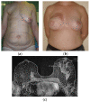
(a) Preoperative design and (b) postoperative image of right mastectomy defect reconstruction with a pedicled TRAM flap and left breast mastopexy. (c) On MRI, the right reconstructed breast consists primarily of fat and the muscle is recognized centrally anteriorly to the chest wall.
Figure 7
Figure 7
Bilateral autologous LD reconstruction. Yellow arrows point to the characteristic tailed aspect of the muscle. Red arrows depict a hypointense circumscribed area with irregular wall enhancement and associated solid-enhancing nodule corresponding to a local relapse of high grade invasive ductal carcinoma, triple negative, with marked central necrosis.
Figure 8
Figure 8
Mediolateral oblique MLO mammography view and ultrasound (US) of the right breast after mastectomy and reconstruction with a single-lumen silicone implant: (a) in the MLO view, the implant appears as a homogenous radiopaque oval mass with smooth borders; (b) on US the implant is anechoic. The intact shell appears as an echogenic line (yellow arrows) and the fibrous capsule is seen as a parallel echogenic line (red arrows).
Figure 9
Figure 9
Normal MRI appearance of an intact single-lumen silicone implant of the reconstructed right breast: (a) in the T1 non-fat-saturated sequence, the silicone has low signal intensity; on T2-weighted images, both non-fat-saturated (b) and fat-saturated (c), the silicone has high signal intensity. Minimal peri-implant fluid is a common normal finding with high signal intensity; (d) in the silicone-selective sequence, the silicone appears with high signal intensity while the peri-implant fluid has low signal intensity.
Figure 10
Figure 10
MRI appearance of double-lumen implant (silicone in the outer lumen, saline in the inner lumen). The red arrow points at the valve used to inflate the inner saline part.
Figure 11
Figure 11
MRI appearance of single-lumen saline implants with radial fold of the right implant and intracapsular rupture of the left implant: (a) axial T2 sequence depicts a line perpendicular to the implant shell line, corresponding to a radial fold, whereas the curvilinear line, parallel to the shell, indicates a subcapsular line, in concordance with an intracapsular rupture; (b) sagittal view confirms the “perpendicular to implant-shell” character of the radial fold of the right implant. Sagittal views can be of help in differentiating radial folds from true intracapsular rupture.
Figure 12
Figure 12
(a) Fat necrosis in the right breast following bilateral mastectomy and implant reconstruction depicted as a complex cystic mass on US. (b) On T1-weighted images, the mass is isointense to fat and (c) has markedly low signal intensity on STIR sequence, known as the “black hole” sign. (d) On subtraction MRI the mass exhibits no enhancement.
Figure 13
Figure 13
A woman with a history of left breast cancer presented with a palpable mass 2 years after bilateral mastectomy and implant reconstruction: (a) on US, the mass was hypoechoic with indistinct and irregular margins; (b) on T1-weighted MR images, the mass is hypointense with an irregular shape and spiculated margins; (c) on DCE MRI, 3 min after contrast administration and (d) subtraction image, the mass is strongly and homogeneously enhancing. The diagnosis was fat necrosis after core biopsy.
Figure 14
Figure 14
MRI appearance of left implant infection. Axial T1-weighted fat-saturation post-contrast view of a left peri-implant inflammatory infusion with peripheral smooth contrast uptake.
Figure 15
Figure 15
Ultrasound appearance of extracapsular rupture. (a) Free silicone in the breast along the upper outer part of the implant, indicating extracapsular rupture. Typical appearance of a marked hyperechoic area with posterior shadowing, referred to as “snowstorm appearance”. (b) “Snowstorm” appearance of an axillary lymph node in another patient. This was an isolated finding, and no rupture was documented on MRI. This was related to silicone gel bleed, i.e., microscopic diffusion of silicone molecules through the semipermeable weakened but intact implant elastomer shell.
Figure 16
Figure 16
MRI appearance of various signs indicative of intracapsular and extracapsular rupture. All images are axial T2-weigthed images from different patients. (a) Salad oil and subcapsular line sign, evocating intracapsular rupture. (b) Isolated salad-oil sign in both implants. This sign is not sufficient per se to validate an intracapsular rupture. (c) Key-hole sign, lasso sign and linguni sign, indicating intracapsular rupture of the right implant. (d) Extracapsular rupture can be safely indicated on this characteristic aspect of extra-implant presence of silicone.
Figure 17
Figure 17
MRI appearance of extracapsular rupture. (a) Axial silicone-only sequence depicts hyperintense free droplets of silicone outside the left implant capsule. (b) Same patient, axial silicone-only sequence through the axillary level depicts marked hyperintense ipsilateral axillary lymph nodes, evocating silicone deposits.
Figure 18
Figure 18
Silicone granuloma associated with peri-implant effusion and mild inflammation. (a) Axial short tau inversion recovery (STIR) sequence depicts hyperintense peri-implant effusion associated with a hypointense nodular component. (b) Axial T1 fat-saturated post-contrast sequence readily depicts the strong enhancement of the nodular component and the mild enhancement of the peri-implant effusion collection. This aspect could evocate a silicone granuloma; nevertheless, a core biopsy was performed to validate this diagnosis.
Figure 19
Figure 19
Breast implant associated atypical large cell lymphoma (BIA-ALCL), presented 10 years after implantation as a peri-implant mass, associated with pronounced edema of the breast and pectoralis major muscle in T2 MRI images (ac). (df) In subtraction images, the mass is irregular with strong contrast enhancement and is invading the pectoral muscle (f). Images are courtesy of Prof. M. Fuchsjäger, Graz, Austria.
Figure 20
Figure 20
BRCA1 carrier. Right mastectomy due to extensive comedo-DCIS and autologous reconstruction. Prophylactic left mastectomy and reconstruction. (a) Maximum intensity projection (MIP) images depict a small enhancing nodule, with a millimetric satellite lesion, both of which are suspicious in this context. (b) Second-look post MRI ultrasound depicts a hypoechoic, taller-than-wider solid nodule with irregular margins. Core biopsy confirmed local relapse of high-grade DCIS.

Similar articles

References

    1. Fefferman M., Nicholson K., Kuchta K., Pesce C., Kopkash K., Yao K. Rates of Bilateral Mastectomy in Patients with Early-Stage Breast Cancer. JAMA Netw. Open. 2023;6:e2251348. doi: 10.1001/jamanetworkopen.2022.51348. - DOI - PMC - PubMed
    1. Sardanelli F., Trimboli R.M., Houssami N., Gilbert F.J., Helbich T.H., Alvarez Benito M., Balleyguier C., Bazzocchi M., Bult P., Calabrese M., et al. Magnetic resonance imaging before breast cancer surgery: Results of an observational multicenter international prospective analysis (MIPA) Eur. Radiol. 2022;32:1611–1623. doi: 10.1007/s00330-021-08240-x. - DOI - PMC - PubMed
    1. Taylor A., Tischkowitz M. Informed decision-making is the key in women at high risk of breast cancer. Eur. J. Surg. Oncol. 2014;40:667–669. doi: 10.1016/j.ejso.2014.02.219. - DOI - PubMed
    1. Alaofi R.K., Nassif M.O., Al-Hajeili M.R. Prophylactic mastectomy for the prevention of breast cancer: Review of the literature. Avicenna J. Med. 2018;8:67–77. doi: 10.4103/ajm.AJM_21_18. - DOI - PMC - PubMed
    1. Kaidar-Person O., Poortmans P., Offersen B.V., Boersma L.J., de Ruysscher D., Noy V., Hermann N., Kuhn T. What are the guidelines for immediate breast reconstruction? Eur. J. Surg. Oncol. 2021;47:1214–1215. doi: 10.1016/j.ejso.2020.03.226. - DOI - PubMed