

Diagnostic Superiority of Dual-Time Point [18F]FDG PET/CT to Differentiate Malignant from Benign Soft Tissue Tumors
- PMID: 37892023
- PMCID: PMC10606132
- DOI: 10.3390/diagnostics13203202
Diagnostic Superiority of Dual-Time Point [18F]FDG PET/CT to Differentiate Malignant from Benign Soft Tissue Tumors
Abstract
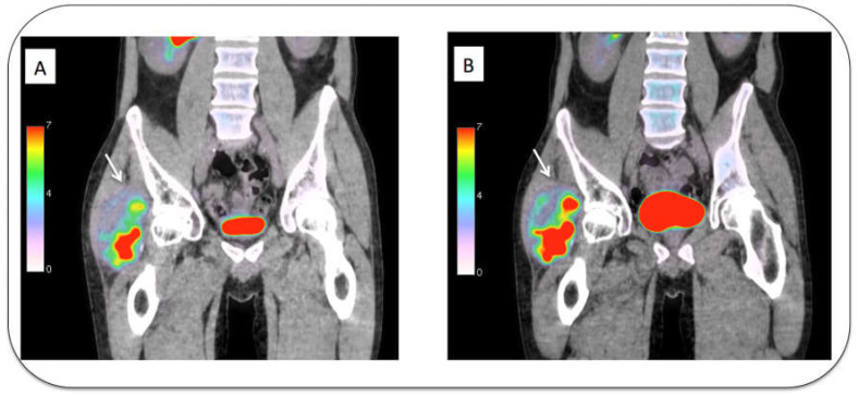
[18F]FDG PET/CT is used in the workup of indeterminate soft tissue tumors (STTs) but lacks accuracy in the detection of malignant STTs. The aim of this study is to evaluate whether dual-time point [18F]FDG PET/CT imaging (DTPI) can be useful in this indication. In this prospective study, [18F]FDG PET/CT imaging was performed 1 h (t1) and 3 h (t2) after injection. Tumor uptake (SUVmax) was calculated at each time point to define a retention index (RI) corresponding to the variation between t1 and t2 (%). Sixty-eight patients were included, representing 20 benign and 48 malignant tumors (including 40 sarcomas). The RI was significantly higher in malignant STTs than in benign STTs (median: +21.8% vs. -2%, p < 0.001). An RI of >14.3% predicted STT malignancy with a specificity (Sp) of 90% and a sensitivity (Se) of 69%. An SUVmaxt1 of >4.5 was less accurate with an Sp of 80% and an Se of 60%. In a subgroup of tumors with at least mild [18F]FDG uptake (SUVmax ≥ 3; n = 46), the RI significantly outperformed the diagnostic accuracy of SUVmax (AUC: 0.88 vs. 0.68, p = 0.01). DTPI identifies malignant STT tumors with high specificity and outperforms the diagnostic accuracy of standard PET/CT.
Keywords: FDG PET/CT; dual-time point acquisition; sarcoma; soft tissue tumor.
Conflict of interest statement
The authors declare no conflict of interest.
Figures


Similar articles
-
Dual-time point 18F-FDG-PET and PET/CT for Differentiating Benign From Malignant Musculoskeletal Lesions: Opportunities and Limitations.Semin Nucl Med. 2017 Jul;47(4):373-391. doi: 10.1053/j.semnuclmed.2017.02.009. Epub 2017 Apr 19. Semin Nucl Med. 2017. PMID: 28583277 Review.
-
More advantages in detecting bone and soft tissue metastases from prostate cancer using 18F-PSMA PET/CT.Hell J Nucl Med. 2019 Jan-Apr;22(1):6-9. doi: 10.1967/s002449910952. Epub 2019 Mar 7. Hell J Nucl Med. 2019. PMID: 30843003
-
Dual-time point 18FDG-PET/CT imaging may be useful in assessing local recurrent disease in high grade bone and soft tissue sarcoma.Nucl Med Rev Cent East Eur. 2016;19(1):22-7. doi: 10.5603/NMR.2016.0005. Nucl Med Rev Cent East Eur. 2016. PMID: 26841376
-
The Clinical Role of Dual-Time-Point (18)F-FDG PET/CT in Differential Diagnosis of the Thyroid Incidentaloma.Nucl Med Mol Imaging. 2014 Jun;48(2):121-9. doi: 10.1007/s13139-013-0247-z. Epub 2013 Dec 6. Nucl Med Mol Imaging. 2014. PMID: 24900152 Free PMC article.
-
Hyperaccumulation of (18)F-FDG in order to differentiate solid pseudopapillary tumors from adenocarcinomas and from neuroendocrine pancreatic tumors and review of the literature.Hell J Nucl Med. 2013 May-Aug;16(2):97-102. doi: 10.1967/s002449910084. Epub 2013 May 20. Hell J Nucl Med. 2013. PMID: 23687644 Review.
Cited by
-
Dual-Time-Point 18F-FDG PET/CT imaging in the diagnosis of colorectal carcinoma or advanced adenoma in patients with fixed focal colorectal 18F-FDG uptake.BMC Cancer. 2025 Apr 22;25(1):755. doi: 10.1186/s12885-025-14129-5. BMC Cancer. 2025. PMID: 40264039 Free PMC article.
References
-
- Katz D., Palmerini E., Pollack S.M. Histology-Driven Treatments for Soft Tissue Sarcoma. Volume 28. American Society of Clinical Oncology Educational Book; Alexandria, VA, USA: 2018. More Than 50 Subtypes of Soft Tissue Sarcoma: Paving the Path for Histology-Driven Treatments; pp. 925–938. - PubMed
-
- Ray-Coquard I., Montesco M.C., Coindre J.M., Dei Tos A.P., Lurkin A., Ranchere-Vince D., Vecchiato A., Decouvelaere A.V., Mathoulin-Pelissier S., Albert S., et al. Sarcoma: Concordance between initial diagnosis and centralized expert review in a population-based study within three European regions. Ann. Oncol. 2012;23:2442–2449. doi: 10.1093/annonc/mdr610. - DOI - PMC - PubMed
LinkOut - more resources
Full Text Sources
