. 2023 Sep 27;13(10):1461.
doi: 10.3390/biom13101461.

Decanoic Acid Rescues Differences in AMPA-Mediated Calcium Rises in Hippocampal CA1 Astrocytes and Neurons in the 5xFAD Mouse Model of Alzheimer's Disease

Affiliations

Decanoic Acid Rescues Differences in AMPA-Mediated Calcium Rises in Hippocampal CA1 Astrocytes and Neurons in the 5xFAD Mouse Model of Alzheimer's Disease

Mina Abghari et al. Biomolecules. .

Abstract

Alzheimer's disease (AD), a devastating neurodegenerative disease characterized by cognitive dysfunctions, is associated with high levels of amyloid beta 42 (Aβ42), which is believed to play a role in cellular damage and signaling changes in AD. Decanoic acid has been shown to be therapeutic in AD. Glutamatergic signaling within neurons and astrocytes of the CA1 region of the hippocampus is critical in cognitive processes, and previous work has indicated deficiencies in this signaling in a mouse model of AD. In this study, we investigated glutamate-mediated signaling by evaluating AMPA-mediated calcium rises in female and male CA1 neurons and astrocytes in a mouse model of AD and examined the potential of decanoic acid to normalize this signaling. In brain slices from 5xFAD mice in which there are five mutations leading to increasing levels of Aβ42, AMPA-mediated calcium transients in CA1 neurons and astrocytes were significantly lower than that seen in wildtype controls in both females and males. Interestingly, incubation of 5xFAD slices in decanoic acid restored AMPA-mediated calcium levels in neurons and astrocytes in both females and males to levels indistinguishable from those seen in wildtype, whereas similar exposure to decanoic acid did not result in changes in AMPA-mediated transients in neurons or astrocytes in either sex in the wildtype. Our data indicate that one mechanism by which decanoic acid could improve cognitive functioning is through normalizing AMPA-mediated signaling in CA1 hippocampal cells.

Keywords: Alzheimer’s disease; calcium; diet; hippocampus; medium-chain fatty acids.


Conflict of interest statement

All authors disclose that they have no relevant financial or non-financial interests to disclose.

Figures

Figure 1
Figure 1
Recordings were performed from astrocytes and neurons in the CA1 region of the hippocampus (A1) Under bright field optics, the structure of the CA1 region of the hippocampus can be seen under 4× magnification in mouse brain slices. (A2) Under 40× magnification and bright field optics, individual cells are apparent. (B1) Representative fluorescent image of cells loaded with the intracellular calcium indicator Fura-2 AM under 380 nm wavelength light is shown. (B2) The same field is shown under 560 nm fluorescent light in which astrocytes loaded with the astrocytic marker sulforhodamine 101 (SR101) can be seen. (B3) In the merged image, cells positive for SR101 and Fura-2 are marked with arrows and determined to be astrocytes. Scalebar in A and B indicates 20 µm. (C) AMPA induces rises in fluorescence in Fura-2 AM loaded cells, which are indirect indicators of increases in calcium as shown in these representative responses from hippocampal CA1 neurons (C1) and CA1 astrocytes (C2) in the hippocampus of the 5xFAD (green and red traces in C1,C2) and WT hippocampus (black traces in C1,C2) when AMPA was applied. The amplitude of the change in %DF/F from baseline induced by AMPA was compared across genotypes in the two cell types.
Figure 2
Figure 2
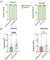
Smaller rises in calcium are elicited in both hippocampal neurons and astrocytes in one-month-old 5xFAD mice compared to rises seen in wildtype (WT) (A) There were no differences in the proportions of neurons or astrocytes responding to AMPA with rises in fluorescence in the CA1 hippocampal region between male and female one-month-old 5xFAD and WT mice (10-22 PND). (B) One-month-old 5xFAD mice exhibited significantly smaller fluorescent changes in response to AMPA than WT in both hippocampal neurons and astrocytes, as shown in the population data. Histograms compare the AMPA-induced fluorescence changes to 10 μM AMPA between the population of 5xFAD and WT neurons (Mann–Whitney test: p < 0.0001) and astrocytes (Mann–Whitney test: p = 0.0097), indicating smaller rises in calcium in both cell types in the 5xFAD. Amplitude of the response is presented as %DF/F in this and subsequent figures. In this and subsequent figures, the number of cells is indicated by n in the x-axis, and the significance level is presented above the bars with asterisks. In this figure, ** p < 0.01 and **** p < 0.0001.
Figure 3
Figure 3
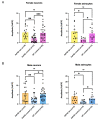
Smaller AMPA-induced rises were elicited in both females and males in neurons and astrocytes in the 5xFAD genotype. There were no differences in either females (A) or males (B) in the proportions of hippocampal neurons or astrocytes responding to AMPA with rises in fluorescence between two-month-old 5xFAD and wildtype (WT) mice. (A,B) Two-month-old female and male 5xFAD mice exhibited significantly smaller amplitude fluorescent changes in response to 10 μM AMPA than WT in both hippocampal neurons (Female: Mann–Whitney test: p = 0.0002; Male: Mann–Whitney test: p = 0.0015) and astrocytes (Female: Mann–Whitney test: p = 0.0053; Male: Mann–Whitney test: p = 0.0077) as shown in the population data. The significance level is presented above the bars with asterisks denoting ** p < 0.01 and *** p < 0.001.
Figure 4
Figure 4
The proportion of neurons and astrocytes that responded to AMPA and the amplitude of the responses did not differ between females or males irrespective of genotype. (A) The amplitude of the AMPA-induced calcium rises in neurons and astrocytes in the 5xFAD genotype and the proportion of each cell type that responded to AMPA with rises in calcium did not differ between females and males. (B) Similarly, the proportion of neurons and astrocytes that responded to AMPA in the WT and the amplitude of the change in fluorescence did not differ between females and males. ns denotes no significance.
Figure 5
Figure 5
Decanoic acid resulted in significantly larger AMPA-mediated transients in the 5xFAD genotype in neurons and astrocytes in both females and males. (A) While the proportion of neurons and astrocytes responding to AMPA in slices that had been incubated in decanoic acid did not differ from control conditions, the amplitude of AMPA-mediated calcium rises was significantly greater in both cell types in slices exposed to decanoic acid from the female (neurons: Mann–Whitney test: p = 0.0003; astrocytes: Mann–Whitney test: p = 0.0038). (B) The amplitude of AMPA-mediated rises in calcium was also greater in neurons and astrocytes from the 5xFAD male that had been preincubated in decanoic acid (neurons: Mann–Whitney test: p = 0.0216; astrocytes: Mann–Whitney test: p = 0.0127). The significance level is presented above the bars with asterisks denoting * p < 0.05, ** p < 0.01, and *** p < 0.001.
Figure 6
Figure 6
There was no difference between females and males in the amplitude of the response to AMPA in neurons or astrocytes following incubation in decanoic acid nor in the proportion responding. (A) The proportion of neurons responding and the amplitude of response to AMPA in slices incubated in decanoic acid did not differ between the female and male 5xFAD genotype. Further, there was no difference between females and males in the relative fold change in amplitude from control conditions in the 5xFAD neurons when decanoic acid was present (Two-way ANOVA, neurons: p = 0.9793). (B) The response proportion and the amplitude of the response to AMPA in presence of decanoic acid also did not differ in astrocytes between females and males, and there was no difference in the relative fold change in amplitude in presence of decanoic acid when compared to control conditions in the 5xFAD (Two-way ANOVA, Astrocytes: p = 0.3534). ns indicates no significance.
Figure 7
Figure 7
The effect of decanoic acid in altering AMPA-mediated calcium rises was not seen in neurons or astrocytes in either females or male wildtype (WT). (A) Neither the proportion responding nor the amplitude of response to AMPA differed in WT female neurons between responses seen in conditions when decanoic acid was present when compared to responses seen in control conditions. (B) Similarly, there was no effect of decanoic acid on the proportion responding or the amplitude of the response to AMPA in the male WT when compared to control conditions. ns indicates no significance.
Figure 8
Figure 8
Decanoic acid rescues the reduction in AMPA-mediated calcium seen in neurons and astrocytes of the 5xFAD genotype and restores the amplitude to that seen in the wildtype (WT) in both sexes. (A) There was no difference between the amplitude of AMPA-mediated calcium transients in neurons and astrocytes of the female 5xFAD genotype when incubated in decanoic acid when compared to transients seen in the WT. (B) There was also no difference in the amplitude of the AMPA-mediated calcium transients elicited in neurons and astrocytes of slices from the male 5xFAD animal that were incubated in decanoic acid when compared to transients seen in the WT. The significance level is presented above the bars with asterisks denoting * p < 0.05, ** p < 0.01, and *** p < 0.001 with ns indicating no significance.

References

    1. Guan R., Wen X., Liang Y., Xu D., He B., Feng X. Trends in Alzheimer’s Disease Research Based upon Machine Learning Analysis of PubMed Abstracts. Int. J. Biol. Sci. 2019;15:2065–2074. doi: 10.7150/ijbs.35743. - DOI - PMC - PubMed
    1. Bailey T.L., Rivara C.B., Rocher A.B., Hof P.R. The nature and effects of cortical microvascular pathology in aging and Alzheimer’s disease. Neurol. Res. 2004;26:573–578. doi: 10.1179/016164104225016272. - DOI - PubMed
    1. Barnes L.L., Wilson R.S., Bienias J.L., Schneider J.A., Evans D.A., Bennett D.A. Sex differences in the clinical manifestations of Alzheimer disease pathology. Arch. Gen. Psychiatry. 2005;62:685–691. doi: 10.1001/archpsyc.62.6.685. - DOI - PubMed
    1. Dumitrescu L., Barnes L.L., Thambisetty M., Beecham G., Kunkle B., Bush W.S., Gifford K.A., Chibnik L.B., Mukherjee S., De Jager P.L., et al. Sex differences in the genetic predictors of Alzheimer’s pathology. Brain. 2019;142:2581–2589. doi: 10.1093/brain/awz206. - DOI - PMC - PubMed
    1. Chene G., Beiser A., Au R., Preis S.R., Wolf P.A., Dufouil C., Seshadri S. Gender and incidence of dementia in the Framingham Heart Study from mid-adult life. Alzheimers Dement. 2015;11:310–320. doi: 10.1016/j.jalz.2013.10.005. - DOI - PMC - PubMed

Publication types

Substances