Review
. 2023 Oct 20;13(10):1555.
doi: 10.3390/biom13101555.

Navigating the ERK1/2 MAPK Cascade

Affiliations
Review

Navigating the ERK1/2 MAPK Cascade

Ana Martin-Vega et al. Biomolecules. .

Abstract

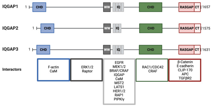
The RAS-ERK pathway is a fundamental signaling cascade crucial for many biological processes including proliferation, cell cycle control, growth, and survival; common across all cell types. Notably, ERK1/2 are implicated in specific processes in a context-dependent manner as in stem cells and pancreatic β-cells. Alterations in the different components of this cascade result in dysregulation of the effector kinases ERK1/2 which communicate with hundreds of substrates. Aberrant activation of the pathway contributes to a range of disorders, including cancer. This review provides an overview of the structure, activation, regulation, and mutational frequency of the different tiers of the cascade; with a particular focus on ERK1/2. We highlight the importance of scaffold proteins that contribute to kinase localization and coordinate interaction dynamics of the kinases with substrates, activators, and inhibitors. Additionally, we explore innovative therapeutic approaches emphasizing promising avenues in this field.

Keywords: ERK1/2; MAPKs; cancer; inhibitors; scaffold; therapies.


Conflict of interest statement

The authors declare no conflict of interest.

Figures

Figure 1
Figure 1
Activation of MEK by different upstream activators causes diverse outcomes. MEK can be activated by different MAP3Ks in a stimulus- and cell type-specific manner. The canonical pathway involves signal transduction through RAF. The synthesis of MOS induces oocyte maturation by direct phosphorylation of MEK and consequent activation of ERK. Upon stimulation, the kinase Tpl2/Cot is released from a ternary complex and is stabilized by phosphorylation of specific residues. The signaling module Tpl2/Cot-MEK-ERK is important in inflammation and immune response. MEKK1 is an upstream regulator of stress-responsive kinases, and it is also able to phosphorylate MEK1/2 to induce ERK activation in inflammatory settings. MAST1 initial or induced expression leads to cisplatin resistance.
Figure 2
Figure 2
ERK2 structure. The ribbon diagram shows the two-domain structure of ERK2. Activation of the kinase occurs upon phosphorylation of the TEY motif in the activation loop (red), which lies in front of the active site. Phosphorylation causes a small domain closure, relative to other protein kinases. Reorganization of loop L16 (blue), part of a C-terminal extension that wraps up the back of the kinase, causes repositioning of alpha helix C (αC, magenta), allowing formation of a salt bridge between the glutamate in the helix to the essential lysine in beta strand 3 in the interior of the active site. The acidic residues that make up the CD domain are shown in orange. The FXF binding site lies between the MAPK insert (green) and the kinase core. Interactions through these two docking motifs may provoke conformational changes in ERK2 [115,116,117,118].
Figure 3
Figure 3
ERK negative feedback loops. ERK phosphorylates MEK1 at T292, thus impeding dimerization with MEK2 and PAK1-mediated phosphorylation of MEK1 in S298. ERK also phosphorylates T386 in MEK1 and the homolog residue in MEK2, T394. In CRAF, there are five ERK phospho-sites throughout the protein as follows: S29 and S43 in the N-terminus, S642 at the C-terminus, and S289, S296 and S301 at the flexible hinge between the regulatory and catalytic domains. The result is impaired localization at the plasma membrane, loss of CRAF–RAS interaction, and enhanced access for PP2A facilitating CRAF dephosphorylation. In BRAF, ERK phosphorylates S151 next to the RAS binding domain (RBD) of RAF, impairing RAS–BRAF interaction. ERK phosphorylates T401, S750 and T753 in BRAF, interfering with BRAF–CRAF dimerization. Pathway activation induces phosphorylation of KSR1 on T260, T274, S320 and S443 by ERK impairing dimerization with RAF and localization in the plasma membrane. ERK directly phosphorylates SOS on S1132, S1167, S1178 and S1193 and indirectly in S1134 and S1161 through RSK2. ERK phosphorylates the EGFR on T693 in the mature form of the receptor.
Figure 4
Figure 4
Schematic of the RAF and KSR protein structures. Domain organization of RAF consists of a RAS binding domain (RBD) followed by a cysteine-rich domain (CRD), both in the N-terminal regulatory region of the protein, and the C-terminal part is a kinase domain. BRAF presents an additional sequence at its N-terminal region, the BRAF specific sequence (BRS). KSR1 and KSR2 are composed of the same five conserved areas, CA1-CA5 [333]. CA1 to CA4 comprise the N-terminal region and constitute the regulatory domain, while CA5 is the pseudokinase region at the C-terminus. CA1 has a coiled-coil fused to a sterile α-motif (CC-SAM, not present in RAF) implicated in KSR1 localization at membrane ruffles [339]; this is not as clear for KSR2 since it has been considerably less studied. CA1, together with CA5, participates in B–RAF binding in a CC-SAM-independent manner [261,340]. CA2 is a proline-rich region, and it is believed to function as an SH3 binding domain. CA3, a cysteine-rich domain (CRD), is a lipid binding domain also involved in KSR subcellular localization, and it is essential to target KSR to the plasma membrane upon RAS activation [341,342,343,344]. CA4 is a serine/threonine-rich region which includes the FXFP docking motif for ERK binding [126,133,345]. Lastly, the CA5 kinase domain contains the MEK [346] and RAF [347] binding domains. Colors indicate structural similarities between family members including the cysteine-rich domain (CRD), serine/threonine-rich domain (S/TRD) and kinase domain.
Figure 5
Figure 5
Schematic of IQGAP1/2 and 3 domain organization and interactors. IQGAPs are divided into five conserved domains, from N- to C-terminus. CHD, calponin homology domain; WW domain, a poly-proline protein–protein domain with two conserved Trp (W) residues; IQ domain, with four repeats (three for IQGAP2) of isoleucine- and glutamine-containing motifs; GRD, GAP-related domain; and RGCT, RAS GAP C-terminus domain. Some representative binding partners for each domain are organized and displayed in the color-coded boxes.

Similar articles

Cited by

References

    1. Cooper J.A., Bowen-Pope D.F., Raines E., Ross R., Hunter T. Similar Effects of Platelet-Derived Growth Factor and Epidermal Growth Factor on the Phosphorylation of Tyrosine in Cellular Proteins. Cell. 1982;31:263–273. doi: 10.1016/0092-8674(82)90426-3. - DOI - PubMed
    1. Ray L.B., Sturgill T.W. Characterization of Insulin-Stimulated Microtubule-Associated Protein Kinase. Rapid Isolation and Stabilization of a Novel Serine/Threonine Kinase from 3T3-L1 Cells. J. Biol. Chem. 1988;263:12721–12727. doi: 10.1016/S0021-9258(18)37813-X. - DOI - PubMed
    1. Sturgill T.W., Ray L.B., Erikson E., Maller J.L. Insulin-Stimulated MAP-2 Kinase Phosphorylates and Activates Ribosomal Protein S6 Kinase II. Nature. 1988;334:715–718. doi: 10.1038/334715a0. - DOI - PubMed
    1. Boulton T.G., Yancopoulos G.D., Gregory J.S., Slaughter C., Moomaw C., Hsu J., Cobb M.H. An Insulin-Stimulated Protein Kinase Similar to Yeast Kinases Involved in Cell Cycle Control. Science. 1990;249:64–67. doi: 10.1126/science.2164259. - DOI - PubMed
    1. Boulton T.G., Cobb M.H. Identification of Multiple Extracellular Signal-Regulated Kinases (ERKs) with Antipeptide Antibodies. Mol. Biol. Cell. 1991;2:357–371. doi: 10.1091/mbc.2.5.357. - DOI - PMC - PubMed

Publication types