Review
. 2023 Oct 20;12(20):6653.
doi: 10.3390/jcm12206653.

Advances in Therapies to Treat Neonatal Hypoxic-Ischemic Encephalopathy

Affiliations
Review

Advances in Therapies to Treat Neonatal Hypoxic-Ischemic Encephalopathy

Amaresh K Ranjan et al. J Clin Med. .

Abstract

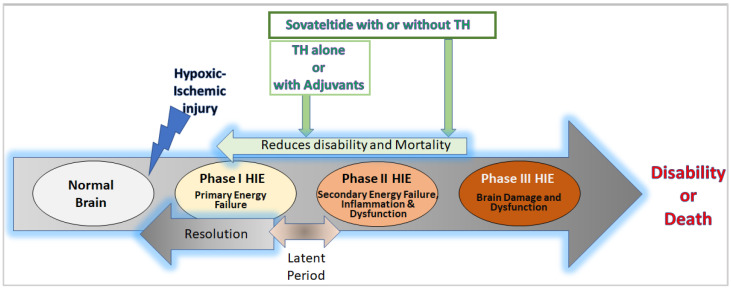
Neonatal hypoxic-ischemic encephalopathy (HIE) is a condition that results in brain damage in newborns due to insufficient blood and oxygen supply during or after birth. HIE is a major cause of neurological disability and mortality in newborns, with over one million neonatal deaths occurring annually worldwide. The severity of brain injury and the outcome of HIE depend on several factors, including the cause of oxygen deprivation, brain maturity, regional blood flow, and maternal health conditions. HIE is classified into mild, moderate, and severe categories based on the extent of brain damage and resulting neurological issues. The pathophysiology of HIE involves different phases, including the primary phase, latent phase, secondary phase, and tertiary phase. The primary and secondary phases are characterized by episodes of energy and cell metabolism failures, increased cytotoxicity and apoptosis, and activated microglia and inflammation in the brain. A tertiary phase occurs if the brain injury persists, characterized by reduced neural plasticity and neuronal loss. Understanding the cellular and molecular aspects of the different phases of HIE is crucial for developing new interventions and therapeutics. This review aims to discuss the pathophysiology of HIE, therapeutic hypothermia (TH), the only approved therapy for HIE, ongoing developments of adjuvants for TH, and potential future drugs for HIE.

Keywords: IRL-1620; PMZ 1620; cerebral palsy; endothelin B receptors; hypoxia; ischemia; neonates; neurogenesis; oxidative stress; perinatal asphyxia; regeneration; sovateltide; stem/progenitor cells.


Conflict of interest statement

A.G. is a Pharmazz, Inc. employee and has issued and pending patents related to the studies described in this review. A.K.R. is a Pharmazz Inc. employee and declares no other competing interest.

Figures

Figure 1
Figure 1
Diagrammatic representation of the phases of hypoxic-ischemic encephalopathy (HIE). After hypoxic-ischemic injury in the normal neonatal brain (white), phase I of HIE (yellow) includes primary energy failure in the neural tissue. The phase I energy failure has a latent period (bi-directional arrow) ranging from 6 h to 15 h (depending upon the severity of the injury), which is a transition period with chances of resolving the issue of primary energy failure due to restoration of blood flow by alternative reperfusion events in the body. Hence, the latent period is also considered the best time for starting a therapy or intervention for treating HIE. If the primary energy failure is unresolved, it leads to phase II of HIE (light orange) with secondary energy failure, overwhelming inflammation, and neural dysfunction. The phase II HIE events are highly devastating and could lead to the phase III HIE (orange) with damaged brain tissue and brain dysfunction, which cause disability or death in HIE patients. At cellular and molecular levels, the primary phase of HIE involves ATP deprivation, abnormal calcium influx, excitotoxicity, and necrosis in neurovascular cells. The secondary phase is marked with oxidative stress following reperfusion, microglial activation, inflammation, excitotoxicity, and cell death (apoptosis/necrosis). The tertiary phase is characterized by decreased neural cell plasticity and a reduced number of neurons (not shown in the diagram). Currently, available interventions for HIE, e.g., TH and adjuvants, are known to act during the latent period (green arrow) and help in reducing the severity of secondary energy failure and would decrease brain damage and dysfunction. However, no evidence of regeneration or repair in the ischemic/hypoxic damaged neural tissue has been reported after treatment with TH and adjuvants. On the other hand, sovateltide (green arrows) has demonstrated mitigation of secondary energy failure and enhancement of neuronal regeneration and repair in the ischemic/hypoxic brain of the HIE rat model and the ischemic stroked brain of the rat and human. Therefore, we anticipate that sovateltide could be developed as a new “First in Class” therapeutic or adjuvant to TH for treating HIE.

References

    1. Lawn J.E., Cousens S., Zupan J., Lancet Neonatal Survival Steering Team 4 million neonatal deaths: When? Where? Why? Lancet. 2005;365:891–900. doi: 10.1016/S0140-6736(05)71048-5. - DOI - PubMed
    1. Allen K.A., Brandon D.H. Hypoxic Ischemic Encephalopathy: Pathophysiology and Experimental Treatments. Newborn Infant Nurs. Rev. 2011;11:125–133. doi: 10.1053/j.nainr.2011.07.004. - DOI - PMC - PubMed
    1. Bruschettini M., Romantsik O., Moreira A., Ley D., Thébaud B. Stem cell-based interventions for the prevention of morbidity and mortality following hypoxic-ischaemic encephalopathy in newborn infants. Cochrane Database Syst. Rev. 2020;8:Cd013202. doi: 10.1002/14651858.CD013202. - DOI - PMC - PubMed
    1. Andersen M., Andelius T.C.K., Pedersen M.V., Kyng K.J., Henriksen T.B. Severity of hypoxic ischemic encephalopathy and heart rate variability in neonates: A systematic review. BMC Pediatr. 2019;19:242. doi: 10.1186/s12887-019-1603-7. - DOI - PMC - PubMed
    1. Yokomaku D., Numakawa T., Numakawa Y., Suzuki S., Matsumoto T., Adachi N., Nishio C., Taguchi T., Hatanaka H. Estrogen enhances depolarization-induced glutamate release through activation of phosphatidylinositol 3-kinase and mitogen-activated protein kinase in cultured hippocampal neurons. Mol. Endocrinol. 2003;17:831–844. doi: 10.1210/me.2002-0314. - DOI - PubMed

LinkOut - more resources