. 2023 Oct 11;10(10):1178.
doi: 10.3390/bioengineering10101178.

Walking with a Posterior Cruciate Ligament Injury: A Musculoskeletal Model Study

Affiliations

Walking with a Posterior Cruciate Ligament Injury: A Musculoskeletal Model Study

Lucia Donno et al. Bioengineering (Basel). .

Abstract

The understanding of the changes induced in the knee's kinematics by a Posterior Cruciate Ligament (PCL) injury is still rather incomplete. This computational study aimed to analyze how the internal loads are redistributed among the remaining ligaments when the PCL is lesioned at different degrees and to understand if there is a possibility to compensate for a PCL lesion by changing the hamstring's contraction in the second half of the swing phase. A musculoskeletal model of the knee joint was used for simulating a progressive PCL injury by gradually reducing the ligament stiffness. Then, in the model with a PCL residual stiffness at 15%, further dynamic simulations of walking were performed by progressively reducing the hamstring's force. In each condition, the ligaments tension, contact force and knee kinematics were analyzed. In the simulated PCL-injured knee, the Medial Collateral Ligament (MCL) became the main passive stabilizer of the tibial posterior translation, with synergistic recruitment of the Lateral Collateral Ligament. This resulted in an enhancement of the tibial-femoral contact force with respect to the intact knee. The reduction in the hamstring's force limited the tibial posterior sliding and, consequently, the tension of the ligaments compensating for PCL injury decreased, as did the tibiofemoral contact force. This study does not pretend to represent any specific population, since our musculoskeletal model represents a single subject. However, the implemented model could allow the non-invasive estimation of load redistribution in cases of PCL injury. Understanding the changes in the knee joint biomechanics could help clinicians to restore patients' joint stability and prevent joint degeneration.

Keywords: PCL injury; knee joint biomechanics; knee ligaments; musculoskeletal modeling.


Conflict of interest statement

The authors declare no conflict of interest.

Figures

Figure 1
Figure 1
Schematic overview of the numerical research procedure.
Figure 2
Figure 2
The walking model simulating the gait cycle (from right to left). Gait cycles events (heel strikes and toe off) are referred to the left lower limb, including the detailed knee joint model.
Figure 3
Figure 3
Springs with non-linear characteristics represented the ligaments connecting femur and tibia. On the left, Lateral Collateral Ligament and anterior and posterior lateral bundles of fibrous capsule. In the middle, the extensor mechanism, superficial and deep bundles of Medial Collateral Ligament and medial portion of fibrous capsule. On the right, Anterior and Posterior Cruciate Ligaments.
Figure 4
Figure 4
Workflow of the study.
Figure 5
Figure 5
Mean increment of posterior tibial displacement calculated in the second half of the swing phase resulting from each simulated condition. In red, the polynomial regression function. Points to the right of the dashed line correspond to the most critical conditions.
Figure 6
Figure 6
Tension of PCL in the simulated conditions (a). Anterior–posterior displacement of the tibia (b) during the gait cycle in the six simulated conditions. Values are positive for the anterior displacement. The gray vertical line refers to the toe–off event, marking the end of the stance phase and the beginning of the swing phase.
Figure 7
Figure 7
Tension of deep Medial Collateral Ligament (a), superficial Medial Collateral Ligament (b), Lateral Collateral Ligament (c) along the gait cycle in the six simulated conditions. The gray vertical line refers to the toe-off event, marking the end of the stance phase and the beginning of the swing phase.
Figure 8
Figure 8
Tibial–femoral contact force in the second half of the swing phase, resulting from the six simulated conditions.
Figure 9
Figure 9
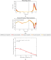
Progressive reduction of hamstring force (a). Anterior–posterior displacement of the tibia (b) during the gait cycle in the five simulated conditions. Values are positive for the anterior displacement. Dashed curve represents the intact knee condition. The gray vertical line refers to the toe–off event, marking the end of the stance phase and the beginning of the swing phase. Posterior tibial displacement average reduction with respect to “PCL15%-Ham100%” condition calculated in the second half of the swing phase (c), resulting from each simulated condition. In red, the polynomial regression function.
Figure 10
Figure 10
Tension of deep Medial Collateral Ligament (a), superficial Medial Collateral Ligament (b), Lateral Collateral Ligament (c) along the gait cycle in the five simulated conditions. Dashed curve represents the intact knee condition. The gray vertical line refers to the toe-off event, marking the end of the stance phase and the beginning of the swing phase.
Figure 11
Figure 11
Tibial–femoral contact force in the second half of the swing phase, resulting from the five simulated conditions. Dashed curve represents the intact knee condition.

References

    1. Majewski M., Susanne H., Klaus S. Epidemiology of Athletic Knee Injuries: A 10-Year Study. Knee. 2006;13:184–188. doi: 10.1016/j.knee.2006.01.005. - DOI - PubMed
    1. Hassebrock J.D., Gulbrandsen M.T., Asprey W.L., Makovicka J.L., Chhabra A. Knee Ligament Anatomy and Biomechanics. Sports Med. Arthrosc. Rev. 2020;28:80–86. doi: 10.1097/JSA.0000000000000279. - DOI - PubMed
    1. Grood E.S., Stowers S.F., Noyes F.R. Limits of Movement in the Human Knee. Effect of Sectioning the Posterior Cruciate Ligament and Posterolateral Structures. J. Bone Jt. Surg. 1988;70:88–97. doi: 10.2106/00004623-198870010-00014. - DOI - PubMed
    1. Logterman S.L., Wydra F.B., Frank R.M. Posterior Cruciate Ligament: Anatomy and Biomechanics. Curr. Rev. Musculoskelet. Med. 2018;11:510–514. doi: 10.1007/s12178-018-9492-1. - DOI - PMC - PubMed
    1. Gollehon D.L., Torzilli P.A., Warren R.F. The Role of the Posterolateral and Cruciate Ligaments in the Stability of the Human Knee. A Biomechanical Study. J. Bone Jt. Surg. 1987;69:233–242. doi: 10.2106/00004623-198769020-00010. - DOI - PubMed