. 2023 Oct 1;11(10):2692.
doi: 10.3390/biomedicines11102692.

YAP/ACSL4 Pathway-Mediated Ferroptosis Promotes Renal Fibrosis in the Presence of Kidney Stones

Affiliations

YAP/ACSL4 Pathway-Mediated Ferroptosis Promotes Renal Fibrosis in the Presence of Kidney Stones

Lei Li et al. Biomedicines. .

Abstract

The potential association between calcium oxalate stones and renal fibrosis has been extensively investigated; however, the underlying mechanisms remain unclear. Ferroptosis is a novel form of cell death characterized by iron-dependent lipid peroxidation and regulated by acyl coenzyme A synthase long-chain family member 4 (ACSL4). Yes-associated protein (YAP), a transcriptional co-activator in the Hippo pathway, promotes ferroptosis by modulating ACSL4 expression. Nevertheless, the involvement of YAP-ACSL4 axis-mediated ferroptosis in calcium oxalate crystal deposition-induced renal fibrosis and its molecular mechanisms have not been elucidated. In this study, we investigated ACSL4 expression and ferroptosis activation in the kidney tissues of patients with calcium oxalate stones and in mice using single-cell sequencing, transcriptome RNA sequencing, immunohistochemical analysis, and Western blot analysis. In vivo and in vitro experiments demonstrated that inhibiting ferroptosis or ACSL4 mitigated calcium oxalate crystal-induced renal fibrosis. Furthermore, YAP expression was elevated in the kidney tissues of patients with calcium oxalate stones and in calcium oxalate crystal-stimulated human renal tubular epithelial cell lines. Mechanistically, in calcium oxalate crystal-stimulated human renal tubular epithelial cell lines, activated YAP translocated to the nucleus and enhanced ACSL4 expression, consequently inducing cellular ferroptosis. Moreover, YAP silencing suppressed ferroptosis by downregulating ACSL4 expression, thereby attenuating calcium oxalate crystal-induced renal fibrosis. Conclusively, our findings suggest that YAP-ACSL4-mediated ferroptosis represents an important mechanism underlying the induction of renal fibrosis by calcium oxalate crystal deposition. Targeting the YAP-ACSL4 axis and ferroptosis may therefore hold promise as a potential therapeutic approach for preventing renal fibrosis in patients with kidney stones.

Keywords: YAP–ACSL4 axis; calcium oxalate crystal; ferroptosis; kidney stone; renal fibrosis.


Conflict of interest statement

The authors declare that they have no competing interest.

Figures
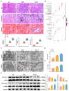
Figure 1
Figure 1
Ferroptosis is activated in mice with CaOx kidney stones. (A) HE, Von Kossa and Masson staining results to evaluate tubular damage, CaOx crystals and collagen fibrillation deposition (n = 5). The scale bar represents 50 µm. (B) GO enrichment analysis. (C) TEM showed shrunken mitochondria with outer membrane ruptured (indicated by red arrows) in mice with CaOx kidney stones. The scale bar represents 1 µm. (D,E) BUN and Scr levels of kidney tissue (n = 5). (F) Western blot analysis showed the expressions of SLC7A11, GPX4, fibronectin, and α-SMA in kidney tissues and quantification by densitometry. Data were presented as mean ± SD; * p < 0.05, ** p < 0.01, *** p < 0.001 vs. control group.
Figure 2
Figure 2
Inhibition of ferroptosis alleviates renal fibrosis. (AE) Measurement of the levels of BUN, Scr, GSH, CAT, and MDA in the serum of mice (n = 5). (F) HE, Von Kossa and Masson staining results to evaluate tubular damage, CaOx crystals and collagen fibrillation deposition (n = 5). The scale bar represents 50 µm. (G) Immunofluorescence staining analysis of SLC7A11, GPX4 and α-SMA expressions in mouse kidney tissues and semi-quantitative analysis (n = 5). The scale bar represents 50 µm. (H) Western blot analysis showed the expressions of SLC7A11, GPX4, fibronectin and α-SMA in kidney tissues and quantification by densitometry. Data were presented as mean ± SD; ** p < 0.01, *** p < 0.001 vs. control group; # p < 0.05, ## p < 0.01 vs. Gly group.
Figure 3
Figure 3
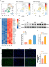
ACSL4 expression is upregulated in CaOx kidney stones. (A) Single-cell sequencing database of ACSL4 expression in normal subjects and patients with CKD. (B) RNA sequencing analysis of the transcriptomes of control and Gly group mice, red represents highly expressed genes and blue represents low expressed genes (n = 3). (C) Western blot analysis showed the expressions of ACSL4 expression in kidney tissues and quantification by densitometry (n = 5). (D,E) qPCR and immunofluorescence analysis of ACSL4 expression in the kidney of normal and kidney stone patients and semi-quantitative analysis (n = 6). The scale bar represents 50 µm. Data were presented as mean ± SD; ** p < 0.01, *** p < 0.001 vs. control group.
Figure 4
Figure 4
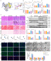
ACSL4-induced ferroptosis promotes renal fibrosis in mice with CaOx kidney stones. (A) Overall docking plot of abemaciclib and ACSL4. (B) Body weight changes in mice (n = 5). (CG) Measurement of the levels of BUN, Scr, GSH, CAT, and MDA in the serum of mice (n = 5). (H) HE, Von Kossa, Masson, and Perls staining to assess tubular damage, CaOx crystals, collagen fibrillation and iron deposition (n = 5). The scale bar represents 50 µm. (I) Western blot analysis showed the expressions of ACSL4, P53, SLC7A11, GPX4, fibronectin, and α-SMA in mouse kidney tissues and quantification by densitometry. (J) Immunofluorescence staining analysis of ACSL4, SLC7A11, GPX4, and α-SMA expressions in mouse kidney tissues and semi-quantitative analysis (n = 5). The scale bar represents 50 µm. (K) TEM revealed mitochondrial damage (indicated by red arrows) in mice with CaOx kidney stones. The scale bar represents 1 µm. Data were presented as mean ± SD; * p < 0.05, ** p < 0.01, *** p < 0.001 vs. control group; # p < 0.05, ## p < 0.01 vs. Gly group.
Figure 5
Figure 5
ACSL4-induced ferroptosis promotes COM-induced fibrosis in HK-2 cells. (A) Western blot analysis showed the expressions of ACSL4, GPX4, fibronectin and α-SMA after stimulation of HK-2 cells with different concentrations of COM and quantification by densitometry. (B) Cell viability of HK-2 under different concentrations of COM stimulation. * p < 0.05, ** p < 0.01, *** p < 0.001 vs. control group. (C) Western blots validated ACSL4-siRNA knockdown efficiency. ** p < 0.01 vs. COM group. (D) Western blot analysis showed the expressions of P53, SLC7A11, GPX4, fibronectin and Collagen I after knockdown of ACSL4 and quantification by densitometry. (E) Immunofluorescence analysis of SLC7A11, GPX4 and α-SMA expressions in HK-2 cells after knockdown of ACSL4 and semi-quantitative analysis. The scale bar represents 50 µm. (F) Measurement of lipid peroxidation in HK-2 cells by the C11 BODIPY581/591 fluorescent probe. The scale bar represents 50 µm. * p < 0.05, ** p < 0.01, *** p < 0.001 vs. Con-siRNA group; # p < 0.05, ## p < 0.01, ### p < 0.001 vs. Con-siRNA + COM group. Data were presented as mean ± SD.
Figure 6
Figure 6
YAP is involved in regulating the expression of ACSL4 transcript levels, and inhibition of YAP inhibits ACSL4-mediated ferroptosis and reverses renal fibrosis. (A) Western blots validated YAP-siRNA knockdown efficiency. * p < 0.05 vs. COM group.(B) Western blot analysis showed the expressions of ACSL4, SLC7A11, GPX4, fibronectin, and Collagen I after knockdown of YAP and quantification by densitometry. (C) Measurement of changes in intracellular ROS in HK-2 cells after knockdown of YAP by flow cytometry. (D) Immunofluorescence analysis of ACSL4, SLC7A11, and GPX4 expressions in HK-2 cells following knockdown of YAP and semi-quantitative analysis. The scale bar represents 50 µm. * p < 0.05, ** p < 0.01, *** p < 0.001 vs. Con-siRNA group; # p < 0.05, ## p < 0.01 vs. Con-siRNA + COM group. (E) Western blot analysis showed the expressions of SLC7A11, GPX4, fibronectin, and Collagen I expressions after overexpression of YAP, with or without ACSL4-siRNA and quantification by densitometry. * p < 0.05, ** p < 0.01 vs. COM group; # p < 0.05 vs. ACSL4-siRNA + COM group; ns, no statistical significance. Data were presented as mean ± SD.
Figure 7
Figure 7
Inhibition of YAP mediates the downregulation of ACSL4 expression which, in turn, inhibits CaOx crystal-induced ferroptosis and renal fibrosis.

References

    1. Singh P., Harris P.C., Sas D.J., Lieske J.C. The genetics of kidney stone disease and nephrocalcinosis. Nat. Rev. Nephrol. 2022;18:224–240. doi: 10.1038/s41581-021-00513-4. - DOI - PubMed
    1. Zisman A.L. Effectiveness of Treatment Modalities on Kidney Stone Recurrence. Clin. J. Am. Soc. Nephrol. 2017;12:1699–1708. doi: 10.2215/CJN.11201016. - DOI - PMC - PubMed
    1. Khan S.R., Pearle M.S., Robertson W.G., Gambaro G., Canales B.K., Doizi S., Traxer O., Tiselius H.-G. Kidney stones. Nat. Rev. Dis. Primers. 2016;2:16008. doi: 10.1038/nrdp.2016.8. - DOI - PMC - PubMed
    1. Lu H., Sun X., Jia M., Sun F., Zhu J., Chen X., Chen K., Jiang K. Rosiglitazone Suppresses Renal Crystal Deposition by Ameliorating Tubular Injury Resulted from Oxidative Stress and Inflammatory Response via Promoting the Nrf2/HO-1 Pathway and Shifting Macrophage Polarization. Oxid. Med. Cell Longev. 2021;2021:5527137. doi: 10.1155/2021/5527137. - DOI - PMC - PubMed
    1. Khan S.R., Canales B.K., Dominguez-Gutierrez P.R. Dominguez-Gutierrez PR. Randall’s plaque and calcium oxalate stone formation: Role for immunity and inflammation. Nat. Rev. Nephrol. 2021;17:417–433. doi: 10.1038/s41581-020-00392-1. - DOI - PubMed

LinkOut - more resources