. 2023 Oct 16;11(10):2801.
doi: 10.3390/biomedicines11102801.

Identifying Potent Nonsense-Mediated mRNA Decay Inhibitors with a Novel Screening System

Affiliations

Identifying Potent Nonsense-Mediated mRNA Decay Inhibitors with a Novel Screening System

Julie Carrard et al. Biomedicines. .

Abstract

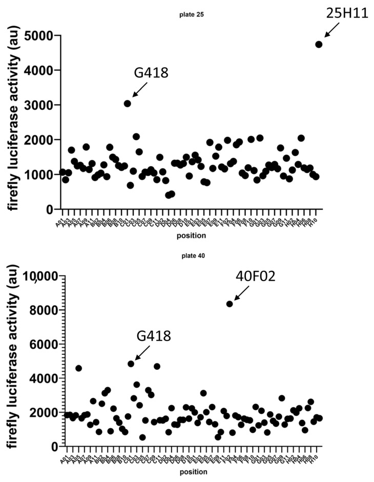
Nonsense-mediated mRNA decay (NMD) is a quality control mechanism that degrades mRNAs carrying a premature termination codon. Its inhibition, alone or in combination with other approaches, could be exploited to develop therapies for genetic diseases caused by a nonsense mutation. This, however, requires molecules capable of inhibiting NMD effectively without inducing toxicity. We have built a new screening system and used it to identify and validate two new molecules that can inhibit NMD at least as effectively as cycloheximide, a reference NMD inhibitor molecule. These new NMD inhibitors show no cellular toxicity at tested concentrations and have a working concentration between 6.2 and 12.5 µM. We have further validated this NMD-inhibiting property in a physiopathological model of lung cancer in which the TP53 gene carries a nonsense mutation. These new molecules may potentially be of interest in the development of therapies for genetic diseases caused by a nonsense mutation.

Keywords: genetic disease; nonsense mutation; nonsense-mediated mRNA decay; small molecules; therapy.


Conflict of interest statement

The authors declare no conflict of interest.

Figures

Figure 1
Figure 1
Identification via screening of two new potential NMD inhibitors. Scatter plot of luciferase activities measured in the different wells. Luciferase activity measurements related to incubation with molecules 1a and 2a are indicated by arrows. G418-related luciferase activity is also shown. G418 serves as positive control. The higher the luciferase activity, the greater the NMD-inhibiting potential of the studied compound.
Figure 2
Figure 2
The one-plasmid screening system for identifying NMD inhibitors. (A) Schematic representation of the construct used to generate an mRNA subject to NMD because of the presence of an intron in the 3′UTR. Exons are represented by rectangles and introns by lines. The 5′UTR and 3′UTR are in red. The firefly luciferase reading frame is in purple. The AUG initiation codon and the STOP codon are indicated. (B) Verification of proper construct splicing. Analysis of firefly luciferase DNA amplification from the DNA by radioactive PCR (rightmost lane) and from the mRNA by radioactive RT-PCR. The different isoforms are indicated to the right of the gel. (C) Bar plot showing the extent of NMD inhibition (luciferase activity) measured in U2OS cells transfected with the construct described in A and incubated with derivatives of molecule 1a (left) or molecule 2a (right). (D) Minimal structural skeleton of 1a-family derivatives liable to promote NMD-inhibiting activity.
Figure 3
Figure 3
Compared NMD inhibition efficacies of different confirmed NMD inhibitors and molecules 1a and 2a. U2OS cells were transfected with the construct depicted in Figure 2A. Each molecule was tested at 0, 0.4, 0.8, 1.6, 3.1, 6.2, 12.5, 25, 50, 100, 200, and 400 µM to establish a dose–response curve. The results presented are representative of two independent experiments.
Figure 4
Figure 4
Molecules 1a and 2a show no significant toxicity at the doses tested. Adenylate kinase activity was measured in the culture supernatant of 16HBE14o- cells after 24 h of exposure to different concentrations of each molecule separately (working concentration, half the working concentration, and twice the working concentration). Cycloheximide was used at 0.5 mg/mL as a positive control. The results shown are representative of two independent experiments. p-values were calculated with Student’s t-test (* < 0.05). (red bar). Green bar corresponds to the no treatment condition (0).
Figure 5
Figure 5
Molecules 1a and 2a inhibit NMD of endogenous p53 mRNA in Calu-6 cells. Calu-6 cells were incubated with molecule 1a, molecule 2a, DMSO, or cycloheximide (CHX) as positive control at 0.5 mg/mL. The upper panel shows a quantitative RT-PCR analysis gel, and the lower panel shows the ratio of p53 mRNA to GAPDH mRNA, normalized with respect to the results obtained with DMSO. The results are representative of two independent experiments.

References

    1. Mort M., Ivanov D., Cooper D.N., Chuzhanova N.A. A meta-analysis of nonsense mutations causing human genetic disease. Hum. Mutat. 2008;29:1037–1047. doi: 10.1002/humu.20763. - DOI - PubMed
    1. Yi Z., Sanjeev M., Singh G. The Branched Nature of the Nonsense-Mediated mRNA Decay Pathway. Trends Genet. 2021;37:143–159. doi: 10.1016/j.tig.2020.08.010. - DOI - PMC - PubMed
    1. Nogueira G., Fernandes R., Garcia-Moreno J.F., Romao L. Nonsense-mediated RNA decay and its bipolar function in cancer. Mol. Cancer. 2021;20:72. doi: 10.1186/s12943-021-01364-0. - DOI - PMC - PubMed
    1. Sun L., Mailliot J., Schaffitzel C. Nonsense-Mediated mRNA Decay Factor Functions in Human Health and Disease. Biomedicines. 2023;11:722. doi: 10.3390/biomedicines11030722. - DOI - PMC - PubMed
    1. Popp M.W., Maquat L.E. Nonsense-mediated mRNA Decay and Cancer. Curr. Opin. Genet. Dev. 2018;48:44–50. doi: 10.1016/j.gde.2017.10.007. - DOI - PMC - PubMed