. 2023 Oct 19;11(10):2836.
doi: 10.3390/biomedicines11102836.

Ultrasound-Driven Healing: Unleashing the Potential of Chondrocyte-Derived Extracellular Vesicles for Chondrogenesis in Adipose-Derived Stem Cells

Affiliations

Ultrasound-Driven Healing: Unleashing the Potential of Chondrocyte-Derived Extracellular Vesicles for Chondrogenesis in Adipose-Derived Stem Cells

Yikai Wang et al. Biomedicines. .

Abstract

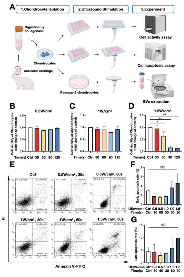
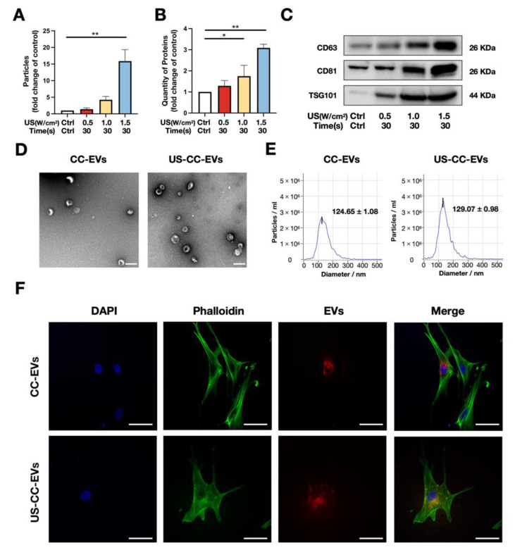
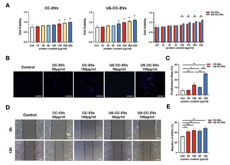
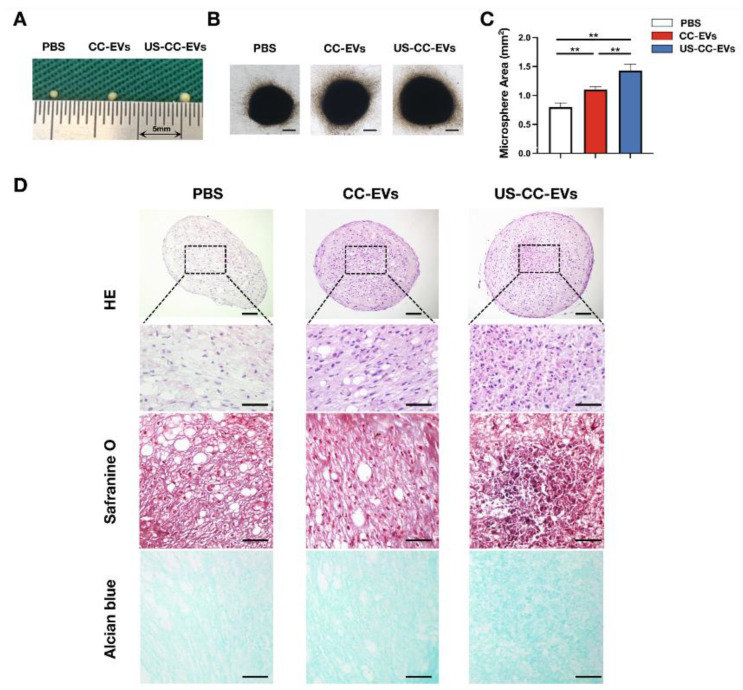
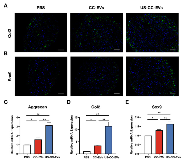
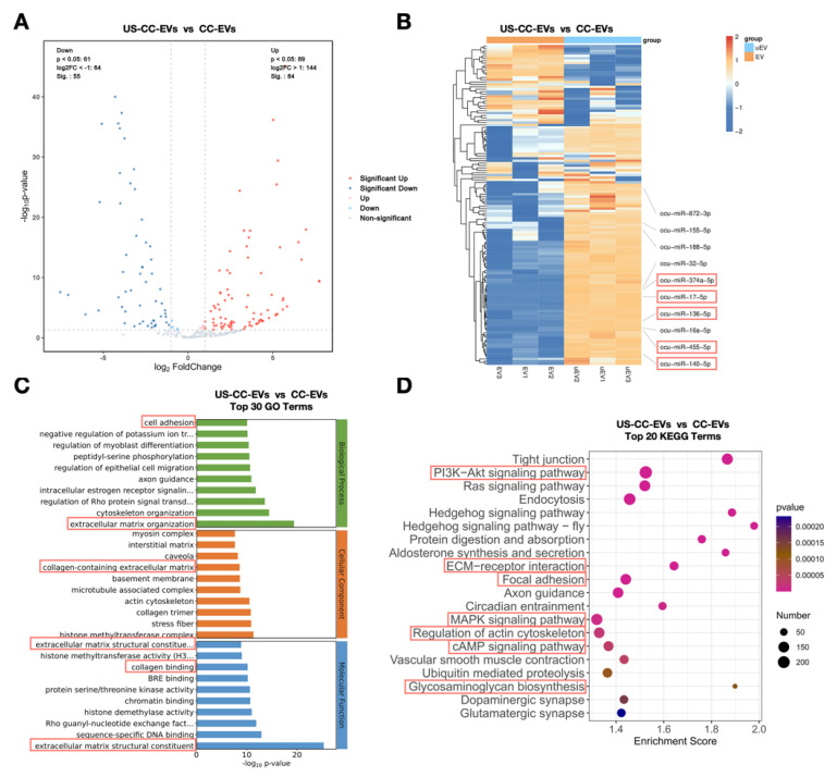
Repairing cartilage defects represents a significant clinical challenge. While adipose-derived stem cell (ADSC)-based strategies hold promise for cartilage regeneration, their inherent chondrogenic potential is limited. Extracellular vesicles (EVs) derived from chondrocytes (CC-EVs) have shown potential in enhancing chondrogenesis, but their role in promoting chondrogenic differentiation of ADSCs remains poorly understood. Moreover, the clinical application of EVs faces limitations due to insufficient quantities for in vivo use, necessitating the development of effective methods for extracting significant amounts of CC-EVs. Our previous study demonstrated that low-intensity ultrasound (LIUS) stimulation enhances EV secretion from mesenchymal stem cells. Here, we identified a specific LIUS parameter for chondrocytes that increased EV secretion by 16-fold. CC-EVs were found to enhance cell activity, proliferation, migration, and 21-day chondrogenic differentiation of ADSCs in vitro, while EVs secreted by chondrocytes following LIUS stimulation (US-CC-EVs) exhibited superior efficacy. miRNA-seq revealed that US-CC-EVs were enriched in cartilage-regeneration-related miRNAs, contributing to chondrogenesis in various biological processes. In conclusion, we found that CC-EVs can enhance the chondrogenesis of ADSCs in vitro. In addition, our study introduces ultrasound-driven healing as an innovative method to enhance the quantity and quality of CC-EVs, meeting clinical demand and addressing the limited chondrogenic potential of ADSCs. The ultrasound-driven healing unleashes the potential of CC-EVs for chondrogenesis possibly through the enrichment of cartilage-regeneration-associated miRNAs in EVs, suggesting their potential role in cartilage reconstruction. These findings hold promise for advancing cartilage regeneration strategies and may pave the way for novel therapeutic interventions in regenerative medicine.

Keywords: adipose-derived stem cells; cartilage regeneration; chondrocyte-derived extracellular vesicles; chondrogenesis; low-intensity ultrasound.


Conflict of interest statement

The authors declare no conflict of interest.

Figures

Figure 1
Figure 1
Optimization of low-intensity ultrasound parameters. (A) Illustration of the experiment. (BD) Chondrocyte viability after different LIUS stimulation parameters on day 3 using Cell Counting Kit-8 (n = 3). (E) Chondrocyte apoptosis following LIUS stimulation at 48 h detected by flow cytometry. (F,G) Analysis of chondrocyte apoptosis; (F) early apoptosis rate (n = 3); (G) late apoptosis rate (n = 3). * p < 0.05; ** p < 0.01; NS: no significant difference.
Figure 2
Figure 2
Low-intensity ultrasound stimulation promoted chondrocyte extracellular release. (A) Particle numbers of EVs following different ultrasound stimulation parameters detected by NTA (n = 3). (B) Protein quantity of EVs following different ultrasound stimulation parameters detected by BCA protein assay (n = 3). (C) Western blotting for EV-related markers CD63, CD81, and TSG101. (D) Morphological characterization of the EVs derived from chondrocytes (CC-EVs) without ultrasound stimulation and EVs derived from chondrocytes with 1.5 W/cm2 and 30 s ultrasound stimulation (US-CC-EVs) via transmission electron microscopy; scale bar = 200 nm; (E) Particle diameter distribution of CC-EVs (median 124.65 ± 1.08 nm) and US-CC-EVs (median 129.07 ± 0.98 nm) detected by NTA; (F) Uptake of CC-EVs and US-CC-EVs in ADSCs detected by confocal microscopy; scale bar = 50 μm. NTA: nanoparticle tracking analysis; * p < 0.05; ** p < 0.01.
Figure 3
Figure 3
CC-EVs and US-CC-EVs promoted cell activity, proliferation, and migration of ADSCs. (A) Cell viability of ADSCs incubated with different concentrations of CC-EVs and US-CC-EVs using Cell Counting Kit-8 (n = 3). (B) Cell proliferation as determined by the EdU assay; EdU-positive ADSCs (green) and cell nucleus (blue) were observed using fluorescence microscopy. Scale bar = 500 μm. (C) ADSCs proliferation rate (n = 4). (D) Wound healing assay of ADSCs observed using a light microscope; scale bar = 200 μm. (E) ADSCs migration rate (n = 3). * p < 0.05; ** p < 0.01.
Figure 4
Figure 4
CC-EVs and US-CC-EVs promoted the chondrogenic differentiation of ADSCs. (A) Macroscopic photo. (B) Microscopic photo; scale bar = 250 μm. (C) Area measured by a light microscope using ImageJ (n = 3). (D) Histological assessment by Hematoxylin and Eosin (H&E), Safranine O, Alcian blue staining at 21 days of chondrogenic differentiation in micromass culture of ADSCs; scale bar = 200 μm (first row); scale bar = 100 μm (last three rows). ** p < 0.01.
Figure 5
Figure 5
CC-EVs and US-CC-EVs promoted the chondrogenic differentiation of ADSCs. (A,B) Immunofluorescence images of Col2 and Sox9 protein accumulation; scale bar = 200 μm. (CE) mRNA expression levels of Aggrecan, Col2, and Sox9 were measured by qPCR at 21 days of chondrogenic differentiation in micromass culture of ADSCs. * p < 0.05; ** p < 0.01.
Figure 6
Figure 6
US-CC-EVs exhibited an enrichment of miRNAs associated with chondrogenic differentiation after low-intensity ultrasound stimulation. (A) Volcano plot of differentially expressed miRNAs between US-CC-EVs and CC-EVs (n = 3). Red dots: significantly upregulated miRNAs; blue dots: significantly downregulated miRNAs. (B) Hierarchical clustering analysis of differentially expressed miRNAs (|log2foldchange| > 1 and q-value < 0.05; unreported miRNAs were omitted) between US-CC-EVs and CC-EVs. Rows represent miRNAs; columns represent individual replicates. The top ten miRNAs with the high fold change are annotated, and miRNAs associated with chondrogenic differentiation are highlighted. (C) Gene Ontology (GO) analysis of differentially expressed genes predicted based on the differentially expressed miRNAs. Terms associated with chondrogenic differentiation are highlighted. (D) Kyoto Encyclopedia of Genes and Genomes (KEGG) pathway analysis of differentially expressed genes predicted based on the differentially expressed miRNAs. Terms associated with chondrogenic differentiation are highlighted.

Similar articles

Cited by

References

    1. Lories R.J., Luyten F.P. The Bone-Cartilage Unit in Osteoarthritis. Nat. Rev. Rheumatol. 2011;7:43–49. doi: 10.1038/nrrheum.2010.197. - DOI - PubMed
    1. Muthu S., Korpershoek J.V., Novais E.J., Tawy G.F., Hollander A.P., Martin I. Failure of Cartilage Regeneration: Emerging Hypotheses and Related Therapeutic Strategies. Nat. Rev. Rheumatol. 2023;19:403–416. doi: 10.1038/s41584-023-00979-5. - DOI - PubMed
    1. Bartlett W., Skinner J.A., Gooding C.R., Carrington R.W.J., Flanagan A.M., Briggs T.W.R., Bentley G. Autologous chondrocyte implantation versus matrix-induced autologous chondrocyte implantation for osteochondral defects of the knee: A Prospective, Randomised Study. J. Bone Jt. Surg. Br. Vol. 2005;87-B:640–645. doi: 10.1302/0301-620X.87B5.15905. - DOI - PubMed
    1. Liu Y., Dzidotor G., Le T.T., Vinikoor T., Morgan K., Curry E.J., Das R., McClinton A., Eisenberg E., Apuzzo L.N., et al. Exercise-Induced Piezoelectric Stimulation for Cartilage Regeneration in Rabbits. Sci. Transl. Med. 2022;14:eabi7282. doi: 10.1126/scitranslmed.abi7282. - DOI - PubMed
    1. Armiento A.R., Stoddart M.J., Alini M., Eglin D. Biomaterials for Articular Cartilage Tissue Engineering: Learning from Biology. Acta Biomater. 2018;65:1–20. doi: 10.1016/j.actbio.2017.11.021. - DOI - PubMed

LinkOut - more resources