

Attenuation of Immunogenicity in MOG-Induced Oligodendrocytes by the Probiotic Bacterium Lactococcus Sp. PO3
- PMID: 37893449
- PMCID: PMC10608413
- DOI: 10.3390/medicina59101731
Attenuation of Immunogenicity in MOG-Induced Oligodendrocytes by the Probiotic Bacterium Lactococcus Sp. PO3
Abstract
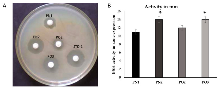
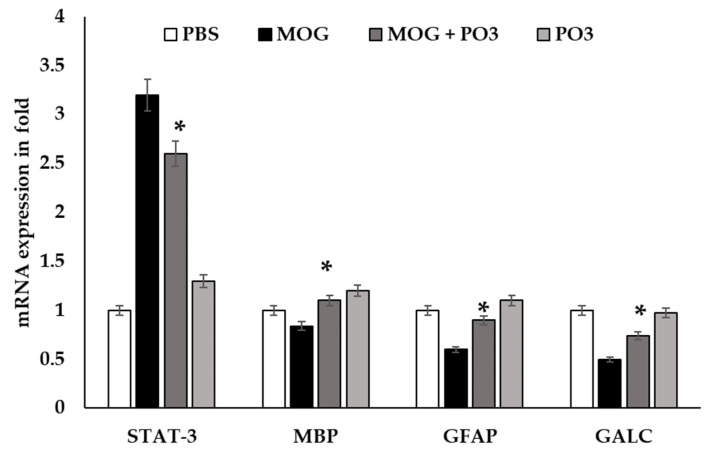
Background and Objectives: Milk is healthy and includes several vital nutrients and microbiomes. Probiotics in milk and their derivatives modulate the immune system, fight inflammation, and protect against numerous diseases. The present study aimed to isolate novel bacterial species with probiotic potential for neuroinflammation. Materials and Methods: Six milk samples were collected from lactating dairy cows. Bacterial isolates were obtained using standard methods and were evaluated based on probiotic characteristics such as the catalase test, hemolysis, acid/bile tolerance, cell adhesion, and hydrophobicity, as well as in vitro screening. Results: Nine morphologically diverse bacterial isolates were found in six different types of cow's milk. Among the isolates, PO3 displayed probiotic characteristics. PO3 was a Gram-positive rod cell that grew in an acidic (pH-2) salty medium containing bile salt and salinity (8% NaCl). PO3 also exhibited substantial hydrophobicity and cell adhesion. The sequencing comparison of the 16S rRNA genes revealed that PO3 was Lactococcus raffinolactis with a similarity score of 99.3%. Furthermore, PO3 was assessed for its neuroanti-inflammatory activity on human oligodendrocyte (HOG) cell lines using four different neuroimmune markers: signal transducer and activator of transcription (STAT-3), myelin basic protein (MBP), glial fibrillary acidic protein (GFAP), and GLAC in HOG cell lines induced by MOG. Unlike the rest of the evaluated neuroimmune markers, STAT-3 levels were elevated in the MOG-treated HOG cell lines compared to the untreated ones. The expression level of STAT-3 was attenuated in both PO3-MOG-treated and only PO3-treated cell lines. On the contrary, in PO3-treated cell lines, MBP, GFAP, and GLAC were significantly expressed at higher levels when compared with the MOG-treated cell lines. Conclusions: The findings reported in this article are to be used as a foundation for further in vivo research in order to pave the way for the possible use of probiotics in the treatment of neuroinflammatory diseases, including multiple sclerosis.
Keywords: Lactococcus; cow’s milk; neuroinflammatory diseases; oligodendrocyte; probiotics.
Conflict of interest statement
The authors declare no conflict of interest.
Figures


Similar articles
-
In vitro and genetic screening of probiotic properties of lactic acid bacteria isolated from naturally fermented cow-milk and yak-milk products of Sikkim, India.World J Microbiol Biotechnol. 2022 Jan 6;38(2):25. doi: 10.1007/s11274-021-03215-y. World J Microbiol Biotechnol. 2022. PMID: 34989904
-
Functional probiotics of lactic acid bacteria from Hu sheep milk.BMC Microbiol. 2020 Jul 28;20(1):228. doi: 10.1186/s12866-020-01920-6. BMC Microbiol. 2020. PMID: 32723292 Free PMC article.
-
Bioprophylactic potential of novel human colostrum probiotics via apoptotic induction of colon cancer cells and cell immune activation.Biomed Pharmacother. 2022 May;149:112871. doi: 10.1016/j.biopha.2022.112871. Epub 2022 Mar 29. Biomed Pharmacother. 2022. PMID: 35364380
-
Molecular identification of mesophilic and psychrotrophic bacteria from raw cow's milk.Food Microbiol. 2009 Apr;26(2):228-31. doi: 10.1016/j.fm.2008.09.005. Epub 2008 Oct 11. Food Microbiol. 2009. PMID: 19171267
-
Functional Characterization of Lactobacillus plantarum Isolated from Cow Milk and the Development of Fermented Coconut and Carrot Juice Mixed Beverage.Curr Microbiol. 2023 Mar 15;80(5):139. doi: 10.1007/s00284-023-03258-4. Curr Microbiol. 2023. PMID: 36920622
Cited by
-
Blood Microbiome Analysis Reveals Biomarkers of Treatment Response in Drug-Naïve Patients with First-Episode Psychosis: A Pilot Study.Microorganisms. 2025 Aug 19;13(8):1935. doi: 10.3390/microorganisms13081935. Microorganisms. 2025. PMID: 40871439 Free PMC article.
References
-
- Houbad K., Bekada A.M.A., Homrani A., Djellid Y. Phenotypic and Genotypic Characterization of Lactococci Isolated from Different Kinds of Raw Milk (Goat, Cow, Sheep and Camel) Ukr. J. Ecol. 2022;12:45–60.
-
- Maślak E., Złoch M., Arendowski A., Sugajski M., Janczura I., Rudnicka J., Walczak-Skierska J., Buszewska-Forajta M., Rafińska K., Pomastowski P., et al. Isolation and Identification of Lactococcus lactis and Weissella cibaria Strains from Fermented Beetroot and an Investigation of Their Properties as Potential Starter Cultures and Probiotics. Foods. 2022;11:2257. doi: 10.3390/foods11152257. - DOI - PMC - PubMed
-
- Baig D.N., Mehnaz S. Probiotic Bacteria and Postbiotic Metabolites: Role in Animal and Human Health. Springer; Singapore: 2021. An Overview of Dairy Microflora; pp. 101–137.
-
- Abushelaibi A., Al-Mahadin S., El-Tarabily K., Shah N.P., Ayyash M. Characterization of Potential Probiotic Lactic Acid Bacteria Isolated from Camel Milk. LWT. 2017;79:316–325. doi: 10.1016/j.lwt.2017.01.041. - DOI
MeSH terms
Substances
Grants and funding
LinkOut - more resources
Full Text Sources
Miscellaneous
