. 2023 Sep 28;11(10):2422.
doi: 10.3390/microorganisms11102422.

SARS-CoV-2 Rapid Antigen Test Based on a New Anti-Nucleocapsid Protein Monoclonal Antibody: Development and Real-Time Validation

Affiliations

SARS-CoV-2 Rapid Antigen Test Based on a New Anti-Nucleocapsid Protein Monoclonal Antibody: Development and Real-Time Validation

Fabiana Fioravante Coelho et al. Microorganisms. .

Abstract

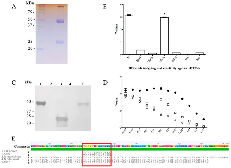
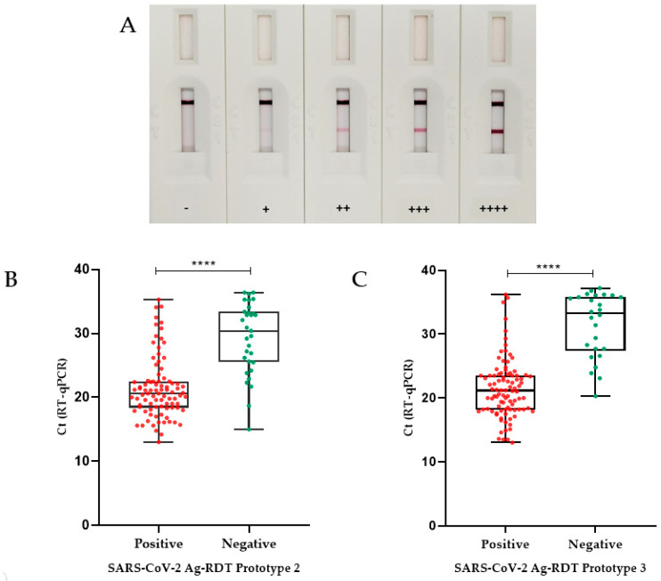
SARS-CoV-2 diagnostic tests have become an important tool for pandemic control. Among the alternatives for COVID-19 diagnosis, antigen rapid diagnostic tests (Ag-RDT) are very convenient and widely used. However, as SARS-CoV-2 variants may continuously emerge, the replacement of tests and reagents may be required to maintain the sensitivity of Ag-RDTs. Here, we describe the development and validation of an Ag-RDT during an outbreak of the Omicron variant, including the characterization of a new monoclonal antibody (anti-DTC-N 1B3 mAb) that recognizes the Nucleocapsid protein (N). The anti-DTC-N 1B3 mAb recognized the sequence TFPPTEPKKDKKK located at the C-terminus of the N protein of main SARS-CoV-2 variants of concern. Accordingly, the Ag-RDT prototypes using the anti-DTC-N 1B3 mAB detected all the SARS-CoV-2 variants-Wuhan, Alpha, Gamma, Delta, P2 and Omicron. The performance of the best prototype (sensitivity of 95.2% for samples with Ct ≤ 25; specificity of 98.3% and overall accuracy of 85.0%) met the WHO recommendations. Moreover, results from a patients' follow-up study indicated that, if performed within the first three days after onset of symptoms, the Ag-RDT displayed 100% sensitivity. Thus, the new mAb and the Ag-RDT developed herein may constitute alternative tools for COVID-19 point-of-care diagnosis and epidemiological surveillance.

Keywords: Ag-RDT development; IgG2b monoclonal antibody; SARS-CoV-2; diagnosis; follow-up study; nucleocapsid (N) antigen; validation.


Conflict of interest statement

The authors declare no conflict of interest.

Figures

Figure 1
Figure 1
Anti-DTC-N 1B3 mAb purification and characterization. (A) The 12% SDS/PAGE anti-DTC-N 1B3 mAb profile stained with Coomassie Blue after the G protein affinity column purification. (B) Anti-DTC-N 1B3 mAb isotyping analysis. C96 MaxiSorp ELISA microtiter plates coated with 10 µg/mL of recombinant rDTC-N or 1 µg/mL of IgG1, 2a, 2b, IgG3, IgA, and IgM, incubated with 100 µL supernatants of anti-DTC-N 1B3 mAb and with rat anti-mouse kappa conjugated with horseradish (1:1000). The asterisk (*) indicates statistically significant differences as compared to all the other isotypes of IgG, and IgA and IgM subclasses (p ≤ 0.0001). (C) Immunobloting analysis. Nitrocellulose membrane containing 10 µg of (1) DTC-N (SARS-CoV-2); (2) N-terminus protein N (SARS-CoV-2); (3) C-terminus protein N (SARS-CoV-2); (4) DENV-2 NS1 recombinant protein; (5) full-length control protein N (SARS-CoV-2). Membrane was probed with the anti-DTC-N 1B3 mAb (1:100) at a concentration of 870 µg/mL and goat anti-mouse IgG conjugated with peroxidase (1:5000). (D) Anti-DTC-N 1B3 recognition patter. The 10 µg/mL rDTC-N, heated-treated [100 °C for 10 min (□), DTT-treated (◇), or intact (●) were used as solid phase-bound antigens. The anti-DTC-N 1B3 mAb was serially diluted (log2) from an initial concentration of 0.007 μg/mL. (E) Alignments of the anti-DTC-N 1B3 mAb epitope with N protein sequences. The epitope sequence recognized by anti-DTC-N 1B3 mAb was aligned with protein N partial sequences, comprised between amino acids 352 and 434 from SARS-CoV-2, SARS, and with sequences obtained for the full-length N and N-terminal recombinant proteins.
Figure 2
Figure 2
Validation of Ag-RDT prototypes based on RT-qPCR. (A) Illustrative figures of Ag-RDT results. Figures show tests with different intensities of reaction as detected by the Ag-RDT. According to the signal strength of the positive samples on the test line (T), the results were classified as + (low intensity), ++ (moderate to low intensity), +++ (moderate to high intensity), and ++++ (high intensity). In the absence of the appearance of a test line (T), the sample was considered negative (−). (B) Box plot analysis of RT-qPCR and Ag-RDT prototype 2. RT-qPCR Ct values (for positive samples only) as compared with positive and negative results obtained with prototype 2 Ag-RDT. The median Ct value found for the false-negative samples on SARS-CoV-2 Ag-RDT (30.4) was significantly different from the median Ct value found for the positive samples on SARS-CoV-2 Ag-RDT (20.6). (C) Box plot analysis of RT-qPCR Ct value (positive only) compared with prototype 3 Ag-RDT results. The median Ct value found for the false-negative samples on SARS-CoV-2 Ag-RDT (33.2) was significantly different from the median Ct value found for the positive samples on SARS-CoV-2 Ag-RDT (21.1) (p < 0.0001 was indicated by ****). Boxplot analysis (2B and 2D) was performed by applying the Mann–Whitney test, using the GraphPad Prism 8.0.1 software. The line across the box is the median. The whiskers represent all points showing minimum to maximum quartiles.

Similar articles

Cited by

  • SARS-CoV-2 Genomic Surveillance from Community-Distributed Rapid Antigen Tests, Wisconsin, USA.
    Emmen IE, Vuyk WC, Lail AJ, Wolf S, O'Connor EJ, Dalvie R, Bhasin M, Virdi A, White C, Hassan NR, Richardson A, VanSleet G, Weiler A, Rounds-Dunn S, Van Horn K, Gartler M, Jorgenson J, Spelman M, Ottosen S, Minor NR, Wilson N, Friedrich TC, O'Connor DH. Emmen IE, et al. Emerg Infect Dis. 2025 May;31(13):61-69. doi: 10.3201/eid3113.241192. Emerg Infect Dis. 2025. PMID: 40359069 Free PMC article.

References

    1. WHO Coronavirus (COVID-19) Dashboard. [(accessed on 4 September 2023)]. Available online: https://covid19.who.int/
    1. Rahman S., Rahman M.M., Miah M., Begum M.N., Sarmin M., Mahfuz M., Hossain M.E., Rahman M.Z., Chisti M.J., Ahmed T. COVID-19 Reinfections among Naturally Infected and Vaccinated Individuals. Sci. Rep. 2022;12:1438. doi: 10.1038/s41598-022-05325-5. - DOI - PMC - PubMed
    1. Giovanetti M., Fonseca V., Wilkinson E., Tegally H., San E.J., Althaus C.L., Xavier J., Nanev Slavov S., Viala V.L., Ranieri Jerônimo Lima A. Replacement of the Gamma by the Delta Variant in Brazil: Impact of Lineage Displacement on the Ongoing Pandemic. Virus Evol. 2022;8:veac024. doi: 10.1093/ve/veac024. - DOI - PMC - PubMed
    1. Naveca F.G., Nascimento V., Souza V., Corado A.L., Nascimento F., Silva G., Mejía M.C., Brandão M.J., Costa Á., Duarte D. Spread of Gamma (P. 1) Sub-Lineages Carrying Spike Mutations Close to the Furin Cleavage Site and Deletions in the N-Terminal Domain Drives Ongoing Transmission of SARS-CoV-2 in Amazonas, Brazil. medRxiv. 2021;10:e02366-21. doi: 10.1128/spectrum.02366-21. - DOI - PMC - PubMed
    1. Baral P., Bhattarai N., Hossen M.L., Stebliankin V., Gerstman B.S., Narasimhan G., Chapagain P.P. Mutation-Induced Changes in the Receptor-Binding Interface of the SARS-CoV-2 Delta Variant B. 1.617. 2 and Implications for Immune Evasion. Biochem. Biophys. Res. Commun. 2021;574:14–19. doi: 10.1016/j.bbrc.2021.08.036. - DOI - PMC - PubMed

LinkOut - more resources