Review
. 2023 Oct 20;15(20):5077.
doi: 10.3390/cancers15205077.

The Prediction of Biological Features Using Magnetic Resonance Imaging in Head and Neck Squamous Cell Carcinoma: A Systematic Review and Meta-Analysis

Affiliations
Review

The Prediction of Biological Features Using Magnetic Resonance Imaging in Head and Neck Squamous Cell Carcinoma: A Systematic Review and Meta-Analysis

Hedda J van der Hulst et al. Cancers (Basel). .

Abstract

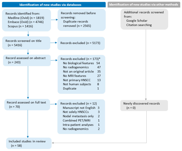
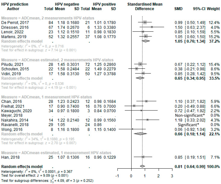
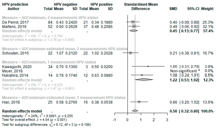
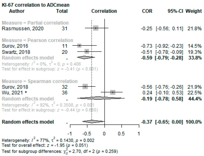
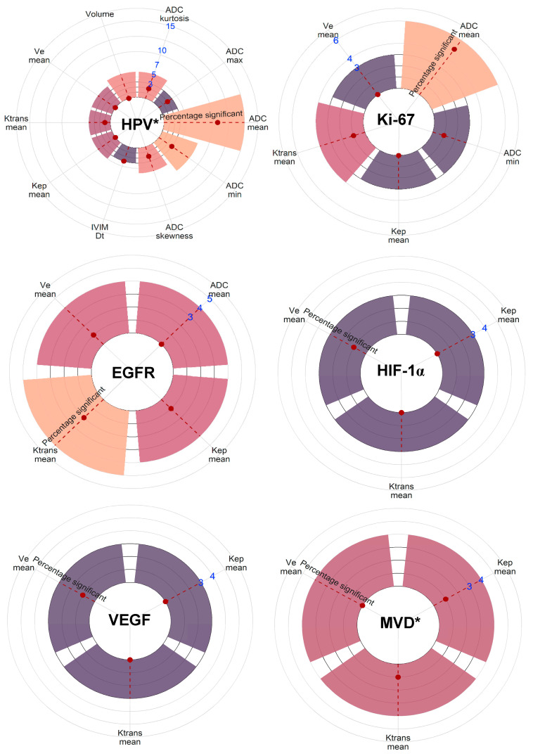
Magnetic resonance imaging (MRI) is an indispensable, routine technique that provides morphological and functional imaging sequences. MRI can potentially capture tumor biology and allow for longitudinal evaluation of head and neck squamous cell carcinoma (HNSCC). This systematic review and meta-analysis evaluates the ability of MRI to predict tumor biology in primary HNSCC. Studies were screened, selected, and assessed for quality using appropriate tools according to the PRISMA criteria. Fifty-eight articles were analyzed, examining the relationship between (functional) MRI parameters and biological features and genetics. Most studies focused on HPV status associations, revealing that HPV-positive tumors consistently exhibited lower ADCmean (SMD: 0.82; p < 0.001) and ADCminimum (SMD: 0.56; p < 0.001) values. On average, lower ADCmean values are associated with high Ki-67 levels, linking this diffusion restriction to high cellularity. Several perfusion parameters of the vascular compartment were significantly associated with HIF-1α. Analysis of other biological factors (VEGF, EGFR, tumor cell count, p53, and MVD) yielded inconclusive results. Larger datasets with homogenous acquisition are required to develop and test radiomic-based prediction models capable of capturing different aspects of the underlying tumor biology. Overall, our study shows that rapid and non-invasive characterization of tumor biology via MRI is feasible and could enhance clinical outcome predictions and personalized patient management for HNSCC.

Keywords: DCE; DWI; HIF-1α; HNSCC; HPV status; Ki-67 proliferation marker; MRI; radiogenomics.


Conflict of interest statement

The authors declare no conflict of interest.

Figures

Figure 1
Figure 1
Flow diagram of the study selection and exclusion process. No radiogenomics: Studies without analyses of biological features compared with MRI features. Combined PET/MRI: Studies focusing on combined PET/MRI parameters rather than stand-alone MRI parameters. Intra-patient analyses: Studies are limited to analyzing multiple biopsies within the same patient. * Studies can be excluded for more than one reason.
Figure 2
Figure 2
Forest plot of the mean ADC values with HPV status. * Limited data available and as reported in reference. Abbreviations: SD = standard deviation; SMD = standard mean difference; and CI = confidence interval [34,35,44,45,54,57,69,29,36,43,47,52,55,70,38].
Figure 3
Figure 3
Forest plot of the minimal ADC values with HPV status. * Limited data available and as reported in reference. Abbreviations: SD = standard deviation; SMD = standard mean difference; CI = confidence interval [34,45,57,43,47,52,38].
Figure 4
Figure 4
Forest plot of the correlation of mean ADC values with the Ki-67 proliferation index split using the correlation test applied. * This cohort consists solely of nasopharyngeal squamous cell carcinoma [60,61,65,63,71].
Figure 5
Figure 5
Visual representation of the number of reporting studies and the level of significance of all MRI parameters sorted by biological factor. MRI parameters are included when three or more studies have reported on associations with the specific metric and the biological factor. The count of studies is indicated by the blue numbers. The statistical significance as reported in the listed studies is indicated by the red dot on the red-striped line within the bars. * As grouped for all biological measurements defining the factor. Note: p53 lacked enough comparable studies.

References

    1. Becker M., Zaidi H. Imaging in head and neck squamous cell carcinoma: The potential role of PET/MRI. Br. J. Radiol. 2014;87:20130677. doi: 10.1259/bjr.20130677. - DOI - PMC - PubMed
    1. Leemans C.R., Snijders P.J.F., Brakenhoff R.H. The molecular landscape of head and neck cancer. Nat. Rev. Cancer. 2018;18:269–282. doi: 10.1038/nrc.2018.11. - DOI - PubMed
    1. Canning M., Guo G., Yu M., Myint C., Groves M.W., Byrd J.K., Cui Y. Heterogeneity of the Head and Neck Squamous Cell Carcinoma Immune Landscape and Its Impact on Immunotherapy. Front. Cell Dev. Biol. 2019;7:52. doi: 10.3389/fcell.2019.00052. - DOI - PMC - PubMed
    1. Stransky N., Egloff A.M., Tward A.D., Kostic A.D., Cibulskis K., Sivachenko A., Kryukov G.V., Lawrence M.S., Sougnez C., McKenna A., et al. The mutational landscape of head and neck squamous cell carcinoma. Science. 2011;333:1157–1160. doi: 10.1126/science.1208130. - DOI - PMC - PubMed
    1. Nishida N., Yano H., Nishida T., Kamura T., Kojiro M. Angiogenesis in cancer. Vasc. Health Risk Manag. 2006;2:213–219. doi: 10.2147/vhrm.2006.2.3.213. - DOI - PMC - PubMed