. 2023 Oct 12;28(20):7052.
doi: 10.3390/molecules28207052.

Endothelialization of Whey Protein Isolate-Based Scaffolds for Tissue Regeneration

Affiliations

Endothelialization of Whey Protein Isolate-Based Scaffolds for Tissue Regeneration

Hatice Genç et al. Molecules. .

Abstract

Background: Whey protein isolate (WPI) is a by-product from the dairy industry, whose main component is β-lactoglobulin. Upon heating, WPI forms a hydrogel which can both support controlled drug delivery and enhance the proliferation and osteogenic differentiation of bone-forming cells. This study makes a novel contribution by evaluating the ability of WPI hydrogels to support the growth of endothelial cells, which are essential for vascularization, which in turn is a pre-requisite for bone regeneration.

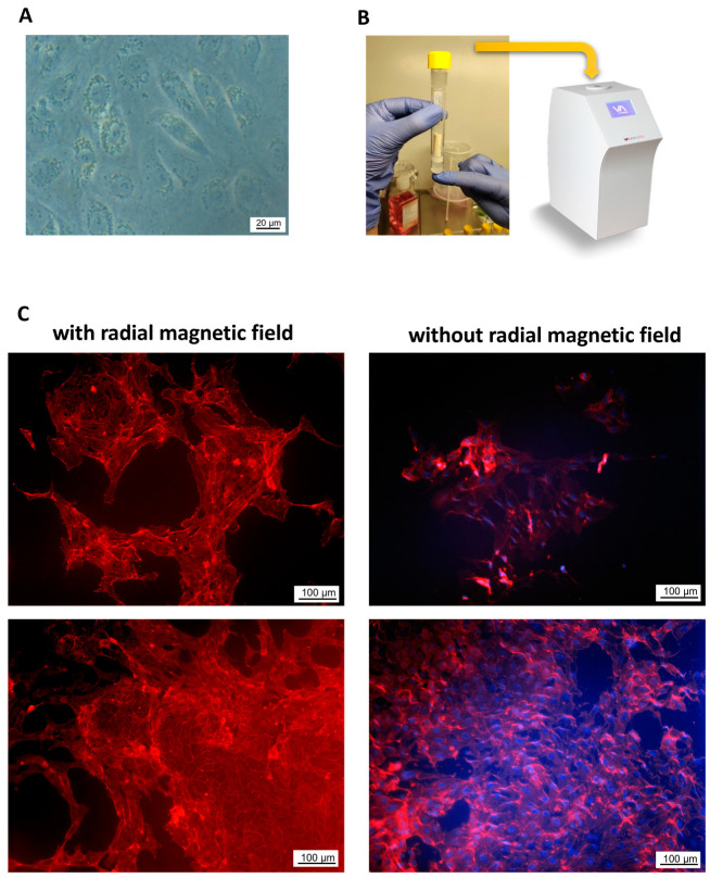
Methods: In this study, the proliferation and antioxidant levels in human umbilical vascular endothelial cells (HUVECs) cultured with WPI supplementation were evaluated using real-time cell analysis and flow cytometry. Further, the attachment and growth of HUVECs seeded on WPI-based hydrogels with different concentrations of WPI (15%, 20%, 30%, 40%) were investigated.

Results: Supplementation with WPI did not affect the viability or proliferation of HUVECs monitored with real-time cell analysis. At the highest used concentration of WPI (500 µg/mL), a slight induction of ROS production in HUVECs was detected as compared with control samples, but it was not accompanied by alterations in cellular thiol levels. Regarding WPI-based hydrogels, HUVEC adhered and spread on all samples, showing good metabolic activity. Notably, cell number was highest on samples containing 20% and 30% WPI.

Conclusions: The demonstration of the good compatibility of WPI hydrogels with endothelial cells in these experiments is an important step towards promoting the vascularization of hydrogels upon implantation in vivo, which is expected to improve implant outcomes in the future.

Keywords: 3D cell seeding; endothelial cell compatibility; hydrogels; thiol levels; tubular scaffolds; whey protein.


Conflict of interest statement

The authors declare no conflict of interest.

Figures

Figure A1
Figure A1
Morphology of primary HUVECs grown on WPI hydrogels for 7 days.
Figure 1
Figure 1
Effects of WPI supplementation on primary HUVECs. Graph shows the results of impedance-based real-time monitoring of Cell Index, reflecting cell number, attachment and viability. HUVECs were treated with the indicated concentrations of WPI and monitored for 48 h after WPI administration (n = 3 independent experiments, each with hexaplicate samples). Control, medium only; ns, not significant.
Figure 2
Figure 2
Flow cytometric analysis of (A) HUVEC viability, (B) intracellular reactive oxygen species (ROS) levels and (C) cellular thiol levels. HUVECs seeded in 24-well plates were supplemented with WPI at indicated concentrations. The flow cytometric analyses were performed after 2 h, 6 h or 24 h of incubation. Graphs show data from n = 3 independent experiments, each with triplicate samples. * p < 0.05, *** p < 0.001 versus control (without WPI); # p < 0.05, ### p < 0.001: 50µg/mL WPI versus 500 µg/mL WPI at respective time points.
Figure 3
Figure 3
Endothelial cell morphology (A) and metabolic activity (B) after 1 or 7 days of culture on flat WPI scaffolds. To visualize cell morphology, HUVECs grown on WPI hydrogels were stained with rhodamine phalloidin (F-actin fibers) and Hoechst (nuclei). Metabolic activity was measured prior to cell fixation using WST-8 assay, based on the extracellular reduction of WST-8 by NADH produced in the mitochondria and resulting in an orange-colored formazan which dissolved directly into the culture medium. Graph shows mean ± SD of n = 3 independent experiments, *** p < 0.001, * p < 0.05.
Figure 4
Figure 4
SEM images of freeze-dried WPI-20% hydrogel discs (A) and distribution of pore sizes in hydrogel (B). Images taken at different magnifications show hydrogel surface structure and porosity. The analysis of pore sizes (number of counts = 1392) shows that above 60% of pores were smaller than 3 µm in size.
Figure 5
Figure 5
Endothelial cell morphology 1, 3 or 7 days after seeding on tubular WPI-20% scaffolds using a rolling mixer. The produced WPI tubes were filled with primary HUVECs and placed on a roller mixer for 40 min to allow cell attachment. Afterwards, scaffolds were incubated for up to 7 days to observe the endothelial cell morphology. The lower magnification image (×10) shows the whole luminal surface of the scaffold. Cells were stained with rhodamine phalloidin (F-actin fibers) and Hoechst (nuclei). Representative images of n = 3 independent experiments are shown.
Figure 6
Figure 6
Magnetic cell seeding on tubular WPI-20% scaffolds. HUVECs were pre-incubated with SPIONs to render them magnetically responsive and were seeded on the lumens of the vertically positioned scaffolds using a radial magnetic force. (A) Light microscopy image of endothelial cells loaded with SPIONLA; (B) magnetic cell seeding set-up; (C) example cell-covered areas after 7 days of culture. Left panel: HUVECs seeded using radial magnetic field; right panel: HUVECs seeded without radial magnetic field. Representative images of n = 3 independent experiments are shown.
Figure 7
Figure 7
SEM images showing the surfaces of (A) hydrogel disc and (B) tube lumen of WPI-20% scaffolds.
Figure 8
Figure 8
Preparation of WPI-20% tubes. (A) Tube-in-tube set-up; (B) The hydrogel form after autoclaving; (C) The resulting 3D tubular scaffold.

References

    1. Chai Q., Jiao Y., Yu X. Hydrogels for Biomedical Applications: Their Characteristics and the Mechanisms behind Them. Gels. 2017;3:6. doi: 10.3390/gels3010006. - DOI - PMC - PubMed
    1. Ilic-Stojanovic S., Nikolic L., Cakic S. A Review of Patents and Innovative Biopolymer-Based Hydrogels. Gels. 2023;9:556. doi: 10.3390/gels9070556. - DOI - PMC - PubMed
    1. Pina S., Oliveira J.M., Reis R.L. Natural-based nanocomposites for bone tissue engineering and regenerative medicine: A review. Adv. Mater. 2015;27:1143–1169. doi: 10.1002/adma.201403354. - DOI - PubMed
    1. Sanz-Horta R., Matesanz A., Gallardo A., Reinecke H., Jorcano J.L., Acedo P., Velasco D., Elvira C. Technological advances in fibrin for tissue engineering. J. Tissue Eng. 2023;14:20417314231190288. doi: 10.1177/20417314231190288. - DOI - PMC - PubMed
    1. Lu Y., Ye W., Kang W., Wang S., Zhu Z., Chen X., Li J. Wound-Healing Material with Antibacterial and Antioxidant Functions, Constructed Using Keratin, Hyperbranched Polymers, and MnO2. ACS Appl. Mater. Interfaces. 2023;15:29841–29853. doi: 10.1021/acsami.3c03237. - DOI - PubMed