. 2023 Oct 17;28(20):7139.
doi: 10.3390/molecules28207139.

Revealing Allosteric Mechanism of Amino Acid Binding Proteins from Open to Closed State

Affiliations

Revealing Allosteric Mechanism of Amino Acid Binding Proteins from Open to Closed State

Quanshan Shi et al. Molecules. .

Abstract

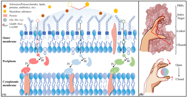
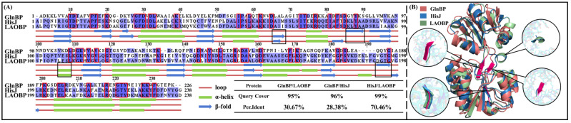
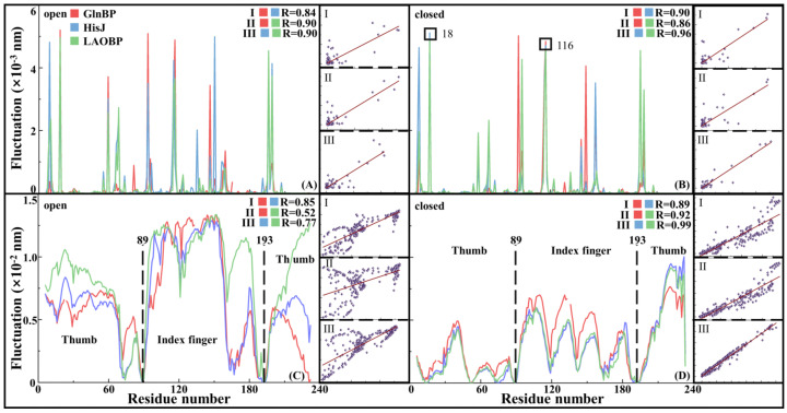
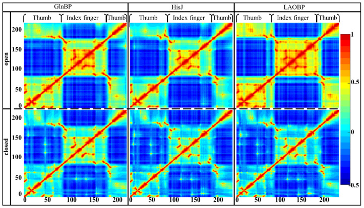
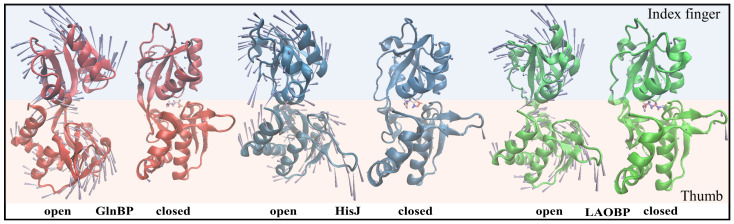
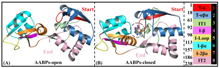
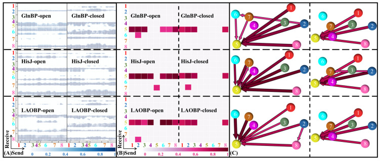
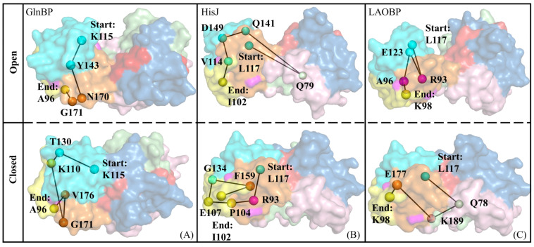
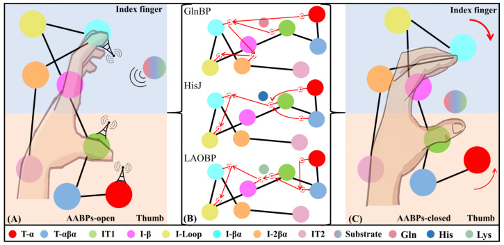
Amino acid binding proteins (AABPs) undergo significant conformational closure in the periplasmic space of Gram-negative bacteria, tightly binding specific amino acid substrates and then initiating transmembrane transport of nutrients. Nevertheless, the possible closure mechanisms after substrate binding, especially long-range signaling, remain unknown. Taking three typical AABPs-glutamine binding protein (GlnBP), histidine binding protein (HisJ) and lysine/arginine/ornithine binding protein (LAOBP) in Escherichia coli (E. coli)-as research subjects, a series of theoretical studies including sequence alignment, Gaussian network model (GNM), anisotropic network model (ANM), conventional molecular dynamics (cMD) and neural relational inference molecular dynamics (NRI-MD) simulations were carried out. Sequence alignment showed that GlnBP, HisJ and LAOBP have high structural similarity. According to the results of the GNM and ANM, AABPs' Index Finger and Thumb domains exhibit closed motion tendencies that contribute to substrate capture and stable binding. Based on cMD trajectories, the Index Finger domain, especially the I-Loop region, exhibits high molecular flexibility, with residues 11 and 117 both being potentially key residues for receptor-ligand recognition and initiation of receptor allostery. Finally, the signaling pathway of AABPs' conformational closure was revealed by NRI-MD training and trajectory reconstruction. This work not only provides a complete picture of AABPs' recognition mechanism and possible conformational closure, but also aids subsequent structure-based design of small-molecule oncology drugs.

Keywords: Gaussian network model; allosteric mechanism; amino acid binding protein; anisotropic network model; neural relational inference molecular dynamics.


Conflict of interest statement

The authors declare no conflict of interest, financial or otherwise.

Figures

Figure 1
Figure 1
Transmembrane transport of nutrients (A) and allostery of periplasmic binding protein (B) in Gram-negative bacteria. Nutrients pass through the outer membrane of Gram-negative bacteria (I), enter the periplasmic space through channel proteins(II), are recognized by periplasmic binding proteins (III), and are transferred to the inner membrane (IV).
Figure 2
Figure 2
Sequence comparison and secondary structure comparison between the GlnBP, HisJ and LAOBP systems (A), red boxes indicate highly conserved residues in the three systems as well as comparison of their 3D structures. Light purple indicates that residues at the same location differ between protein sequences; Conversely, dark purple means that the residues are identical. (B). Black squares and round boxes represent regions with large secondary and 3D structural differences between GlnBP and other proteins, respectively. (Query cover: the degree of match between multiple sequences; Per.Ident: the consistency of the sequence alignment).
Figure 3
Figure 3
Comparison of fast (A,B) and slow (C,D) motion modes for the open (A,C) and closed (B,D) states.
Figure 4
Figure 4
Motion correlation calculated by GNM for the GlnBP/HisJ/LAOBP monomers and substrate-bound complexes. Correlation values range from −0.5 to 1, where warm and cool colors represent the positive and negative correlations, respectively.
Figure 5
Figure 5
Functional motion patterns for the open and closed states of GlnBP/HisJ/LAOBP via ANM analysis. The length and direction of cone are used to characterize motion amplitude and direction, respectively. Light blue and light orange for Index Finger and Thumb region, respectively. Red, blue, and green respectively represent GlnBP, HisJ and LAOBP.
Figure 6
Figure 6
Convergence analysis of cMD trajectories for the GlnBP-open/GlnBP-closed, HisJ-open/HisJ-closed and LAOBP-open/LAOBP-closed systems. (A) RMSD distribution versus simulation time; (B) RMSF distribution at residual level; (C) The correlations of RMSF values between the closed and open states.
Figure 7
Figure 7
Domain partitions of the AABPs’ open (A) and closed (B) states, including eight parts, T-α, T-αβα, IT1, I-β, I-Loop, I-βα, I-2βα and IT2. For the convenience of subsequent domain interaction analysis, the last column of numbers 1–8 is used to represent the eight parts mentioned above.
Figure 8
Figure 8
Distribution of residue interactions among learned edges (A)/domains (B) in six AABPs cMD simulations. (C) The interaction graph is mapped from the learned edges, where node thickness and arrow direction respectively indicate interaction strength and the direction of signal transmission. 1. T-α; 2. T-αβα; 3. IT1; 4. I-β; 5. I -Loop; 6. I-βα; 7. I-2βα; 8. IT2.
Figure 9
Figure 9
The most likely pathways mediating inter-domain allosteric communications in AABPs. GlnBP (A), HisJ (B), and LAOBP (C) mediate interdomain signal communication pathways in the open and closed states.
Figure 10
Figure 10
Possible allosteric mechanisms of AABPs. (A) The initial state of the AABPs’ open and closed substrate systems; (B) signaling between protein domains; (C) the closure of active pocket in the AABPs’ closed states.

Similar articles

References

    1. Bladen H.A., Mergenhagen S.E. Ultrastructure of Veillonella and morphological correlation of an outer membrane with particles associated with endotoxic activity. J. Bacteriol. 1964;88:1482–1492. doi: 10.1128/jb.88.5.1482-1492.1964. - DOI - PMC - PubMed
    1. Ames G.F.-L. Bacterial periplasmic transport systems: Structure, mechanism, and evolution. Annu. Rev. Biochem. 1986;55:397–425. doi: 10.1146/annurev.bi.55.070186.002145. - DOI - PubMed
    1. Brown K. Formation of aromatic amino acid pools in Escherichia coli K-12. J. Bacteriol. 1970;104:177–188. doi: 10.1128/jb.104.1.177-188.1970. - DOI - PMC - PubMed
    1. Gerstein M., Lesk A.M., Chothia C. Structural mechanisms for domain movements in proteins. Biochemistry. 1994;33:6739–6749. doi: 10.1021/bi00188a001. - DOI - PubMed
    1. Neu H.C., Heppel L.A. The release of enzymes from Escherichia coli by osmotic shock and during the formation of spheroplasts. J. Biolog. Chem. 1965;240:3685–3692. doi: 10.1016/S0021-9258(18)97200-5. - DOI - PubMed

LinkOut - more resources