. 2023 Oct 11;24(20):15096.
doi: 10.3390/ijms242015096.

Comparison of Methods for Quantifying Extracellular Vesicles of Gram-Negative Bacteria

Affiliations

Comparison of Methods for Quantifying Extracellular Vesicles of Gram-Negative Bacteria

Chanel A Mosby et al. Int J Mol Sci. .

Abstract

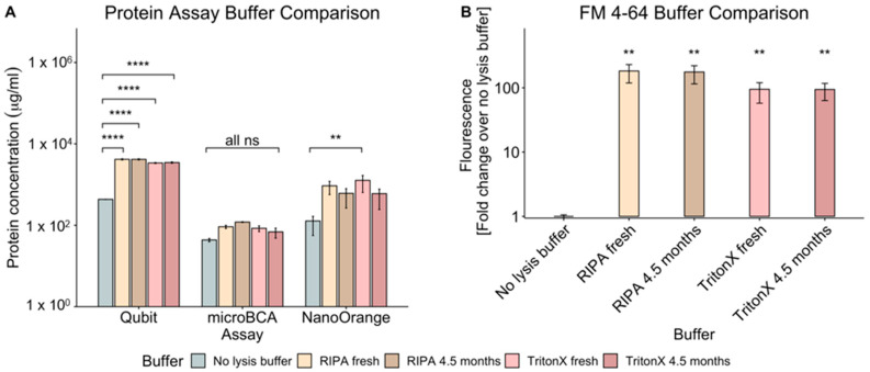
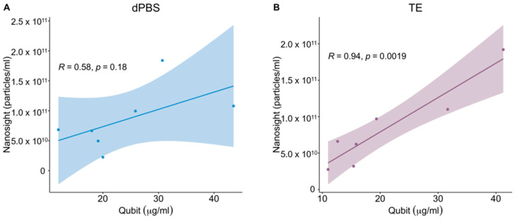
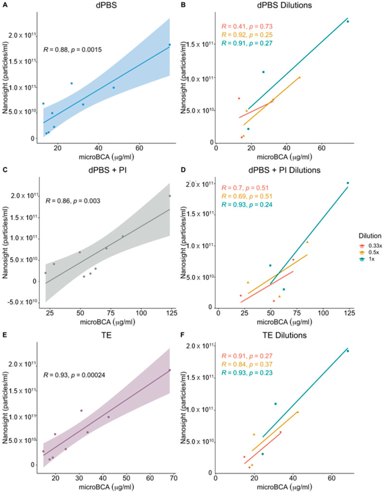
There are a variety of methods employed by laboratories for quantifying extracellular vesicles isolated from bacteria. As a result, the ability to compare results across published studies can lead to questions regarding the suitability of methods and buffers for accurately quantifying these vesicles. Within the literature, there are several common methods for vesicle quantification. These include lipid quantification using the lipophilic dye FM 4-64, protein quantification using microBCA, Qubit, and NanoOrange assays, or direct vesicle enumeration using nanoparticle tracking analysis. In addition, various diluents and lysis buffers are also used to resuspend and treat vesicles. In this study, we directly compared the quantification of a bacterial outer membrane vesicle using several commonly used methods. We also tested the impact of different buffers, buffer age, lysis method, and vesicle diluent on vesicle quantification. The results showed that buffer age had no significant effect on vesicle quantification, but the lysis method impacted the reliability of measurements using Qubit and NanoOrange. The microBCA assay displayed the least variability in protein concentration values and was the most consistent, regardless of the buffer or diluent used. MicroBCA also demonstrated the strongest correlation to the NTA-determined particle number across a range of vesicle concentrations. Overall, these results indicate that with appropriate diluent and buffer choice, microBCA vs. NTA standard curves could be generated and the microBCA assay used to estimate the particle number when NTA instrumentation is not readily available.

Keywords: FM 4-64; NTA; NanoOrange; Qubit; bacterial extracellular vesicle; microBCA; nanoparticle tracking analysis; outer membrane vesicles; vesicle quantification.


Conflict of interest statement

The authors declare no conflict of interest.

Figures

Figure 1
Figure 1
Impact of lysis buffer type and age on OMV protein and lipid quantification. OMVs isolated from E. cloacae were resuspended in dPBS with PI and then treated with lysis buffers of varying age and composition before analysis with protein assays (Qubit, microBCA, or NanoOrange) or with the lipophilic dye, FM 4-64. (A) Comparison of protein concentration readings of OMVs using Qubit, microBCA, or NanoOrange assays and with a no-lysis-buffer control, RIPA, or TritonX of 4.5 months of age or fresh. The statistical significance was found using Tukey’s pairwise comparison tests after a two-way ANOVA. (B) Comparison of fresh or 4.5-month-old RIPA or Triton-X buffers and a no-lysis-buffer control on the emitted fluorescence on OMVs incubated with FM 4-64 lipophilic dye. t-tests were used for pairwise comparisons to determine statistical significance. For ease of visualization, the asterisks show the result of different buffers against the no lysis buffer control for the FM 4-64 graph. Asterisks represent adjusted p values: **, p  ≤  0.01; ****, p  ≤  0.0001. ns = not significant Error bars display SEM, n  =  4.
Figure 2
Figure 2
Impact of diluent on OMV quantification. (A) OMVs isolated from E. cloacae were resuspended in different diluents (dPBS, dPBS + protease inhibitor (PI), or TE buffer) prior to protein quantification via the Qubit or microBCA assays as well as particle count on the Nanosight. Protein quantification values in μg/mL for the Qubit and microBCA assays can be read on the left y-axis, while the particle counts in particles/mL can be found on the right y-axis for Nanosight readings. (B) The same diluents were run alone without any OMVs for the two protein assays (Qubit and microBCA). Statistical significance was found using Tukey’s pairwise comparison tests after a two-way ANOVA. Asterisks represent adjusted p values: *, p  ≤  0.05; **, p  ≤  0.01; ***, p  ≤  0.001. Error bars display SEM, n  =  3.
Figure 3
Figure 3
Correlation between NTA and Qubit measurement of OMVs in differing buffers. OMVs isolated from E. cloacae were resuspended in either (A) dPBS or (B) TE. The OMVs were then analyzed using Qubit for protein quantification and with NTA for particle count. A linear regression line and 95% confidence interval are displayed on the plots as well as the Pearson’s correlation coefficient. n = 3 biological replicates (2 technical replicates for each experiment).
Figure 4
Figure 4
Correlation between NTA and microBCA measurement of OMV samples in differing buffers. OMVs isolated from E. cloacae were resuspended in (A) dPBS, (C) dPBS + PI, or (E) TE before serial dilution for 1:1, 1:2, and 1:3 dilutions with the respective diluents, (B,D,F). The OMVs were then analyzed using microBCA for protein quantification and with NTA for particle count. A linear regression line and 95% confidence interval are displayed on the plots as well as the Pearson’s correlation coefficient. n = 3 biological replicates (3 technical replicates for each experiment).

Similar articles

Cited by

References

    1. Toyofuku M., Nomura N., Eberl L. Types and origins of bacterial membrane vesicles. Nat. Rev. Microbiol. 2019;17:13–24. doi: 10.1038/s41579-018-0112-2. - DOI - PubMed
    1. Bhar S., Edelmann M.J., Jones M.K. Characterization and proteomic analysis of outer membrane vesicles from a commensal microbe, Enterobacter cloacae. J. Proteom. 2021;231:103994. doi: 10.1016/j.jprot.2020.103994. - DOI - PubMed
    1. Mosby C.A., Edelmann M.J., Jones M.K. Murine Norovirus Interaction with Enterobacter cloacae Leads to Changes in Membrane Stability and Packaging of Lipid and Metabolite Vesicle Content. Microbiol. Spectr. 2023;11:e0469122. doi: 10.1128/spectrum.04691-22. - DOI - PMC - PubMed
    1. Sartorio M.G., Valguarnera E., Hsu F.F., Feldman M.F. Lipidomics Analysis of Outer Membrane Vesicles and Elucidation of the Inositol Phosphoceramide Biosynthetic Pathway in Bacteroides thetaiotaomicron. Microbiol. Spectr. 2022;10:e0063421. doi: 10.1128/spectrum.00634-21. - DOI - PMC - PubMed
    1. Gerritzen M.J.H., Martens D.E., Wijffels R.H., Stork M. High throughput nanoparticle tracking analysis for monitoring outer membrane vesicle production. J. Extracell. Vesicles. 2017;6:1333883. doi: 10.1080/20013078.2017.1333883. - DOI - PMC - PubMed

MeSH terms

LinkOut - more resources