

Gene Augmentation of CHM Using Non-Viral Episomal Vectors in Models of Choroideremia
- PMID: 37894906
- PMCID: PMC10607001
- DOI: 10.3390/ijms242015225
Gene Augmentation of CHM Using Non-Viral Episomal Vectors in Models of Choroideremia
Abstract
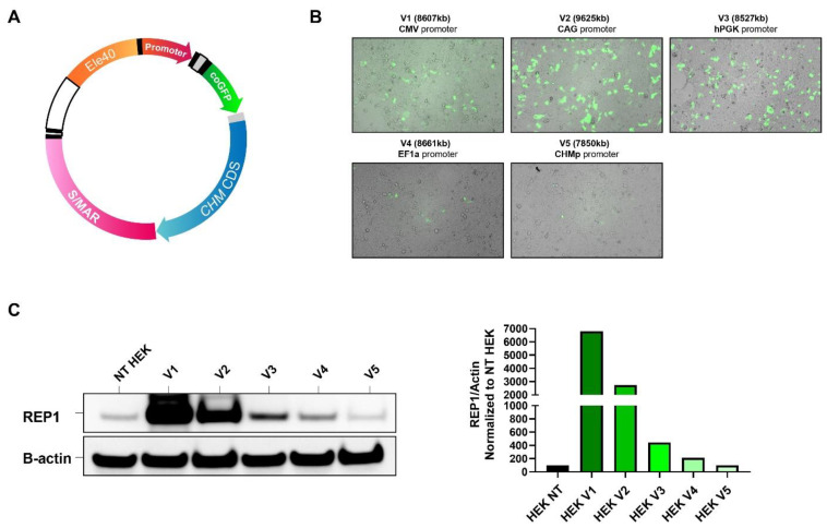
Choroideremia (CHM) is an X-linked chorioretinal dystrophy leading to progressive retinal degeneration that results in blindness by late adulthood. It is caused by mutations in the CHM gene encoding the Rab Escort Protein 1 (REP1), which plays a crucial role in the prenylation of Rab proteins ensuring correct intracellular trafficking. Gene augmentation is a promising therapeutic strategy, and there are several completed and ongoing clinical trials for treating CHM using adeno-associated virus (AAV) vectors. However, late-phase trials have failed to show significant functional improvements and have raised safety concerns about inflammatory events potentially caused by the use of viruses. Therefore, alternative non-viral therapies are desirable. Episomal scaffold/matrix attachment region (S/MAR)-based plasmid vectors were generated containing the human CHM coding sequence, a GFP reporter gene, and ubiquitous promoters (pS/MAR-CHM). The vectors were assessed in two choroideremia disease model systems: (1) CHM patient-derived fibroblasts and (2) chmru848 zebrafish, using Western blotting to detect REP1 protein expression and in vitro prenylation assays to assess the rescue of prenylation function. Retinal immunohistochemistry was used to investigate vector expression and photoreceptor morphology in injected zebrafish retinas. The pS/MAR-CHM vectors generated persistent REP1 expression in CHM patient fibroblasts and showed a significant rescue of prenylation function by 75%, indicating correction of the underlying biochemical defect associated with CHM. In addition, GFP and human REP1 expression were detected in zebrafish microinjected with the pS/MAR-CHM at the one-cell stage. Injected chmru848 zebrafish showed increased survival, prenylation function, and improved retinal photoreceptor morphology. Non-viral S/MAR vectors show promise as a potential gene-augmentation strategy without the use of immunogenic viral components, which could be applicable to many inherited retinal disease genes.
Keywords: S/MAR; choroideremia; inherited retinal disease; non-viral gene therapy.
Conflict of interest statement
The authors declare no conflict of interest.
Figures


Similar articles
-
CHM/REP1 cDNA delivery by lentiviral vectors provides functional expression of the transgene in the retinal pigment epithelium of choroideremia mice.J Gene Med. 2012 Mar;14(3):158-68. doi: 10.1002/jgm.1652. J Gene Med. 2012. PMID: 22228595
-
Functional rescue of REP1 following treatment with PTC124 and novel derivative PTC-414 in human choroideremia fibroblasts and the nonsense-mediated zebrafish model.Hum Mol Genet. 2016 Aug 15;25(16):3416-3431. doi: 10.1093/hmg/ddw184. Epub 2016 Jun 21. Hum Mol Genet. 2016. PMID: 27329764
-
Choroideremia: molecular mechanisms and therapies.Trends Mol Med. 2022 May;28(5):378-387. doi: 10.1016/j.molmed.2022.02.011. Epub 2022 Mar 24. Trends Mol Med. 2022. PMID: 35341685 Review.
-
Pathogenicity of a novel missense variant associated with choroideremia and its impact on gene replacement therapy.Hum Mol Genet. 2017 Sep 15;26(18):3573-3584. doi: 10.1093/hmg/ddx244. Hum Mol Genet. 2017. PMID: 28911202
-
Adeno-Associated Viral Gene Therapy for Inherited Retinal Disease.Pharm Res. 2019 Jan 7;36(2):34. doi: 10.1007/s11095-018-2564-5. Pharm Res. 2019. PMID: 30617669 Free PMC article. Review.
Cited by
-
A Simple Nonviral Method to Generate Human Induced Pluripotent Stem Cells Using SMAR DNA Vectors.Genes (Basel). 2024 Apr 30;15(5):575. doi: 10.3390/genes15050575. Genes (Basel). 2024. PMID: 38790204 Free PMC article.
-
In Vivo Selection of S/MAR Sequences to Favour AAV Episomal Maintenance in Dividing Cells.Int J Mol Sci. 2024 Nov 27;25(23):12734. doi: 10.3390/ijms252312734. Int J Mol Sci. 2024. PMID: 39684442 Free PMC article.
-
Updates on protein-prenylation and associated inherited retinopathies.Front Ophthalmol (Lausanne). 2024 Jul 4;4:1410874. doi: 10.3389/fopht.2024.1410874. eCollection 2024. Front Ophthalmol (Lausanne). 2024. PMID: 39026984 Free PMC article. Review.
-
Oxidative Stress, Inflammation and Altered Glucose Metabolism Contribute to the Retinal Phenotype in the Choroideremia Zebrafish.Antioxidants (Basel). 2024 Dec 23;13(12):1587. doi: 10.3390/antiox13121587. Antioxidants (Basel). 2024. PMID: 39765914 Free PMC article.
References
MeSH terms
Substances
Grants and funding
LinkOut - more resources
Full Text Sources
Molecular Biology Databases
Research Materials
