Review
. 2023 Oct 16;24(20):15234.
doi: 10.3390/ijms242015234.

The Role of the Oxidative State and Innate Immunity Mediated by TLR7 and TLR9 in Lupus Nephritis

Affiliations
Review

The Role of the Oxidative State and Innate Immunity Mediated by TLR7 and TLR9 in Lupus Nephritis

Raquel Echavarria et al. Int J Mol Sci. .

Abstract

Lupus nephritis (LN) is a severe complication of systemic lupus erythematosus (SLE) and is considered one of the leading causes of mortality. Multiple immunological pathways are involved in the pathogenesis of SLE, which makes it imperative to deepen our knowledge about this disease's immune-pathological complexity and explore new therapeutic targets. Since an altered redox state contributes to immune system dysregulation, this document briefly addresses the roles of oxidative stress (OS), oxidative DNA damage, antioxidant enzymes, mitochondrial function, and mitophagy in SLE and LN. Although adaptive immunity's participation in the development of autoimmunity is undeniable, increasing data emphasize the importance of innate immunity elements, particularly the Toll-like receptors (TLRs) that recognize nucleic acid ligands, in inflammatory and autoimmune diseases. Here, we discuss the intriguing roles of TLR7 and TLR9 in developing SLE and LN. Also included are the essential characteristics of conventional treatments and some other novel and little-explored alternatives that offer options to improve renal function in LN.

Keywords: DNA damage; antioxidants; lupus nephritis; mitochondrial function; mitophagy; oxidative stress; systemic lupus erythematosus.


Conflict of interest statement

The authors declare no conflict of interest.

Figures

Figure 1
Figure 1
Factors in SLE development. Microvascular inflammation with the development of autoantibodies, pathogenic immune complexes, and autoreactive T cell activation against ubiquitous nuclear antigens are characteristic of SLE. Oxidative stress (OS), antioxidant capacity, DNA damage and repair, mitochondrial function, mitophagy, and innate immunity mechanisms mediated by the toll-like receptors TLR7 and TLR9 influence the induction of systemic autoimmunity and tissue damage.
Figure 2
Figure 2
Oxidative stress, neutrophils, and NETs in APS and SLE. Neutrophils’ immune functions, including NETosis, are primarily mediated by ROS. Neutrophil–platelet interactions augment ROS production and ease NET release. Autoantibodies against phospholipids and DNA are typical of APS and SLE, and components released by NETs often contribute to the antigenic load.
Figure 3
Figure 3
LN and its damage mechanism. Glomerular damage in LN features endothelial injury, glomerular membrane damage, and podocytes dysfunction mediated by autoantibodies, immune complexes, complement system activation, inflammation, autophagy, NETs, and redox imbalances. In patients with LN, a robust self-renewal and repair system of the glomerular filtration membrane results in instability regarding the degrees of renal injury and proteinuria. In early LN, it is possible to repair pathological damage to the filtration membrane caused by complement proteins and cytokines through autoregulation. However, tissue damage cannot be repaired as the disease progresses, leading to severe proteinuria, increased risk of cardiovascular disease, and high mortality.
Figure 4
Figure 4
Influence of OS, DNA damage, antioxidants, mitochondrial function and mitophagy on the development of LN. Oxidation leads to organic damage characteristic of SLE, especially in LN. Increases in LPO due to OS, limited antioxidant defense, and low NO availability contribute to the injury observed in LN. The balance between oxidation and DNA repair is altered in patients with LN, accumulating the marker of oxidative DNA damage (8-OHdG), genomic instability, and cellular dysfunction. Additionally, metabolic abnormalities influence the nature and state of activation of the kidney’s immune cell infiltrate, highlighting the importance of mitochondrial function in developing diseases such as LN through Danger-Associated Molecular Patterns (DAMPs), TLR activation, and the production of autoantibodies. Finally, aberrant or defective mitophagy is central to the pathology of LN as it facilitates the release of mitochondrial antigens, promotes their processing by major histocompatibility complex (MHC) molecules, and contributes to mitochondrial dysfunction.
Figure 5
Figure 5
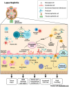
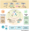
Participation of innate immunity and TLRs in the development of LN. Some TLRs, such as TLR3, TLR7, and TLR9, are in the endosomal compartment, where they detect endogenous ligands such as RNA, DNA, hypo-methylated self-DNA within immune complexes, chromatin released from NETs, oxidized mitochondrial nucleoids, and other chromatin formats. Dendritic and B cells can process antigens and present antigens to T cells. In LN, TLR7 and TLR9 allow the persistent activation of dendritic and B cells by autoantigens, thereby promoting autoantibody production, systemic autoimmunity, and glomerulonephritis mediated by immune complexes. In addition, the nucleic acid component of the immune complexes also activates intra renal inflammation via TLR7 and TLR9 in intra renal macrophages, resulting in the activation of glomerular endothelium and mesangial cells that are characteristic of LN.
Figure 6
Figure 6
Therapeutic objectives and management of LN in SLE. The goal of therapy for LN was established with a reduction in proteinuria by ≥25% with a stable glomerular filtration rate (GFR, ±10% of baseline) in the first three months after the start of treatment, a reduction in ≥50% proteinuria at six months and a rate of proteinuria <0.5–0.7 g/24 h at 12–24 months (all with stable GFR). Immunosuppressive therapies, immunomodulatory drugs, and vitamin D are currently used to control disease activity, prevent relapses, reduce the use of glucocorticoids, and lessen inflammation and fibrosis. Voclosporin, tacrolimus, rituximab, belimumab, and cyclophosphamide limit damage to target organs, including the kidney, in patients with SLE. However, new therapies focused on reducing the effect of endogenous nucleic acids mediated by TLR7 and TLR9 present promising results in preclinical studies, suggesting that their translation to the clinic could significantly benefit the quality of life, survival, and management of comorbidities in patients with SLE and LN.

References

    1. Smallwood M.J., Nissim A., Knight A.R., Whiteman M., Haigh R., Winyard P.G. Oxidative stress in autoimmune rheumatic diseases. Free. Radic. Biol. Med. 2018;125:3–14. doi: 10.1016/j.freeradbiomed.2018.05.086. - DOI - PubMed
    1. Ball E.M., Bell A.L. Lupus arthritis—Do we have a clinically useful classification? Rheumatology. 2012;51:771–779. doi: 10.1093/rheumatology/ker381. - DOI - PubMed
    1. Kyttaris V.C. Systemic Lupus Erythematosus: From Genes to Organ Damage. Methods Mol. Biol. 2010;662:265–283. doi: 10.1007/978-1-60761-800-3_13. - DOI - PMC - PubMed
    1. Fayyaz A., Igoe A., Kurien B.T., Danda D., James J.A., Stafford H.A., Scofield R.H. Haematological manifestations of lupus. Lupus Sci. Med. 2015;2:e000078. doi: 10.1136/lupus-2014-000078. - DOI - PMC - PubMed
    1. Parodis I., Tamirou F., A Houssiau F. Prediction of prognosis and renal outcome in lupus nephritis. Lupus Sci. Med. 2020;7:e000389. doi: 10.1136/lupus-2020-000389. - DOI - PMC - PubMed